Journal List > J Cerebrovasc Endovasc Neurosurg > v.22(2) > 1144890

Lee, Park, You, Choi, Park, Kim, Kim, Yee, Kim, and Kim: Risk factors of postoperative remote intracerebral hemorrhage after craniotomy for ruptured cerebral aneurysms

Abstract

Objective

We aimed to identify the relation between perioperative cerebrospinal fluid (CSF) drain through lumbar drainage (LD) and development of postoperative (POP) remote intracerebral hemorrhage (rICH) in craniotomy to treat ruptured intracranial aneurysms.

Methods

We retrospectively reviewed consecutive patients who underwent craniotomy for ruptured cerebral aneurysms at the authors’ institution between 1998 and 2004. We subsequently compared the incidence and characteristics of POP rICH between the patients who had a perioperative LD and those who did not. All statistical analyses were conducted using the software package SPSS 19.0 (SPSS Inc., Chicago, IL, USA). A p value of <0.05 was considered statistically significant.

Results

We enrolled 688 patients, of which 80 patients (11.6%) received perioperative LD, and 608 did not. LD and non-LD groups were comparable because although clinical characteristics of the two groups were significantly different considering history of hypertension, timing of surgery, and closed system negative pressure suction drain (SD) placement, none of these three variables was an independent risk factor associated with POP rICH in multivariate analysis. POP rICH incidence was significantly higher in the LD goup (12.5%) than non-LD group (0.8%) (p=0.000) in univariate analysis. LD placement was the only independent risk factor associated with the development of rICH in multivariate logistic regression analysis.

Conclusions

POP rICH incidence was significantly higher in patients who were managed with perioperative LD than in those who did not. LD insertion in craniotomy for ruptured intracranial aneurysm, should be closely monitored to address the occurrence of POP rICH.

INTRODUCTION

Draining the cerebrospinal fluid (CSF) via lumbar drainage (LD) is frequently performed in numerous neurosurgical settings and to clip the ruptured intracranial aneurysm for primarily obtaining appropriate brain relaxation, following which minimizing brain retraction, avoiding bloody CSF from obscuring the operative field. However, many adverse events from CSF drain have been established and reported. These include the Arnold-Chiari syndrome,1)3)24) intracerebral hemorrhage (ICH),4)26) intracranial hypotension,24)28) local and central nervous system infections,3) peripheral nerve injury,3) part of posterior reversible encephalopathy syndrome (PRES),17)32) hydrocephalus (after subarachnoid hemorrhage[SAH]),14) intraspinal epidermoid tumor,23) brain swelling,11)32)34)37) transtentorial brain herniation,24) and sudden death.11)32)
Postoperative (POP) ICH occurrence is one of the most serious complications in almost all neurosurgical procedures. Remote cerebellar hemorrhage was first introduced in 1977 by Yasargil and Yonekawa36) following a revascularization surgery. Additionally among the several types of POP ICH, a remote ICH (rICH) that was responsible for complicating the cerebral aneurysm surgery, was first reported in 1983 by Waga et al.35) Following which, a number of such cases have been published by different authors.2)13)18)20)25)27)31)33) Although numerous hypotheses2)4)5)8)9)12)13)2931)38) attempting to explain the formation of rICH including over-drainage of CSF have been suggested by case reports or literature reviews, none has been able to clearly demonstrate the associated definite underlying pathogenesis.
Though rICH is relatively rare complication, it may result in significant morbidity and mortality.30)31)
Therefore, the immediate goal of this study was to evaluate and demonstrate the most approximate rICH pathogenesis by investigating the relationship between LD and POP rICH specially in the craniotomy for ruptured intracranial aneurysms.

MATERIALS AND METHODS

We retrospectively reviewed the medical records and radiographies of consecutive patients who underwent craniotomy for ruptured cerebral aneurysms at our institute from 1998 to 2004. We excluded cases of unruptured cerebral aneurysms, ruptured aneurysms that were treated with both clipping and extraventricular drainage, endovascular embolization or operated using various skull base approaches including orbitozygomatic, lateral suboccipital, or different types of combined skull base approaches.
We identified age, sex, initial Hunt-Hess and Fischer grades, location of ruptured aneurysm, coagulation profiles, timing of surgery (early or delayed), presence or absence of hypertensive-hypervolemic (H-H) therapeutic intervention, history of hypertension, and LD and closed system negative pressure suction drain (SD) usage in each of the patients included in this study. H-H therapy was conducted in compliance with an internationally recommended protocol.15) Target levels of intracranial pressure, blood pressure (BP), and central venous pressure were also determined by the universal standards,16) along with the POP transcranial Doppler result, and neurological signs and symptoms of the patient. POP rICH diagnosis was confirmed by using either the head computed tomography (CT) or magnetic resonance image (MRI). We then compared the incidence and characteristics of POP rICH between patients who had a perioperative LD and patients who did not, and analyzed variety of potential risk factors of POP rICH via univariate analysis and multivariate logistic regression analysis.
LD duration and the volume of CSF drain were also investigated in patients who had a perioperative LD. Final diagnosis of spontaneous SAH, ruptured cerebral aneurysm, microsurgical techniques, LD and SD management, and perioperative care were performed based on universal standards. The neurosurgeon conducting the procedure solely decided whether LD and suction drainage (SD) needed to be inserted or not, based on each patient’s status including surgical field, initial radiographic findings and the surgeon’s experience. During the study period, we exclusively used a lumbar kit from Integra-NeurosciencesTM (Plainsboro, NJ, USA) to drain of CSF, and Hemovac® (Zimmer Surgical, Inc., Dover, OH, USA) as a SD to drain epidural and/or subgaleal oozing blood and exudates.
For the statistical analysis, commercially available software, statistical package for the social science SPSS 19.0 (SPSS Inc., Chicago, IL, USA) was used. Demographic and clinical data with regard to the LD and rICH were compared using the t-test, [chi]2 test, ANOVA as appropriate. We also conducted multivariate logistic regression analyses upon possible clinical variables. All variables except age were considered as categorical. A p value of <0.05 was considered as statistically significant.

RESULTS

Between 1998 and 2004, total 688 patients were deemed suitable based on inclusion criteria. Of these subjects, 80 patients had perioperative LD. The patients with LD have higher rate of SD placement compared to non-LD group and it was statistically significant (Table 1).

Incidence of POP rICH

LD and POP rICH

Among 688 patients, 41 showed several kinds of POP hemorrhages on POP head CT. Of these patients, 17 had an LD and 24 did not. In the LD group (80 patients [Table 1]), 6 demonstrated tiny ICH in the operative field, and 1 had an intraventricular hemorrhage, and 10 presented POP rICH. Alternatively, in the non-LD group (608 patients [Table 1]), 8 had a minor amount (thinner than the skull table) of epidural hematoma (EDH) under the skull flap, 10 showed small amounts of ICH in the operative site, 1 presented thin EDH on opposite side of craniotomy, and 5 demonstrated POP rICH. All POP EDHs and operative site POP ICHs were not amenable to surgical removal. POP rICH incidence was 12.5% in the LD and 0.8% in the non-LD group. Additionally, there was a statistically significant difference in POP rICH incidence between these two groups (p=0.000) (Table 2).

SD and POP rICH

From the total 688 patients, 355 had an SD. Of these 355, 15 demonstrated POP rICH. Namely, all 15 rICH patients in this study had an SD. Subsequently, the rate of rICH was 4.2% (15/355) in patients with an SD (Table 2).

Simultaneous insertion of an LD and an SD, and POP rICH

Among the entire 688 patients, 77 were treated by using an LD and an SD simultaneously (Table 1). Of these 77 patients, POP rICH developed in 10, then the resulting POP rICH rate was 13.0% (10/77). There was statistically significant difference in of POP rICH incidence between simultaneous use of LD and SD group (10/77, 13%) and non-simultaneous group (5/611, 0.008%) (p=0.000) (Table 1, 2).

Amount of CSF drainage via LD and POP rICH

Mean drained CSF through LD per day was below 50 mL/day in 41 patients, between 50–100 mL/day in 23, and above 100 mL/day in 16. The POP rICHs were developed in 4 patients of <50 mL group (9.8%), 6 of 50–100 mL (26.1%), and none from the >100 mL (0%), in each (Table 3).

Duration of LD and POP rICH

Number of patients that showed POP rICH in relation to LD duration are summarized in Table 3. There was significant difference in rICH frequency in association with the duration of LD (p=0.026) (Table 3).

Prior history of hypertension and POP rICH

Among 688 patients, 238 had a medical history of hypertension and 450 did not (Table 1). Considering 238, 5 patients developed POP rICH and of the remaining 450, 10 patients resulted in POP rICH. Then, the incidences of rICH were 2.1% and 2.2%, respectively (Table 1, 4).

H-H therapy and POP rICH

Among the included 688 patients, 468 underwent prophylactic or therapeutic H-H therapy, irrespective of its duration. Among these 468 patients, 13 showed POP rICH. The rates of rICH were 2.8% and 0.9% in the group of patients who had H-H therapy and who did not, respectively (Table 1, 4).

Multivariate analysis

Higher volume of the subarachnoid blood clot (or Fisher grade) indicated an equally greater risk of developing POP rICH, and this correlation was statistically significant (p=0.046) (Table 4). However, the Fisher grade was not an independent risk factor in the multivariate logistic regression analysis (Table 5). LD was the only independent risk factor associated with the formation of POP rICH in the multivariate analysis. Additionally, SD also was not an independent risk factor (Table 5).

Location of POP rICH

In a group of 80 patients who underwent LD, 9 exhibited POP rICH in the cerebellum (3, ipsilateral to the operative site; 4, contralateral; 2, bilateral) and 1 developed POP rICH at basal ganglia contralateral to the operative side (Table 2). In the group that did not undergo LD, 4 of 608 patients developed POP rICH in the cerebellum (1, ipsilateral to the craniotomy; 1, contralateral; 2, bilateral) and 1 presented POP rICH at the contralateral basal ganglia (Table 2). Furthermore, the location of POP rICH was neither consistent nor statistically significant with regard to the use of LD (p=0.591) (Table 2).

Management and outcome of POP rICH

Table 2 demonstrates POP rICH management methods and their results in 10 of 80 patients who had LD and in 5 out of 608 patients who did not. The temporary neurological deficits were dizziness, sense of imbalance, hemiparesis, tremor, and confusion. And the permanent neurological deficit was hemiparesis. The overall mortality associated with POP rICH was 40.0% (6/15) in this study (Table 2).

DISCUSSION

Although LD has frequently been used in a variety of conditions in neurosurgery, it has been clearly understood that the over-drainage of CSF irrespective of the routes, may induce a plethora of complications4)8)14)21)24)28)30)31) with the rICH being considered the most critical POP event. Fortunately, it has been rare adverse event.12)30)31) Numerous authors and researchers have described the incidence and suggested the pathogenesis of POP rICH in their studies.5)810)13)18)20)22)25)27)29)33)35)36)38) During their descriptions, several causative factors have been proposed, however, due to the low incidence of rICH and the numerous limitations of their reports, the definite pathogenesis and related risk factors associated with POP rICH remain unclear. Subsequently, we aimed to identify if the CSF loss via LD influenced the occurrence of POP rICH, and other risk factors that contributed to the development of POP rICH if any.

Incidence of POP rICH

Although the characteristics of two groups (LD group and non-LD group) in this study were significantly different in the frequency of history of hypertension, timing of surgery and placement of SD, these variables were not independent risk factors in the multivariate analysis (Table 5). Then, we would decide to directly compare the LD group to the non-LD group. Subsequently, POP rICH incidence was significantly higher in the LD group (12.5%) than non-LD group (0.8%) (p=0.000) (Table 2). And the overall incidence of POP rICH was 2.2% (Table 2).
In a review of 4992 intracranial procedures, Kalfas and Little identified that rICH accounted for 18% (7/40) of all POP intracranial hemorrhages.12) Their POP rICH rate was approximately 0.1%. Waga et al.35) in 1983 reported 4 cases of POP rICH after neurosurgical procedures such as microsurgical repair of ruptured cerebral aneurysm in patients with recent SAH. However, they did not specify rICH incidence. In 2001, Friedman et al.10) published that POP rICH occurred in approximately 0.6% of all supratentorial craniotomies and 2.8% of all craniotomies to treat unruptured cerebral aneurysm. In 1996, Lee et al.20) reported that the incidence of POP rICH in 720 ruptured intracranial aneurysms was below 1%, which was lower than our result (2.2%). They claimed that the sudden elevation of BP10)12)35) during the remission period of the cerebral vasospasm was the root cause of the rICH.
POP rICH incidence (2.2%) according to our study is relatively lower than that after direct surgical repair of ruptured and/or unruptured cerebral aneurysms as reported by others.2)10) It is possible that this minor difference may provide an extremely important clue, or at least an incomplete clue, regarding POP rICH pathogenesis. It suggested that although CSF loss was exactly the same amount and/or velocity both during surgery and POP period, the displacement or shrinkage of cerebrum and cerebellum was less in patients with recent SAH than in those with unruptured aneurysm without SAH due to the presence of an irritated and edematous cerebrum and cerebellum by the subarachnoid blood.

Location of POP rICH

Studies have previously demonstrated that certain cases of POP rICH following intracranial aneurysm surgery occurred in the cerebral hemisphere contralateral to the operative site, while other reports suggested that POP rICH typically developed in the contralateral cerebellar hemisphere.2)18)20)31) In 2001, Friedman et al.10) suggested that the typical location of POP rICH was the superior cerebellar folia. A systematic review of literatures on the remote cerebellar hemorrhage after supratentorial procedures, was published by Sturiale et al.31) in 2016, reported that even though their report was confined to the cerebellar hemorrhage, they noted that 55.1% of cerebellar rICH were bilateral, and 21.9% were contralateral to the supratentorial surgery. In our study, 13 out of 15 patients with POP rICH appeared in the cerebellum (Table 2). Then, we assumed that although POP rICH characteristically occurred more often in the cerebellum than in the cerebrum, it did not have any such preference in the laterality.

Pathogenesis of POP rICH

As described before, the overall POP rICH incidence was 2.2% (15/688) in our study and among these POP rICH cases, the incidences were 12.5% (10/80) and 0.8% (5/608) in the patients who had LD and who did not, respectively (Table 2). This result implicated that the loss of CSF through LD might be a crucial causative factor associated with the generation of POP rICH. Moreover, among our study population of 688, 77 patients had an LD and an SD simultaneously. Of these 77, 10 patients presented POP rICH. Subsequently, POP rICH incidence for this group (LD+SD) was 13.0%. Numerically, simultaneous application of an LD and an SD was a more impactive risk factor associated with the formation of POP rICH than using LD or SD alone in this study (Table 1, 2). Typically, the drained liquid material through SD was composed of blood components and CSF. However, it was not possible to isolate CSF from blood components in the clinical practice. Accordingly, we could not quantify the drained CSF via SD. We hypothesized that more rapid and/or larger amount of CSF loss per given time period through LD and SD might happen in this group (LD+SD). If it was so, it might have had a considerable impact on the occurrence of POP rICH. This observation might prove to be critical as conclusion of present study.
Although the initial Fisher grade of patient was not an independent risk factor associated with the formation of POP rICH in the multivariate analysis, we noted that the initial Fisher grade demonstrated a statistically significant correlation with the risk of developing POP rICH in the univariate analysis (Table 4, 5). We presumed the background of this result was that the degree of dysfunction in brain autoregulation at least partially, depends on the volume of subarachnoid blood clot, and this dysfunction might partially at least, generate POP rICH.
Unexpectedly, the authors failed to verify that POP rICH incidence was directly proportional to the amount and/or duration of CSF drain via LD in spite of these two parameters (amount and duration of CSF drain via LD) showed statistically significant correlations in the frequency of POP rICH (Table 3). It was our understanding that this failure was due to the small number of patients involved in this study and the inherent limitations of our retrospective study. We believe that CSF drain velocity per defined unit time via LD or any route has a greater impact on the development of POP rICH, rather than the total amount and/or duration of CSF drain. Furthermore, the amount and/or duration of CSF drain without POP rICH formation might vary greatly among patients. A large scale, well designed multicenter prospective research effort on this subject should be created in the future.
According to relatively early period of case reports on the POP rICH, fluctuation or sudden elevation of BP during the craniotomy, initial POP period, vasospasm, H-H therapy, autoregulatory impairment, and perioperative coagulopathy might potentially cause POP rICH.4)12)13)20)33)35) We did not explore BP as a variable associated with the genesis of POP rICH since medical records showed that no fluctuation or sudden elevation of BP during the above mentioned intervals or periods despite our meticulous examination of the records of the entire 688 included patients. Here, 2 patients without LD proved to have coagulopathy although they did not present POP rICH. Then, even though the number of coagulopathy cases in present study was too insignificant to reach a certain opinion, we believe that after thoroughly examining relevant literatures along with our collective experiences, association between coagulopathy and POP rICH was not evident despite of a few literatures contradicting the same issue.46)10)12)18)31)33)
Alternatively, the consensus that the position of the patient’s head during craniotomy with microsurgery has a certain degree of influence in the development of POP rICH is relatively vague. Compression of internal jugular vein at the transverse process of the first cervical vertebra and resulting intracranial venous hypertension as a consequence of the patient’s head position during the craniotomy may cause POP rICH.29) Although our standard protocol suggest keeping the head position as neutral as possible during the frontotemporal craniotomy, we could not absolutely exclude the chance of surgical head position of patient as an underlying cause of the POP rICH.
Brain sagging as a basic etiology of POP rICH, was introduced following intraoperative and/or perioperative CSF over-drain in craniotomy.4)21) It might be possible that the cerebellar displacement and sagging4)19)21)26) due to the over-loss of CSF leads to transient stretching and angulation of superiorly directed cerebellar veins draining towards the cephalad direction into the deep venous system. Finally, occlusion of these cerebellar veins sufficiently produces cerebellar rICH and/or venous infarction.4)21)22)
Recently, a few authors34) coined and used a new term that intrinsically and potentially had identical pathogenesis as POP rICH, but showed different clinical phenomena from rICH, the POP ‘pseudohypoxic brain swelling’ that presented as consciousness disturbance, brain stem dysfunction, and generalized seizures after an uneventful craniotomy. These authors proposed a common pathogenesis that the diffuse brain swelling, particularly swelling of the deep brain nuclei such as basal ganglia and thalami on head CT or MRI, was ascribed to the over-drain of CSF through the SD. After their introduction of a new concept, a few neurosurgeons have reported such adverse events.11)32)37) In 2019, Katsevman et al.17) experienced a PRES in a patient and reviewed the related literatures on the PRES.7) They admitted CSF over-drainage (CSF hypovolemia) could be one of the several possible causative pathophysiologic mechanisms associated with the occurrence of PRES. If it is so, it is possible that the pseudohypoxic brain swelling and PRES may have a common etiological factor.
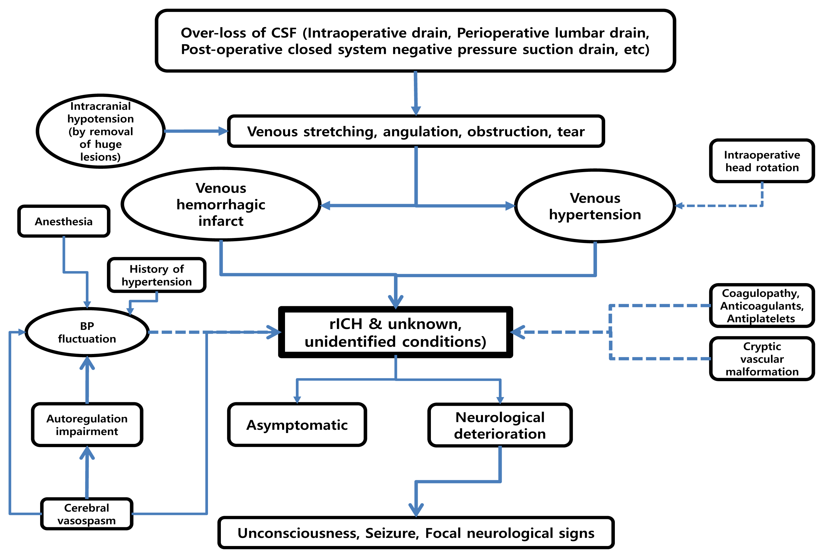
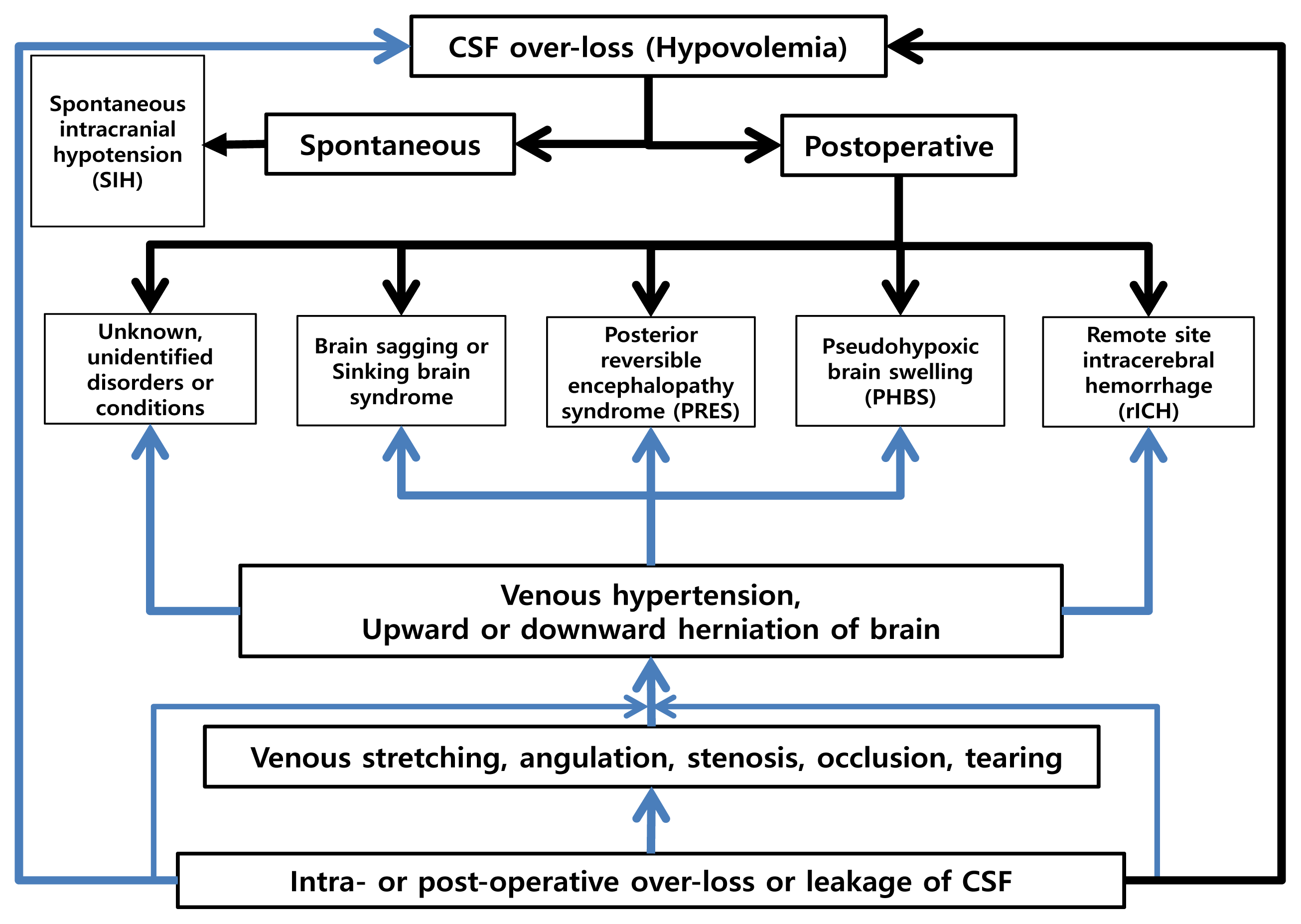
Given the situation we do not have choice but to accept that the unidentified or unknown POP clinical settings including neurosurgical conditions, which can violently disturb homeostasis or normal internal milieu of the brain may occur beyond the known clinical conditions as specified in Fig. 1 and 2, because of the CSF over-loss. Upon further consideration, we understood that the basic and core pathogeneses of POP rICH, brain sag, pseudohypoxic brain swelling and some of PRES might be essentially identical (Fig. 1, 2).26) Additionally according to the authors’ review of the literatures, although a number of potential etiologies might be possible,12)31) the most impactfully, frequently, and continuously suggested key point, was the reduction in intracranial CSF volume (CSF hypovolemia).2)4)810)25)31)38)
Comprehensively, we propose the pathogenesis of POP rICH considering the results of this study and a thorough review of the literatures is shown in Fig. 1 and 2. However, the POP rICH pathogenesis may be more complex than expected.

Limitations of this study

This study has numerous weaknesses and there may be more drawbacks apart from those mentioned below. First, there are several inherent limitations due to the retrospective natures of this study, including an absence of a corresponding control group, missing data that was neither documented nor checked up. Second, we did not subdivide the volume of rICH in the POP rICH patients and the corresponding volume of CSF loss through the LD. However, this two might be associated with each other. Third, we considered and analyzed CSF drain for only the 24 hours (1 day) basis without checking the velocity of CSF drain via LD within more segmented time periods. Fourth, we did not subdivide the patients into age groups which might have an effect on the frequency, appearance, and volume of POP rICH. Finally, we did not consider CSF drain volume via SD, although it was not an independent risk factor associated with the emergence of POP rICH in the multivariate analysis.

CONCLUSIONS

The LD is characteristically used for a variety of neurosurgical procedures including cerebral aneurysm. However, it should be used in only specific and selected conditions, and rapid and/or large amount of CSF drain through LD should be prevented and closely monitored. Additionally, while treating patients with any CSF drain routes, neurosurgeons should be aware of the possibility of POP rICH even after a smooth, uneventful, and successful procedure.

ACKNOWLEDGEMENTS

The authors would like to thank Prof. DP Jang (Hanyang University, Korea) for the contribution of statistical analysis and its interpretation.

Notes

Disclosure

The authors report no conflict of interest concerning the materials or methods used in this study or the findings specified in this paper.

REFERENCES

1. Atkinson JL, Weinshenker BG, Miller GM, Piepgras DG, Mokri B. Acquired Chiari I malformation secondary to spontaneous spinal cerebrospinal fluid leakage and chronic intracranial hypotension syndrome in seven cases. J Neurosurg. 1998; Feb. 88(2):237–42.
crossref
2. Baek IH, Park KY, Lee JW, Huh SK. Remote cerebellar hemorrhage after surgery for an unruptured aneurysm. J Cerebrovasc Endovasc Neruosurg. 2008; Sep. 10(3):454–8.
3. Basauri LT, Concha-Julio E, Selman JM, Cubillos P, Rufs J. Cerebrospinal fluid spinal lumbar drainage: indications, technical tips, and pitfalls. Crit Rev Neurosurg. 1999; Jan. 9(1):21–7.
crossref
4. Borkar SA, Lakshmiprasad G, Sharma BS, Mahapatra AK. Remote site intracranial haemorrhage: a clinical series of five patients with review of literature. Br J Neurosurg. 2013; Dec. 27(6):735–8.
crossref
5. Brisman MH, Bederson JB, Sen CN, Germano IM, Moore F, Post KD. Intracerebral hemorrhage occurring remote from the craniotomy site. Neurosurgery. 1996; Dec. 39(6):1114–1121. discussion 1121–2.
crossref
6. Chang SH, Yang SH, Son BC, Lee SW. Cerebellar hemorrhage after burr hole drainage of supratentorial chronic subdural hematoma. J Korean Neurosurg Soc. 2009; Dec. 46(6):592–5.
crossref
7. Delgado-López PD, Garcés-Pérez G, García-Carrasco J, Alonso-García E, Gómez-Menéndez AI, Martín-Alonso J. Posterior reversible encephalopathy syndrome with status epilepticus following surgery for lumbar stenosis and spondylolisthesis. World Neurosurg. 2018; Aug. 116:309–15.
crossref
8. Doddamani RS, Sawarkar D, Meena RK, Gurjar H, Singh PK, Singh M, et al. Remote cerebellar hemorrhage following surgery for supratentorial lesions. World Neurosurg. 2019; Jun. 126:e351–9.
crossref
9. Friedman JA, Ecker RD, Piepgras DG, Duke DA. Cerebellar hemorrhage after spinal surgery: report of two cases and literature review. Neurosurgery. 2002; Jun. 50(6):1361–3. discussion 1363–4.
crossref
10. Friedman JA, Piepgras DG, Duke DA, McClelland RL, Bechtle PS, Maher CO, et al. Remote cerebellar hemorrhage after supratentorial surgery. Neurosurgery. 2001; Dec. 49(6):1327–40.
crossref
11. Honeybul S. Sudden death after cranioplasty. J Korean Neurosurg Soc. 2016; Mar. 59(2):182–4.
12. Kalfas IH, Little JR. Postoperative hemorrhage: a survey of 4992 intracranial procedures. Neurosurgery. 1988; Sep. 23(3):343–7.
crossref
13. Kang YG, Chung H, Lee SP, Choi KH, Yeo HT, Rhee JK. Remote intracerebral hematoma after supratentorial craniotomy. J Korean Neurosurg Soc. 1996; Sep. 25(9):1910–6.
14. Kasuya H, Shimizu T, Kagawa M. The effect of continuous drainage of cerebrospinal fluid in patients with subarachnoid hemorrhage: a retrospective analysis of 108 patients. Neurosurgery. 1991; Jan. 28(1):56–9.
crossref
15. Kassell NF, Sasaki T, Colohan ART, Nazar G. Cerebral vasospasm following aneurysmal subarachnoid hemorrhage. Stroke. 1985; Jul–Aug. 16(4):562–72.
crossref
16. Kassell NF, Torner JC, Haley EC Jr, Jane JA, Adams HP, Kongable GL, et al. The international cooperative study on the timing of aneurysm surgery. Part 1: Overall management results. J Neurosurg. 1990; Jul. 73(1):18–36.
17. Katsevman GA, Turner RC, Cheyuo C, Rosen CL, Smith MS. Post-partum posterior reversible encephalopathy syndrome requiring decompressive craniectomy: case report and review of the literature. Acta Neurochir (Wien). 2019; Feb. 161(2):217–24.
crossref
18. Kim SH, Lee HK, Moon JK, Kim CH, Choi JH. Remote cerebellar hemorrhage after supratentorial aneurysm surgery; report of 2 cases. J Cerebrovasc Endovasc Neruosurg. 2008; Dec. 10(4):570–4.
19. Komotar RJ, Ransom ER, Mocco J, Zacharia BE, McKhann GM 2nd, Mayer SA, et al. Critical postcraniotomy cerebrospinal fluid hypovolemia: risk factors and outcome analysis. Neurosurgery. 2006; Aug. 59(2):284–90.
20. Lee JW, Yim MB, Lee JC, Son EI, Kim DW, Kim IH. Intracerebral hemorrhage remote from the site of aneurysm surgery. J Korean Neurosurg Soc. 1996; Apr. 25(4):834–41.
21. Liu JKC. Neurologic deterioration due to brain sag after bilateral craniotomy for subdural hematoma evacuation. World Neurosurg. 2018; Jun. 114:90–3.
crossref
22. Mandonnet E, Faivre B, Bresson D, Cornelius J, Guichard JP, Houdart E, et al. Supratentorial craniotomy complicated by an homolateral remote cerebellar hemorrhage and a controlateral perisylvian infarction: case report. Acta Neurochir (Wien). 2010; Jan. 152(1):169–72.
crossref
23. McDonald JV, Klump TE. Intraspinal epidermoid tumors caused by lumbar puncture. Arch Neurol. 1986; Sep. 43(9):936–9.
crossref
24. Motoyama Y, Nakajima T, Takamura Y, Nakazawa T, Wajima D, Takeshima Y, et al. Risk of brain herniation after craniotomy with lumbar spinal drainage: a propensity score analysis. J Neurosurg. 2018; Jun. 1–11.
crossref
25. Papanastassiou V, Kerr R, Adams C. Contralateral cerebellar hemorrhagic infarction after pterional craniotomy: report of five cases and review of the literature. Neurosurgery. 1996; Oct. 39(4):841–51. discussion 851–2.
crossref
26. Park CW. Postoperative cerebrospinal fluid hypovolemia in neurosurgery. J Neurointensive Care. 2018; 1(1):3–6.
crossref
27. Park SJ, Lee MK, Kim DJ. Supratentorial surgery complicated by cerebellar hemorrhage: report of three cases. J Korean Neurosurg Soc. 1997; Jul. 26(7):1011–6.
28. Samadani U, Huang JH, Baranov D, Zager EL, Grady MS. Intracranial hypotension after intraoperative lumbar cerebrospinal fluid drainage. Neurosurgery. 2003; Jan. 52(1):148–51. discussion 151–2.
crossref
29. Seoane E, Rhoton AL Jr. Compression of the internal jugular vein by the transverse process of the atlas as the cause of cerebellar hemorrhage after supratentorial craniotomy. Surg Neurol. 1999; May. 51(5):500–5.
crossref
30. Sturiale CL, Rossetto M, Ermani M, Baro V, Volpin F, Milanese L, et al. Remote cerebellar hemorrhage after spinal procedures (part 2): a systematic review. Neurosurg Rev. 2016; Jul. 39(3):369–76.
crossref
31. Sturiale CL, Rossetto M, Ermani M, Volpin F, Baro V, Milanese L, et al. Remote cerebellar hemorrhage after supratentorial procedures (part 1): a systematic review. Neurosurg Rev. 2016; Oct. 39(4):565–73.
crossref
32. Sviri GE. Massive cerebral swelling immediately after cranioplasty, a fatal and unpredictable complication: report of 4 cases. J Neurosurg. 2015; Nov. 123(5):1188–93.
crossref
33. van Calenbergh F, Goffin J, Plets C. Cerebellar hemorrhage complicating supratentorial craniotomy: report of two cases. Surg Neurol. 1993; Oct. 40(4):336–8.
crossref
34. van Roost D, Thees C, Brenke C, Oppel F, Winkler PA, Schramm J. Pseudohypoxic brain swelling: a newly defined complication after uneventful brain surgery, probably related to suction drainage. Neurosurgery. 2003; Dec. 53(6):1315–26. discussion 1326–7.
crossref
35. Waga S, Shimosaka S, Sakakura M. Intracerebral hemorrhage remote from the site of the initial neurosurgical procedure. Neurosurgery. 1983; Dec. 13(6):662–5.
crossref
36. Yasargil MG, Yonekawa Y. Results of microsurgical extra-intracranial arterial bypass in the treatment of cerebral ischemia. Neurosurgery. 1977; Jul–Aug. 1(1):22–4.
crossref
37. Yokota H, Yokoyama K, Miyamoto K, Nishioka T. Pseudohypoxic brain swelling after elective clipping of an unruptured anterior communicating artery aneurysm. Clin Neurol Neurosurg. 2009; Dec. 111(10):900–3.
crossref
38. You SH, Son KR, Lee NJ, Suh JK. Remote cerebral and cerebellar hemorrhage after massive cerebrospinal fluid leakage. J Korean Neurosurg Soc. 2012; Apr. 51(4):240–3.
crossref

Fig. 1
Here, we demonstrate and summarize potential pathogenesis and progress associated with the development of rICH and unknown, unidentified neurological conditions. The arrows and lines of dotted, and the arrow and relatively thinner line indicate low and moderate levels of possible pathogeneses, respectively. BP, blood pressure; CSF, cerebrospinal fluid; LD, lumbar drainage; SD, suction drainage. rICH, remote intracerebral hemorrhage.
jcen-22-2-53f1.gif
Fig. 2
Suggested pathogenesis of postoperative (POP) remote intracerebral hemorrhage (rICH) and other feasible neurological disorders or phenomena. CSF over-loss (hypovolemia) is the initial event and/or root cause which may trigger diverse neurological phenomena or complications including POP rICH. CSF, cerebrospinal fluid.
jcen-22-2-53f2.gif
Table 1
Demographic and clinical characteristics of patients with an LD and without an LD
Variables LD(+)* LD(−) p value
No. of patients (%) 80 (11.6) 608 (88.4)
Sex (Male:Female ratio) 31:49 202:406 0.326
Age (years) 0.057
 Range 26–67 20–79
 Mean 49.88 52.52
Hypertension history (%) 36 (45.0) 202 (33.2) 0.037
Hunt-Hess grade (%) 0.109
 1 3 (3.7) 52 (8.5)
 2 40 (50) 339 (55.8)
 3 17 (21.3) 116 (19.1)
 4 15 (18.7) 87 (14.3)
 5 5 (6.3) 14 (2.3)
Fischer grade (%) 0.295
 1 1 (1.2) 4 (0.7)
 2 10 (12.5) 123 (20.2)
 3 38 (47.5) 290 (47.7)
 4 31 (38.8) 191 (31.4)
Location of aneurysms 0.811
 Anterior circulation 69 524
 Posterior circulation 1 14
 Multiple 10 70
Coagulopathy 0 2 0.607
Surgery timing (%) 0.021
 Early operation 63 (78.8) 535 (88.0)
 Delayed operation 17 (21.2) 73 (12.0)
Insertion of SD (%) 77 (96.3) 278 (45.7) 0.000
H-H therapy (%) 60 (75) 408 (67.1) 0.155

Values are presented as number (%, if necessary)

* LD(+), Patients who underwent a perioperative lumbar drainage

LD(−), Patients who did not undergo a perioperative lumbar drainage

Operation performed within 72 hours after rupture of cerebral aneurysm

H-H therapy, Hypertensive-Hypervolemic therapy; SD, suction drainage

Table 2
Comparison of clinical characteristics of POP rICH between patients with and without an LD
Variables LD(+)* (n=80) LD(−) (n=608) p value
No. of patients with rICH (%) 10 (12.5) 5 (0.8) 0.000
Location of rICH 0.591
 Cerebellum 9 4
  Contralateral to operative site 3 1
  Ipsilateral to operative site 4 1
  Bilateral hemispheres 2 2
 Cerebral hemisphere 1 1
  Contralateral to operative site 1 1
Treatment 0.143
 Conservative 10 4
 Surgical 0 1
Outcome 0.648
 No residual neurologic deficits 2 1
 Temporary neurologic deficits 4 1
 Permanent neurologic deficits 1 0
 Death 3 3
Suction drainage 0.000
 Yes 10 5
 No 0 0

Values are presented as number (%) if necessary

* LD(+), Patients who underwent a perioperative lumbar drainage

LD(−), Patients who did not undergo a perioperative lumbar drainage

POP, Postoperative; rICH, remote intracerebral hemorrhage

Table 3
Clinical features of patients who had an LD and developed rICH
Features POP rICH (%) (n=10) p value
Amount of drained CSF (%) 0.040
 < 50 mL/day (n=41) 4 (9.8)
 50–100 mL/day (n=23) 6 (26.1)
 >100 mL/day (n=16) 0 (0)
Duration of LD (%) 0.026
 Only during surgery (n=7) 3 (42.9)
 1 day (n=38) 3 (7.9)
 2 days (n=4) 2 (50)
 3 days (n=6) 0 (0)
 4 days (n=5) 0 (0)
 5 days (n=6) 0 (0)
 6 days (n= 5) 2 (40)
 7 days (n=5) 0 (0)
 8 days (n=1) 0 (0)
 9 days (n=3) 0 (0)
Hypertension history 0.089
 Yes 2
 No 8
Lumbar and suction drainage 0.000
 Yes 10
 No 0
Hypertensive-Hypervolemic* 0.089
 Yes 8
 No 2

Values are presented as number (%, if necessary)

* Hypertensive-Hypervolemic, Hypertensive-Hypervolemic therapy

LD, lumbar drainage; POP, postoperative; rICH, remote intracerebral hemorrhage; CSF, cerebrospinal fluid

Table 4
Evaluation of risk factors for postoperative rICH development in this study
Variables rICH(+)* rICH(−) p value
No. of patients (%) 15 673
Sex (Male: Female ratio) 8:7 225:448 0.107
Age (years) 0.433
 Range 28–70 20–79
 Mean 49.87 52.27
Hypertension history (%) 0.057
 Yes 5 233
 No 10 440
Hunt-Hess grade (%) 0.851
 0
 1 1 54
 2 7 372
 3 3 130
 4 3 99
 5 1 18
Fischer grade (%) 0.046
 1 1 4
 2 2 131
 3 8 320
 4 4 218
Coagulopathy 0 2 0.833
Surgery timing (%) 0.977
 Early operation 13 585
 Delayed operation 2 88
H-H therapy (%) 0.089
 Yes 13 455
 No 2 218

Values are presented as number

* rICH(+), patients who developed remote intracerebral hemorrhage

rICH(−), patients who did not develop remote intracerebral hemorrhage

Early Operation, Operation performed within 72 hours after rupture of cerebral aneurysm

H-H, Hypertensive-Hypervolemic

Table 5
Multivariate analysis for the clinical features of remote intracerebral hemorrhage
Variables OR 95% CI p value
Sex (Female/Male) 0.475 0.148–1.525 0.211
Age (years) 0.998 0.958–1.038 0.905
Hunt-Hess Grade 1.183 0.623–2.246 0.607
Fischer Grade 0.477 0.215–1.059 0.248
Surgery timing (Early*/Delayed) 2.083 0.408–10.622 0.377
Hypertension history (No/Yes) 1.208 0.410–3.559 0.732
Suction drainage insertion (No/Yes) 0.000 0.000 0.994
Lumbar drainage insertion (No/Yes) 0.112 0.035–0.356 0.0002
H-H therapy (No/Yes) 0.344 0.074–1.607 0.175

* Early, Operation performed within 72 hours after rupture of cerebral aneurysm

H-H, Hypertensive-Hypervolemic; OR, odd ratio; CI, confidence interval

TOOLS
Similar articles