Abstract
The technique of balloon-assisted tracking (BAT) has been demonstrated in transradial cardio-angiographic procedures. Using three commonly encountered clinical scenarios, we outline the technical details of BAT for managing peripheral and cerebral interventions with challenging vascular access. We describe methods used to overcome vasospasm, stenosis and vascular shelves during interventions for acute ischemic stroke, but these issues are not unique to neuroendovascular cases and the techniques can be applied across all endovascular interventions. We present three acute stroke interventions where anatomic challenges were overcome with the use of endovascular BAT. This article describes a novel application for BAT techniques in endovascular interventions to assist with access in peripheral, cervical and intracranial vessels. These methods can also be used to improve access during diagnostic cerebral angiography. BAT is a useful adjunct when navigating catheters through vasospasm, tortuous anatomy, vascular step-offs or intraluminal plaques.
Go to : 
Intravascular balloons have become important tools during interventional procedures, facilitating safe angioplasty, vessel sacrifice, remodeling assistance during aneurysm coiling, and preventing reflux during embolizations.7–10) Innovations in engineering have resulted in new forms such as detachable balloons, extra-compliant materials, and even dual-lumen balloon-catheters. Although cardiac and body interventionalists have documented some of their experience with balloons in peripheral (mostly related to radial artery) vasculature,6) the rapid expansion of the field of endovascular neurosurgery promises new indications and techniques using endovascular balloons. This article describes three acute stroke interventions complicated by access challenges in peripheral, cervical and intracranial vessels. These techniques can be used during diagnostic or interventional procedures to help ensure safe endovascular access.
Go to : 
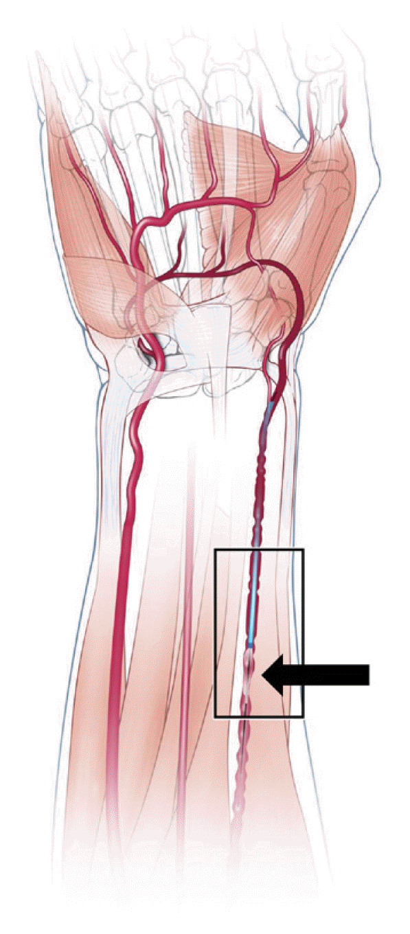
A hypertensive middle-aged patient with a four-hour history of worsening dysarthria, ataxia and new onset atrial fibrillation was found to have an acute basilar artery occlusion and transferred to our comprehensive stroke center for mechanical thrombectomy. During transradial catheterization, the artery demonstrated signs of spasm at the level of the recurrent radial artery immediately proximal to the brachial artery limiting distal access (Fig. 1). Intra-arterial delivery of verapamil and nitroglycerine did not fully resolve the vasospasm.
A 3×10 mm Hyperglide balloon (Medtronic Neurovascular, Irvine, CA, USA) was positioned with several millimeters of the inflated balloon protruding outside the 6 French Neuron Max guide catheter (Penumbra, Alameda, CA, USA). The unit was advanced together over a 0.010 inch Expedion microwire (Medtronic Neurovascular) past the point of obstruction and to the level of the subclavian artery. The balloon was then deflated, withdrawn and replaced by a coaxial system consisting of Catalyst 6 aspiration catheter (Stryker Neurovascular, Fremont, CA, USA) and Velocity microcatheter (Penumbra) over a Synchro-2 microwire (Stryker). Under roadmap guidance, this system was navigated beyond the right vertebral artery into the distal basilar artery where the microcatheter and wire were withdrawn. Complete reperfusion was then obtained with a single pass using contact thromboaspiration, and there were no procedural complications. Hemostasis was obtained using a transradial occlusive device, and no hematoma was identified.
A hypertensive diabetic octogenarian presented with facial asymmetry, aphasia and right hemiplegia, last known well eight hours prior. Computed tomographic angiogram (CTA) demonstrated tandem lesions of the left extracranial internal carotid and the proximal middle cerebral artery (MCA), with a perfusion deficit favorable for thrombectomy.2) An 8F Flowgate2 balloon guide catheter (Stryker) was navigated into the common carotid artery, and angiography showed occlusive mural hematoma of the cervical internal carotid artery. Under roadmap guidance, an initial attempt was made to cross the carotid occlusion using a 0.035 inch Glidewire, which was unsuccessful. A second attempt at distal passage was made using a 0.014 inch Synchro2 pre-shaped microwire through a 0.021 inch Prowler Select Plus microcatheter (Codman Neurovascular, Raynham, MA, USA) within a Catalyst 6 intermediate catheter. Microwire passage was successful, but we were unable to advance any catheters beyond the occlusion due to catheter edges getting caught on the plaque.
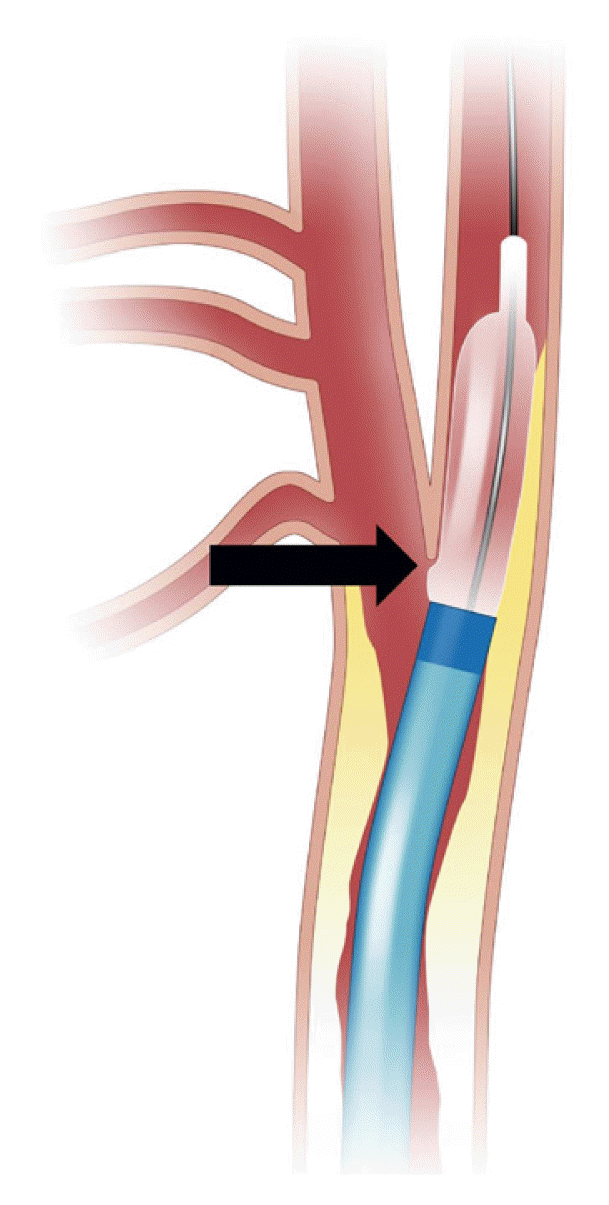
A 4×40 mm Aviator balloon (Cordis, Milpitas, CA, USA) was then introduced over the microwire and navigated across the lesion. Angioplasty was performed after balloon deflation and subsequent angiography demonstrated improvement in the degree of luminal stenosis. We were again unable to track the guide catheter through the lesion, so the balloon was sub-maximally inflated (to 75% of nominal volume) within the carotid plaque. The guide catheter was slowly advanced over the partially inflated balloon into the internal carotid artery, eliminating the “shelf” between the guide catheter and the plaque (Fig. 2). The balloon was then deflated and removed. Subsequent MCA thrombectomy was performed with a stent retriever for complete reperfusion after a single pass. Final angiography of the cervical carotid artery demonstrated stable improvement of the stenosis without dissection or distal emboli noted.
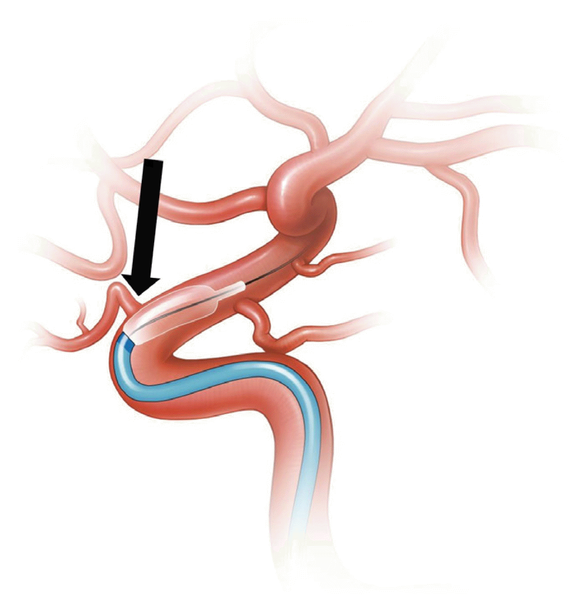
A patient in their sixth decade with a two-hour history of left-sided hemiparesis, neglect and hemibody sensory changes presented to our emergency department for evaluation. A CTA showed occlusion in the right M1 segment of the MCA and the patient was referred for mechanical thrombectomy. Transfemoral access was obtained and an 8F FlowGate2 balloon guide catheter was navigated into the cervical internal carotid artery. A Prowler Select Plus microcatheter was placed within a Catalyst 6 aspiration catheter and advanced over a 0.014 inch Transend microwire (Stryker). The microwire and microcatheter were navigated beyond the MCA occlusion, but in the distal portion of the horizontal cavernous carotid artery, a calcified vascular step-off was encountered that prohibited passage of the aspiration catheter. The microcatheter was removed and replaced by a 0.027 inch Velocity microcatheter and 0.014 inch Synchro-2 microwire, but navigation past the step-off was again unsuccessful. The microcatheter was removed and a 4×7 mm Transform balloon (Stryker) was navigated over the Transend wire immediately proximal to the location of the step-off. The balloon was then partially inflated to approximately 60% of maximal volume, while still partially within the Catalyst 6, and this combination was advanced past the step-off into the distal cavernous carotid artery (Fig. 3). The balloon was deflated and removed, and mechanical thrombectomy was performed with using a combined technique including stent retrieval with local aspiration for complete reperfusion after a single pass. There were no procedural complications.
Go to : 
Vascular access issues can occur at any stage of an endovascular procedure and may limit operator ability to access targeted pathology. Familiarity with balloon types, length, sheath compatibility, and compliance will allow for rapid adaptation of existing devices, minimizing procedural time and cost, and complications. Many vascular issues can be identified from non-invasive imaging pre-operatively and appropriate measures can be taken, including the use of balloon-guide, tapered or more flexible catheters. In larger vessels, there are more “off the shelf” options but in the neurovascular setting, devices that address the delicate regional anatomy have yet to be created. We present adaptive techniques using readily available equipment to surmount commonly encountered challenges across all types of endovascular procedure.
When spasm, stenosis or vascular shelves are encountered, an escalating management approach should be considered prior to attempting BAT. In addition to the costs associated with intravascular balloons, there are several complications that may occur using this technique. Over-inflation of balloon may cause arterial perforation and might also cause rupture of the balloon itself especially if not sized appropriately for the delivery catheter.1) Care should be taken to make certain that balloon preparation is meticulous (free from air) and that the appropriate concentration of radio-opaque solution is used upon inflation.
In cases where arterial spasm is encountered in peripheral vessels, especially during transradial access, medical techniques should first be attempted. Anti-spasm agents, including verapamil and nitroglycerin,4) may be infused through the radial sheath or catheter. Spasm in the extracranial carotid artery is typically catheter-related and will resolve over time by withdrawing or repositioning the catheter more proximally. Pharmacologic agents are not usually required and may not be optimal for carotid injection during many neurointerventions, where blood pressure is of primary concern.
During navigation past stenosis, care must be taken to avoid exacerbating any active atherosclerotic lesion and causing plaque fragmentation that may result in ischemia from distal emboli. Sub-maximal inflation and gentle forward pressure allow smooth passage without disrupting local anatomy. Intravascular step-offs present a navigation challenge. These vascular “shelves” may be due to stenosis, tortuosity or other vascular anomaly and are difficult to predict from non-invasive imaging. In the case of stroke, this peri-operative finding adds to procedural difficulty and can be associated with a shearing or dissection of the vascular wall. Prior to introducing a balloon into the intracranial vasculature, attempts should be made with smaller bore or more flexible catheters or by tracking up a microcatheter that has been positioned or anchored distally.
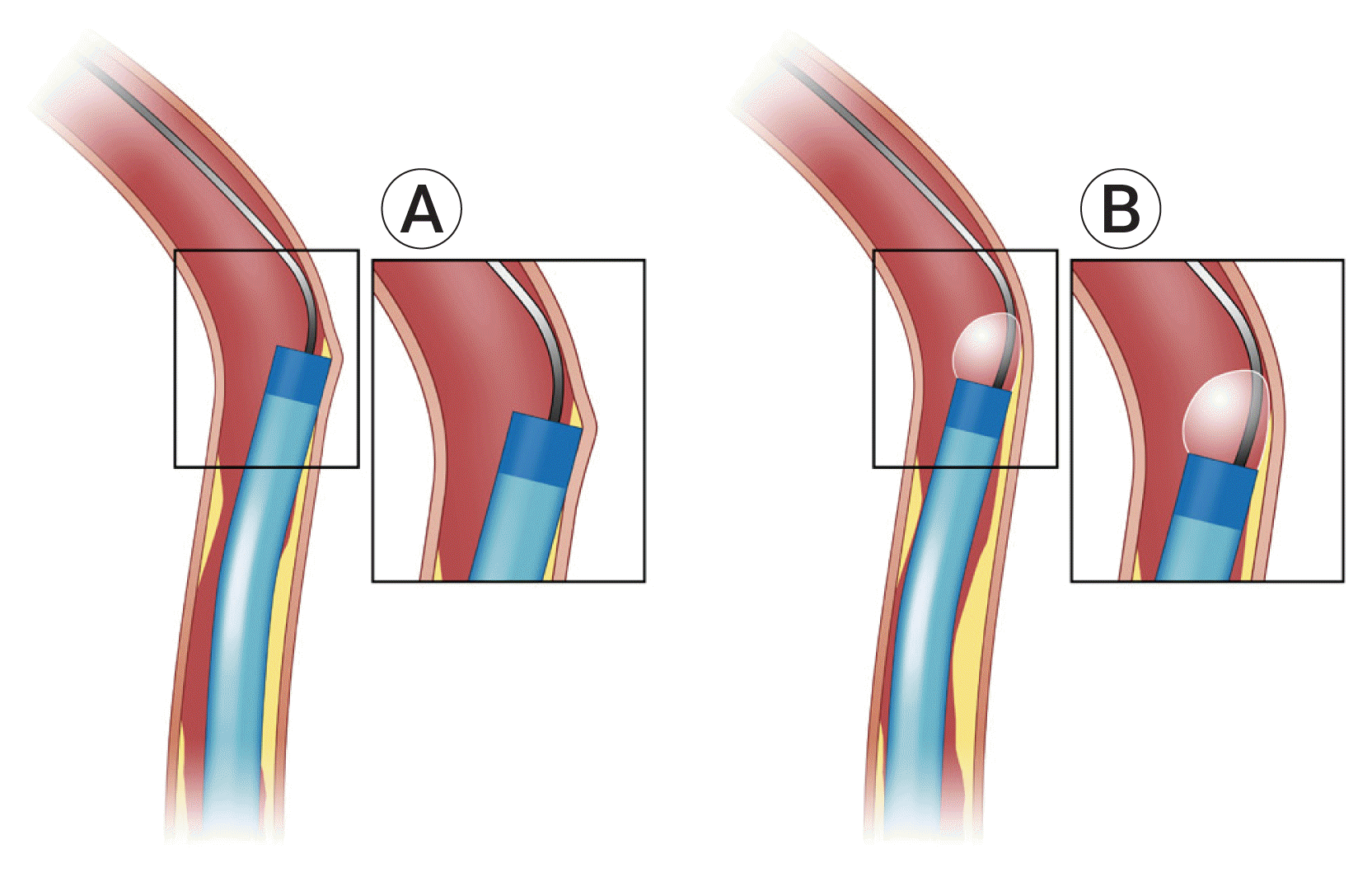
Similar techniques have been previously described in the stroke literature to overcome stenosis and can help prevent the razor-like shearing caused by catheters at these vascular step-off points (Fig. 4).3)5) The methods we outline encompass peripheral, cervical and intracranial vascular anatomy and are adaptable across all micro, guide and diagnostic catheters. Reasonable tracking support can be obtained through most devices with appropriate balloon selection. For radial or intracranial carotid arteries, microcatheter dimensions are the limiting factor and compliant intracranial balloons (Hyperglide, Transform) are likely to be the most helpful. For extracranial carotid challenges, larger noncompliant balloons (such as the Aviator) are required. Standard precautionary and preventive measures outlined above should be taken in every case, including pain relief, sedation, vasodilatation and use of hydrophilic guidewires. Pre-intervention, and in some cases mid-procedural intraarterial vasodilation reduces the risk for vasospasm.
Go to : 
ACKNOWLEDGEMENTS
The authors wish to credit Emma Vought for the technical illustrations in this manuscript.
Go to : 
Notes
Disclosure
The authors report no conflict of interest concerning the materials or methods used in this study or the findings specified in this paper.
Go to : 
REFERENCES
1. Agelaki M, Koutouzis M. Balloon-assisted tracking for challenging transradial percutaneous coronary intervention. Anatol J Cardiol. 2017; Feb. 17(2):E1.

2. Albers GW, Lansberg MG, Kemp S, Tsai JP, Lavori P, Christensen S, et al. A multicenter randomized controlled trial of endovascular therapy following imaging evaluation for ischemic stroke (DEFUSE 3). Int J Stroke. 2017; Oct. 12(8):896–905.

3. Burkhardt JK, Shapiro M, Tanweer O, Litao M, Chancellor B, Raz E, et al. Balloon-assisted tracking technique to overcome intracranial stenosis during thrombectomy for stroke. J Neurointerv Surg. 2019; Mar. 11(3):e1.

4. Mason PJ, Shah B, Tamis-Holland JE, Bittl JA, Cohen MG, Safirstein J, et al. An update on radial artery access and best practices for transradial coronary angiography and intervention in acute coronary syndrome: a scientific statement from the American Heart Association. Circ Cardiovasc Interv. 2018; Sep. 11(9):e000035.

5. Merella P, Lorenzoni G, Casu G. Inside the “Razor Effect”: lessons from optical coherence tomography-what does angiography hide? JACC Cardiovasc Interv. 2019; Feb. 12(4):409–10.
6. Patel T, Shah S, Pancholy S, Rao S, Bertrand OF, Kwan T. Balloon-assisted tracking: a must-know technique to overcome difficult anatomy during transradial approach. Catheter Cardiovasc Interv. 2014; Feb. 83(2):211–20.

7. Pierot L, Cognard C, Anxionnat R, Ricolfi F. CLARITY Investigators. Remodeling technique for endovascular treatment of ruptured intracranial aneurysms had a higher rate of adequate postoperative occlusion than did conventional coil embolization with comparable safety. Radiology. 2011; Feb. 258(2):546–53.

8. Pierot L, Spelle L, Leclerc X, Cognard C, Bonafé A, Moret J. Endovascular treatment of unruptured intracranial aneurysms: comparison of safety of remodeling technique and standard treatment with coils. Radiology. 2009; Jun. 251(3):846–55.

Go to : 


 PDF
PDF Citation
 Print
Print


 XML Download