This article has been
cited by other articles in ScienceCentral.
Abstract
Purpose
Tendon injuries in hand were one of the most frequent injuries caused by trauma. Although consequent tenorrhaphy is frequently conducted, in complete tendon rupture the location of retracted tendon stumps cannot be identified clearly, which causes difficulties in surgical management. Ultrasonography is known to be a fast, accurate, and cost-effective diagnostic method for tendon injury; therefore, the authors studied the usefulness of preoperative ultrasonography in diagnosis and management of tendon injuries in hand.
Methods
Among the 34 patients who had hand tendon injuries and visited between January 2017 and March 2018, retrospective studies were conducted on six patients with unidentified tendon stumps. By identifying the location of the tendon injuries and ruptured tendon stumps through preoperative ultrasonography, the operation was conducted under a predetermined surgical plan. The authors compared the ultrasonography results and the surgical findings.
Results
For diagnosis of the unidentified tendon injuries and location of ruptured tendon stumps, the ultrasonography results and the surgical findings were matched for all six patients. By prior planning of the operation incision line using the preoperative ultrasonography results, the length of the incision site was minimized without additional incision, and the operation time was shortened.
Conclusion
For hand tendon injuries with unidentified tendon stumps, identifying the location of tendon stumps through preoperative ultrasonography is helpful for determining the incision range for the operation. Therefore, preoperative ultrasonography may be useful in the diagnosis and management of tendon injuries in hand.
Go to :

Keywords: Ultrasonography, Tendon injuries, Hand injuries
서론
수부 건 손상은 사지 외상에 의한 손상 중에 가장 빈도가 높은 질환 중 하나이고[
1,
2], 그에 따른 건 봉합술은 자주 시행되는 수술이다. 그 중 완전 건 파열에서는 건의 탄성과 근육의 수축에 따른 단단 수축(end-to-end retraction)으로 인해 파열된 건의 근위부 및 원위부 절단단의 위치를 정확히 알기가 어려우며, 이런 경우 건을 짜내는 방법을 쓰거나(milking), 관절 등을 구부려보거나 수술 시 건의 진행 방향을 따라 기구를 넣어 건의 절단단을 찾는 등 여러 가지 방법을 사용하고 있으나 대부분의 경우 실패하기 쉽다. 결국 수술 시 절단단을 찾을 때까지 계속 추가 절개를 하게 되면서[
3] 추가 절개 횟수가 늘어나, 절개 부위의 길이가 증가되고 수술 시간이 길어진다.
초음파 검사는 건 병리의 진단에 있어 빠르고 정확하면서도 비용 대비 효과적인 진단 방법으로 알려져 있다[
2,
4-
6]. 초음파 검사는 건 손상의 진단을 위해 오래 전부터 사용하던 방법이었지만, 수부 건 손상의 진단에 대하여는 최근에야 연구가 진행되고 있고 특히 수술 전 미리 정확한 수술을 계획할 때 사용함으로써 수술과 치료 과정에 도움이 되는지는 국내 발표가 없었다. 이에 저자들은 수부 건 손상의 진단과 치료에 있어서 수술 전 초음파 검사의 유용성에 대한 경험을 문헌 고찰과 함께 보고하고자 한다.
Go to :

대상 및 방법
2017년 1월부터 2018년 3월까지 방문한 수부 외상 환자들 중 수부 건 손상 환자는 34명이었으며 그 중 절단단이 확인되지 않은 총 6명의 환자만을 대상으로 후향적 연구를 시행하였다. 이학적 검사에서 건 손상이 강하게 의심되나 그 손상 부위를 특정할 수 없는 경우와 완전 건 파열 이후 절단단이 말려들어가 확인되지 않는 경우에 수술 전 초음파 검사를 시행하여 손상 부위와 절단단의 위치를 정확히 파악한 후 수술 절개선의 디자인과 박리 범위 등을 미리 계획하고 수술적 처치를 시행하였다. 수술 전 초음파 검사 결과와 수술 시 육안으로 확인된 소견을 비교 관찰하였다.
환자들의 성비는 남성 5명, 여성 1명이고, 수상 부위는 1수지가 2례, 2, 3, 4, 5수지가 각 1례씩이었다. 수상 건은 신전건이 4례, 굴곡건이 2례였으며, 수상 기전은 열상이 3례, 당겨짐 손상이 1례, 과사용 손상이 1례, 열상에 의한 건 손상으로 건 봉합술 후 건 유착이 1례였다(
Table 1). 5례는 건 봉합술을, 1례는 건 유착 박리술을 시행하였다.
Table 1.
Comparison of ultrasonography results and operative findings
|
Patient |
Sex |
Age (yr) |
Site |
Injured tendon |
Cause |
Comparison of ultrasonography results and operative findings
|
|
Diagnosis |
Location |
|
1 |
Male |
47 |
1st finger |
EPL rupture at wrist level |
Overuse injury |
Matches |
Accurately identified |
|
2 |
Male |
56 |
3rd finger |
FDP rupture at the level of DIP joint area |
Laceration |
Matches |
Accurately identified |
|
3 |
Male |
36 |
1st finger |
EPL rupture at distal phalangeal insertion |
Traction |
Matches |
Accurately identified |
|
4 |
Male |
61 |
2nd finger |
ED rupture at MCP joint area |
Laceration |
Matches |
Accurately identified |
|
5 |
Female |
19 |
5th finger |
FDS, FDP rupture at proximal phalanx |
Laceration |
Matches |
Accurately identified |
|
6 |
Male |
40 |
4th finger |
EDP adhesion at PIP joint area |
Postoperative adhesion |
Matches |
Accurately identified |

모든 초음파 검사는 단일 기관의 단일 영상의학과 전문의에 의하여 iU22 Gray-Scale US System (Philips Healthcare, Eindhoven, Netherlands) 초음파 장비와 5–12 MHz 선형 탐촉자(linear transducer)를 이용하여 시행하였다. 모든 예에서 검사자는 단순 X-ray 사진과 임상 소견을 먼저 접한 후 검사를 진행하였다. 건 손상은 건의 연속성이 선형 패턴으로 끊어져 있는 것을 확인하여 진단하였고, 관절 움직임, 근육 수축, 건 유착, 건 이동에 관련된 사항은 유발조작검사법(provocative maneuvers)을 이용하여 수동적, 능동적 운동을 하며 실시간 검사를 하는 역동적 스캐닝(dynamic scanning)을 통해 정지 사진을 보완하였다.
이 연구는 분당제생병원 기관윤리심의위원회에 의해 심의 및 서면 동의 면제 과제로 승인되었다(No. DMC 2020-07-009).
Go to :

결과
손상 부위를 특정할 수 없는 건 손상의 진단과 완전 파열된 건의 근위부 및 원위부 절단단의 위치에 대한 수술 전 초음파 검사 결과와 수술적 소견이 모든 환자들에서 일치하였다. 따라서 모든 환자들에서 추가 절개 없이 미리 계획된 한 번의 절개선으로 수술을 완료할 수 있었다(
Table 1).
이에 절개 길이를 최소화하고 수술 시간을 단축할 수 있어 이차 수술 손상, 추가 절개로 인한 통증, 그리고 반흔 구축 등과 같은 수술 후 후유증을 최소화할 수 있었으며[
7], 환자가 수술 후 재활 운동에 더 적극적으로 임하게 하여 좀더 빠르고 완전한 기능 회복을 기대할 수 있었다.
1. 증례 1
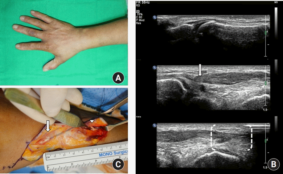
47세 남자 환자로, 배관 수리공이고 파이프 작업 중 파이프를 비틀다가 우측 손 부위에서 ‘뚝’ 하는 소리가 난 후 우측 엄지손가락이 펴지지 않아 내원하였다. 이학적 검사에서 열린 상처는 없었고, 손상 부위를 확인할 수 없는 장무지신근 손상이 의심되었다(
Fig. 1A). 초음파 검사에서 장무지신근의 완전 파열이 발견되었고 원위부 절단단은 손목 부위에서, 근위부 절단단은 아래팔 부위에서 발견되었다(
Fig. 1B). 수술 전 초음파 검사를 바탕으로 최소 길이의 수술 절개선을 계획하고 수술을 실행하였다. 수술적 탐색 결과 파열된 건의 원위부 절단단은 우측 제1중수골 중간 부위에서 발견되었고 근위부 절단단은 아래팔 부위의 근육 이행 부위에서 발견되었는데, 이는 초음파 검사에서 확인하였던 것과 동일한 위치였다(
Fig. 1C). 추가 절개 없이 미리 계획된 한 번의 절개선으로 건 봉합술을 완료할 수 있었다.
 | Fig. 1.Case 1. (A) A patient had pain on wrist without open wound from screwing a tube and could not extend his right thumb. Extensor pollicis longus (EPL) tendon rupture was suspected. (B) Ultrasonography results showed EPL rupture at wrist level. Intact distal insertion of right EPL tendon was seen (top). Diffuse thickening and discrete hypoechoic change with discontinuity of EPL tendon at the level of wrist area (arrow) was seen (middle). Diffuse swelling of proximal portion of EPL tendon at forearm level (brackets) was seen (bottom). (C) Operative findings showed complete EPL tendon rupture. We found ruptured tendon end with small predetermined incision in accordance with the ultrasonography results. The location of the distal end was identified at shaft level of right first metacarpal bone (arrow). The location of the proximal end was identified at forearm level in accordance with ultrasonography (arrowhead). 
|
2. 증례 2
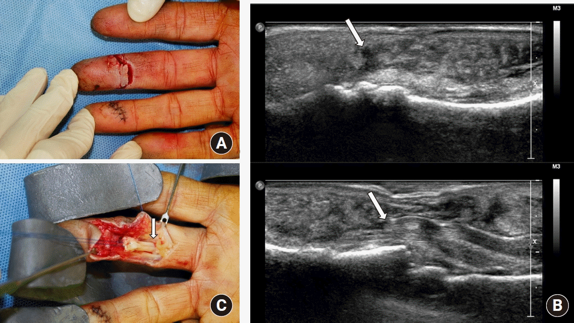
56세 남자 환자로, 스테인리스 물품의 날카로운 모서리에 우측 제 3수지를 수상하여 내원하였다. 이학적 검사에서 원위지의 능동적 굴곡이 불가능했고 원위지 간 관절의 수장부에 수직 방향으로 1.5 cm의 깊은 열상이 있었으며, 심수지굴건의 완전 절단을 확인하였다. 건의 원위부 절단단은 열상부 바로 아래에 있어 확인 가능했으나 건의 근위부 절단단은 당겨져 확인되지 않았다(
Fig. 2A). 초음파 검사에서 심수지굴건의 근위부 절단단은 중위지골의 기저부 근처에서 발견되었다(
Fig. 2B). 수술 전 초음파 검사를 바탕으로 최소 길이의 수술 절개선을 계획하고 수술을 실행하였다. 수술 소견은 수술 전 초음파 검사와 일치하였고(
Fig. 2C), 추가 절개 없이 미리 계획된 한 번의 절개선으로 건 봉합술을 완료할 수 있었다.
 | Fig. 2.Case 2. (A) A patient had 1.5 cm deep laceration at his 3rd finger from aluminum material. He could not flex his distal interphalangeal (DIP) joint of 3rd finger. We could determine complete rupture of flexor digitorum profundus (FDP) tendon through the wound, but we could not identify the location of proximal stump. (B) Ultrasonography results showed soft tissue laceration with rupture of right 3rd finger FDP tendon at the level of DIP joint area. Distal end of ruptured FDP tendon (arrow) was located at the level of DIP joint area (top). Proximal end of ruptured FDP tendon was located at the level of middle phalangeal base (bottom). Triangle shaped hypoechoic shadow, which means dead space in front of ruptured proximal stump, was seen (arrow). (C) Operative findings showed complete FDP tendon rupture. The location of the proximal tendon stump was identified at middle phalangeal base level in accordance with ultrasonography (arrow). This photo was taken after pulling the proximal tendon stump. 
|
Go to :

고찰
건 병리의 진단에 있어 초음파 검사는 빠르고 정확하며 비용 대비 효과적인 진단 방법이고 비침습적이며 위험도가 낮은 영상검사로서[
8], 반복하여 검사하기 용이하고 정상 측과 비교하여 검사하기도 쉬워 비교 역량이 우수하다[
4,
6]. 특히 초음파 탐침과 스크린의 기술 발전에 따라 초음파 영상의 해상도가 증가하면서 공간 분해능이 높아져 근래에는 손가락 건과 같은 작은 건 병리도 파악할 수 있게 되었다[
2,
4,
9].
건 파열의 초음파 검사는 에코 발생도(echogenicity)에 따라 이미지가 달라지며, 일차적 소견으로는 경계가 뚜렷하고(well-marginated), 불연속성 어두운 결손(discreted hypoechoic defects)을 보이고, 이차적 소견으로는 주위 연부 조직의 끼어듦, 굴절 음영(refractile shadow)등을 볼 수 있다고 알려져 있다[
8,
10]. 이에 따라 완전 또는 부분 건 파열 여부, 건 파열의 위치, 파열된 건의 수축 정도, 규모 등을 파악할 수 있으며[
11-
13], 유발조작검사법을 이용한 역동적 스캐닝을 통하여 수동적, 능동적 운동을 하여 완전, 부분 파열 또는 건 유착의 진단이 어려운 경우에도 더 쉽게 진단할 수 있고[
10,
11], 정지 영상에 비해 수술에 필요한 기능적이고 상세한 정보도 얻을 수 있다.
수부 초음파 검사는 외상에 의한 건 손상뿐만 아니라 류마티스 관절염이나 염증성 관절염 등의 염증질환에 의한 건 손상, 또는 등산가 손가락(climber’s finger), 방아쇠 수지(trigger finger) 등의 손가락 도르래(pulley) 손상의 진단에도 효과적으로 사용할 수 있다고 알려져 있다[
11]. 또한 건 병리의 진단에 있어 초음파 검사를 이학적 검사, 수술적 탐색, 자기공명영상(magnetic resonance imaging, MRI)과 비교한 보고에 의하면, 초음파 검사가 이학적 검사보다 민감도와 특이도가 모두 높고 수술적 탐색보다 시간이 덜 소요되며[
6], MRI보다 더 빠르고 정확하며 가격이 저렴하다고 알려져 있다[
4-
6,
10].
수술 전 초음파 검사와 건 손상에 대한 외국의 논문들을 살펴보면, 최근 영상의사와 임상의사에 의해 행해지는 근골격 부위의 초음파 검사 수가 모두 증가하고 있으며[
14], 응급의학과, 수부외과 의사 등 임상의사에 의해 행해지는 초음파 검사들에 대한 연구들에서 임상의사의 초음파 검사 시행 정확도가 높은 수준을 보여주고 있다고 보고하고 있다[
7,
15]. 따라서 어느 정도의 교육과 환경만 갖춰진다면 응급수술 전에도 집도의가 간단히 수술 전 초음파 검사를 시행하여 수술 계획에 도움을 받을 수 있을 것이다.
건 손상 시 초음파 검사를 이용할 때의 주의할 점은 초음파 검사가 가진 본연의 한계로서 술자에 따라 결과가 달라질 수 있다는 것이다. 예를 들어 탐촉자의 각도에 따라 조직의 비등방성에 의해 보이는 비특이적인 간섭들이 건 파열로 보일 수 있다고 알려져 있다[
6,
9]. 이에 따른 위양성률을 줄이기 위해서는 세심한 술기와 많은 경험이 필요할 것이다.
본 연구의 한계점은 단일 기관에서 조사하였기 때문에 표본 수가 적어 통계적 유의성에 대한 연구는 진행할 수 없었다는 점이다. 또한 초음파 검사는 수술 경과 관찰에도 사용할 수 있다고 보고되어 왔고[
4-
6] 저자들의 증례에도 경과 관찰에 대한 사례가 있으나 이에 대한 연구는 폭넓게 진행하지 못한 점도 있다. 따라서 이에 관하여 후속 연구가 필요할 것이다.
Go to :

결론
수부 외상 환자에 대한 이학적 검사에서 건 손상이 강하게 의심되나 그 손상 부위를 특정할 수 없는 경우와 완전 건 파열 이후 절단단이 말려들어가 확인되지 않는 경우, 수술 전 초음파 검사를 통한 절단단 위치 확인이 수술 시 절개 범위를 결정하는 데에 도움이 되었다. 따라서 수부 건 손상의 진단과 치료에서 수술 전 초음파 검사를 유용하게 사용할 수 있을 것이다.
Go to :


 PDF
PDF Citation
 Print
Print


 XML Download