

 PDF
PDF Citation
 Print
Print


INTRODUCTION
1. The principle of treatment of scaphoid nonunion
Untreated scaphoid nonunion progresses to carpal collapse resulting in wrist arthritis and chronic painful disability [1-4]. In addition, since most scaphoid fractures occur in young and active individuals, scaphoid nonunion has the potential to create great functional impairment. Therefore, timely surgical intervention by anatomic restoration of a stable scaphoid architecture and its linkage to adjacent bones is necessary before arthritis sets in. The principle of treatment of scaphoid nonunion is maintaining blood supply, debridement of necrotic bone and scar tissue, exposure of healthy well vascularized cancellous scaphoid bone, fracture reduction, bone grafting and rigid internal stabilization are all critical requirements [5,6]. Various open surgical techniques, such as corticocancellous or cancellous bone graft and various vascularized bone grafting techniques have been developed to treat scaphoid nonunion. The advantage of open treatment and bone grafting for nonunion is that it allows direct visualization of the fracture fragment for the freshening, reduction, and correction of associated deformities. These techniques, however, sometimes lead to increased stiffness, pain and hypertrophic scar of the wrist and hand due to the dissection of wrist capsule and ligament, resulting in unnecessary stripping and can devitalize the fracture site leading to further problems [5,7-9]. A meta-analysis by Merrell et al. [10] noted that there was a 94% union rate following screw fixation, and wedge grafting in 72 unstable nonunion patients as compared with a 77% union rate following Kirschner (K)-wire fixation and wedge grafting in 53 unstable nonunion patients. However, screw fixation with wedge grafting can be technically quite difficult because of the correct placement of screws to fix 3 bone fragments together. These features continuously challenge researchers in the field of medicine to devise new treatment procedures for scaphoid nonunion [11].
2. Wrist arthroscopy/characteristic wrist and scaphoid anatomy
After the introduction of wrist arthroscopy by Chen [12] in 1979, this technique was not accepted as a diagnostic method until the mid-1980s; however, wrist arthroscopical abilities and technologies steadily developed. During the past 25 years, diagnostic wrist arthroscopy became the gold standard. Concurrently, various therapeutic procedures also steadily established their roles and indications. The remarkable development in the past 3 decades has significantly altered the management algorithm of various clinical conditions of the wrist.
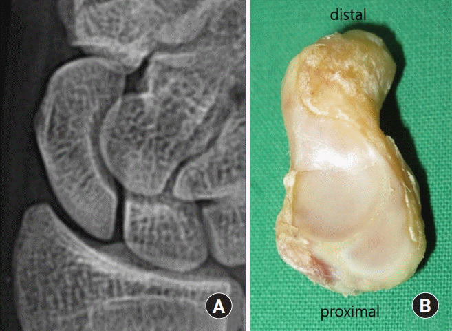
The wrist can be considered as the most complex synovial joint in the human body, consisting of 15 bones with 27 articulation surfaces and numerous ligamentous connections. Among them, the scaphoid is an almost entire intraarticular bone and 80% of the bone is covered by cartilage, which allows only few potential sites for the entrance of perforating vessels. The scaphoid has a tenuous blood supply [13,14]. Moreover, the scaphoid is an important insertion site for ligaments forming carpal joints; it is the key link between the proximal carpal row and distal carpal row, and controls the carpal kinematics. Therefore, preservation of the scaphoid anatomy and vascularity is critical for normal hand and wrist functions [15]. Another outstanding feature of the scaphoid is its shape in the mid-carpal joint, i.e., the gentle curvature of the waist and proximal portion of the scaphoid provides surgeons with a fairly stable and spacious platform and enables them to take down the nonunion site and carry out bone grafting (Fig. 1).
Therefore, if arthroscopy is applied to the scaphoid, ligament injury can be avoided, the scaphoid can be completely assessed through mid-carpal and radio-carpal joint, and the blood supply to the scaphoid can be preserved as much as possible.
Whipple first applied wrist arthroscopy in the treatment of scaphoid fracture and reported the combined advantages of internal fixation and minimal invasive surgical technique [16,17]. Subsequently, many surgeons have used wrist arthroscopy for the treatment of scaphoid fractures [18,19]. Currently, wrist arthroscopy, with its many advantages, is used for treating scaphoid nonunion [8,15,20-23].
In proximal pole nonunion, humpback of dorsal intercalated segment instability (DISI) deformity is uncommon. Moreover, scaphoid nonunion advanced collapse (SNAC) progresses more slowly and there is usually a flat crescent-like smaller bone defect. However, due to the lack of surrounding ligaments or capsules in the nonunion site, and in very proximal fracture, the nonunion site can be directly communicating with the radio-carpal joint. The grafted bone may escape from the graft site, so caution is required during bone grafting. To avoid spillage of bone graft from the nonunion site, only fibrous and necrotic tissue should be removed while preserving the cortical shell as much as possible. Wong and Ho [23] recommended the use of a pediatric urinary catheter for proving a mechanical block, a pediatric size 6 urinary catheter is inserted into the radio-carpal joint through the 3−4 portal and insufflated with saline to block the bone graft going into the radio-carpal joint. Avascular necrosis (AVN) can often arise at the proximal pole nonunion. However, AVN cannot be a contraindication. Instead, a vascularized bone graft can be considered in the event of arthroscopic bone graft failure. No punctate bleeding was observed in the proximal fragment in 3 cases, and we diagnosed these cases as AVN. However, we could achieve bone union in all cases, due to the minimal disturbance of the scaphoid blood supply and the firm impacted cancellous bone graft.
Distal pole nonunion occurs relatively less often. It is more difficult to perform arthroscopic operation because the nonunion site is unstable when the wrist is distended. Moreover, the surgeons should be aware of the difficulty of reduction and fixation of the nonunion site.
3. The roles of arthroscopy in scaphoid nonunion
Arthroscopy in scaphoid nonunion can have diagnostic and therapeutic roles. The diagnostic roles are (1) confirmation of the healing status of the fracture site; (2) assessment of the possibility of healing potential, i.e., the presence of punctate bleeding; (3) assessment of the cartilage status; (4) assessment of associated injuries that account for the symptoms, especially the ulnar side wrist pain; and (5) more comprehensive assessment of combined injuries such as scapho-lunate (SL), luno-triquetral (LT), triangular fibrocartilage complex (TFCC) injuries, and chondral injuries [17,21,22,24]. The therapeutic roles are styloidectomy, carpectomy, and bone grafting, of which arthroscopic assisted bone grafting is most important [22,23].
Go to : 
SURGICAL TECHNIQUE
1. Setup
The operation is performed under general anesthesia with the patient positioned supine on the side of the iliac crest region draped for bone graft harvesting. The arm to be operated is placed in a wrist traction tower and a vertical traction of 4−6 kg force applied through plastic finger trap devices to the middle 3 fingers for joint distraction on a hand table. An arm tourniquet is applied and inflated only if necessary, and a C-arm image intensifier is prepared for the percutaneous scaphoid fracture reduction and K-wires fixation.
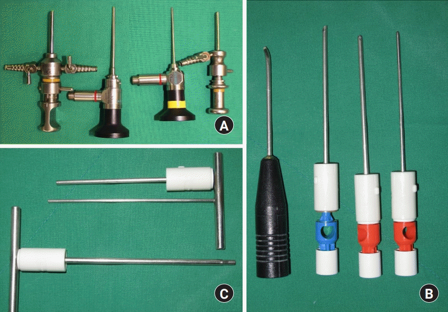
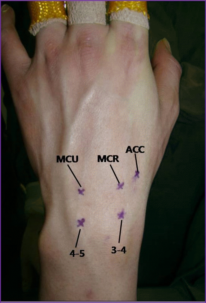
We use a 2.5- or 1.9-mm video arthroscope (CONMED, Utica, NY, USA), 2.0 mm and 2.9 mm shavers, a 3.0-mm burr, and a radiofrequency probe for surgical instruments. We also use 2 custom-made cannulas (3.8 mm and 3.0 mm) and 2 custom-made trocars (3.2 mm and 2.7 mm) for percutaneous bone grafting (Fig. 2). We employ continuously sending irrigation. We make the 3/4 and 4/5 portals for the radio-carpal joint, mid-carpal radial (MCR), and mid-carpal ulna (MCU) and one accessory portal for the mid-carpal joint (Fig. 3). An accessory portal is located mid-way between the MCR and scaphotrapezial trapezoidal (STT) portals. The bone grafting is performed through the mid-carpal joint.
2. Arthroscopic inspection
An inspection is first done on the radio-carpal joint. For a radio-carpal examination, first, the 3/4 portal is established as the viewing portal. The outflow is commonly made at 6U using an 18 gauge needle. From the 3/4 portal, it is possible to look at the dorsal ridge of the scaphoid where capsular reflection is attached, by orienting the arthroscope dorso-radially along the curvature of the scaphoid. When looking downward, the radial styloid process can easily be reached. From there inwards, the palmar capsular ligaments, the interosseous ligament, the TFCC, and articular cartilage should be evaluated systemically. During arthroscopy, carpal ligament injuries were graded using the system described by Geissler et al. [25] and the lesions of the TFCC were classified according to the system described by Palmer [26]. A working portal should not be established over the 4/5 or 6R portal at the beginning of the procedure until the entire ulno-carpal joint area is inspected through the 3/4 portal. This precaution helps to eliminate the dilemma of determining whether a dorsal capsular defect or synovial foldings over the dorso-ulnar corner commonly found at arthroscopy is a pathological lesion or whether the result of capsular intrusion by the trocar set. During arthroscopy, the interosseous ligament, articular cartilage, presence of synovitis, and other un-suspected pathology are closely momitored. Synovectomy is then performed since synovitis may obscure the observation of true cartilage condition. Any accompanying chondral lesions in patient with SNAC should be documented because it affects the prognosis and choice of procedure. Early SNAC changes are not a contraindication for arthroscopic bone grafting. Wong and Ho [23] described arthroscopic radial styloidectomy for the reduction of subsequent impingement in their SNAC patients. However, we hypothesized that, if scaphoid nonunion is properly reduced without the impingement of scaphoid and radial styloid process, progression of arthritis or pain would not occur. While performing arthroscopic bone grafting in our 15 SNAC patients, synovectomy and debridement on the radio-scaphoid joint was thoroughly performed while radial styloidectomy was not. Usually, fracture site cannot be found from the radio-carpal joint except a large gap and very proximal fracture, because the fracture is obscured by the capsular reflection or cartilage shell. We then transfer the arthroscope to the mid-carpal joint. Establishment of the MCU portal as the viewing portal is preferred because the MCR portal may become exceptionally tight in patients with radial side pathology. We identify the presence of any associated injuries and nonunion site (Fig. 4). We then assess the status of the articular cartilage and integrity and stability of the SL and LT joints. Before takedown of nonunion site, a repetitive synovectomy, debridement, and vaporization of the mid-carpal joint should be done to obtain a clear view.
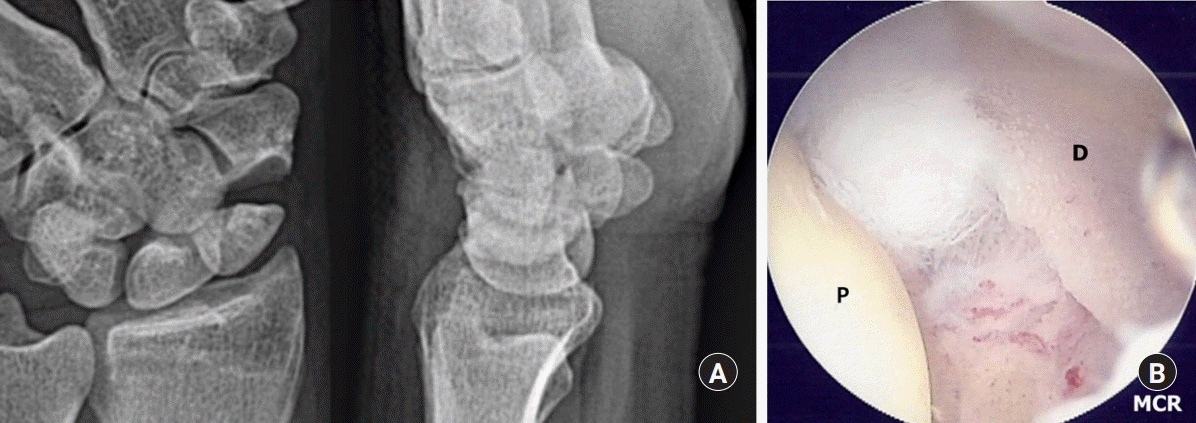 | Fig. 4.A 46-year-old male patient with nonunion of the left scaphoid fracture. Preoperative left wrist plain scaphoid. (A) A view showing nonunion at the waist of the scaphoid. (B) Same patient’s left wrist, mid-carpal arthroscopy image of scaphoid nonunion site shows large gap and sclerotic margins of both fragments. P, proximal fragment; D, distal fragment; MCR, mid-carpal radial.
|
3. Takedown of nonunion site
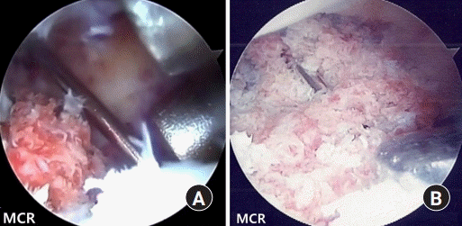
Takedown of nonunion site is carried out through the mid-carpal joint with the MCU as the viewing portal. The fracture gap is directly palpated with a probe inserted through the MCR portal. If a frank bone defect is encountered, both ends of the nonunion site are debrided and burred to remove all fibrotic tissue and sclerotic bone by switching the burr and shaver to the MCR and accessory portals until healthy looking cancellous bone with punctate bleeding is visible. Any intact cartilage around the nonunion site should be preserved for better graft containment. Since we do not use a tourniquet, a punctate bleeding can be seen during the debridement. Both ends are inspected for punctate bleeding with inflow irrigation fluid immediately stopped when no distinct bone bleeding is observed, despite the sufficient removal of necrotic bone (Fig. 5A). The cases are diagnosed as AVN when no punctate bleeding is observed in the proximal fragment according to Green’s bone viability test [27] (Fig. 5B).
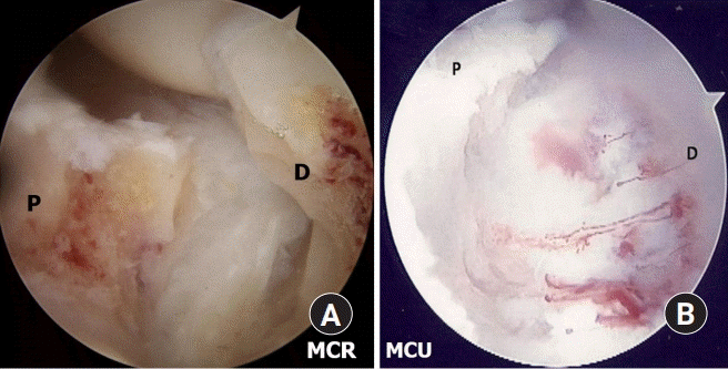 | Fig. 5.Left wrist, mid-carpal arthroscopy images of nonunion site of 2 patients after debridement. (A) Showing punctate bleeding from the proximal and distal fragments. (B) Showing no punctate bleeding from the proximal fragment. P, proximal fragment; D, distal fragment; MCR, mid-carpal radial; MCU, mid-carpal ulnar.
|
4. Reduction of scaphoid and provisional fixation
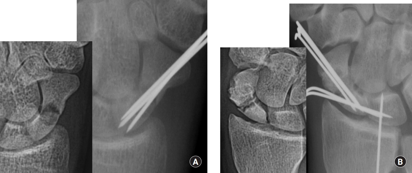
After sufficient debridement and removal of necrotic bone, the 2 fragments of the nonunion should be mobile enough for subsequent reduction. The distal fragment is reduced to the proximal fragment under the C-arm image intensifier by traction, gentle passive ulnar deviation, hypersupination and extension of the wrist with a surgical towel placed under the forearm and a 1.2 mm K-wire is inserted percutaneously from the tubercle of the scaphoid to the proximal pole for provisional scaphoid fixation (Fig. 6). The position of the K-wire is then confirmed and the integrity and stability of SL joint under arthroscopic view rechecked before proceeding to the bone graft. In the presence of a DISI deformity and extended lunate, the wrist is first flexed to realign the extended lunate with the radius for deformity correction. The radio-lunate (RL) joint is then transfixed with a percutaneous 1.2 mm K-wire inserted from the dorsal distal radius [28]. The scaphoid is then percutaneously fixed with a 1.2 mm K-wire (Fig. 7). It is advantageous to place a K-wire on the volar side as much as possible so that the pin does not block the placement and impaction of the bone graft. For the reduction of scaphoid in cases of early staged scaphoid nonunion, the reduction could be performed under arthroscope control. However, in cases of late staged nonunion, the gap is usually large and the appearance of gaps after debridement is variable; therefore, reduction under arthroscope control is difficult and deformity correction may not be possible. Therefore, reduction of scaphoid was performed under the C-arm image intensifier, and the reduction of lateral margin of the scaphoid was endeavored. However, care should be taken because reduction under the C-arm image intensifier can cause overcorrection of the scaphoid length. In this condition, it may cause limitation of radial deviation.
 | Fig. 6.After preparing the bone graft, we reduce the scaphoid with traction, gentle passive ulnar deviation, hypersupination and extension of the wrist with a surgical towel placed under the forearm and a 1.2 mm Kirschner-wire is inserted percutaneously from the tubercle of the scaphoid to the proximal pole for provisional scaphoid fixation.
|
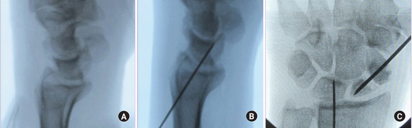 | Fig. 7.(A) In the presence of a dorsal intercalated segmental instability deformity and extended lunate, the wrist is first flexed to realign the extended lunate with the radius for deformity correction. (B) The radio-lunate joint is then transfixed with a percutaneous 1.2-mm Kirschner-wire inserted from the dorsal distal radius. (C) We then percutaneously fix the scaphoid with a 1.2-mm Kirschner-wire.
|
5. Bone grafting at the nonunion site
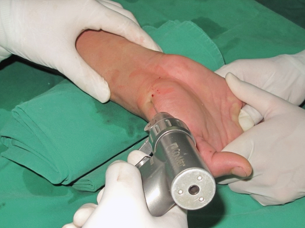
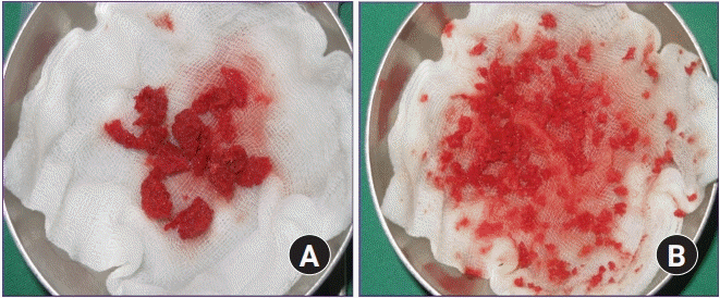
After preparing the bone grafting, cancellous bone graft is harvested from the iliac crest instead of distal radius using an open approach through a small incision. The quality of the iliac bone is superior. The volume of the harvested bone graft has to be at least 3 to 5 times that of the defect because the graft needs to be tightly compressed into the defect to increase the strength of the graft. The bone graft is then cut into small chips using scissors (Fig. 8).
For bone grafting, an arthroscope is introduced in the MCU portal to continuously show the nonunion site, a custom-made 3.8 mm cannula is introduced to the nonunion site through the MCR portal, and cancellous chip bone is delivered to the entrance of the cannula. The bone graft is packed with a 3.2-mm trocar until a satisfactory volume of graft is achieved (Fig. 9). The tourniquet can be inflated and continuous inflow maintained to enable visual ability during the graft impaction process. A custom-made 3.0 mm cannula is introduced into the accessory portal for the outflow of irrigation fluid. After completely filling the defect, routine surveillance of the joint is carried out to detect the need for removing any spilled bone graft material. The saline of the mid-carpal joint is then aspirated and 1 mL of fibrin glue (Greenplast Kit; Green Cross, Yongin, Korea) is routinely injected onto the surface of the graft substance. After arthroscopy, the wrist is taken out of traction to allow the natural compression from the capitate onto the graft.
 | Fig. 9.(A) Left wrist, mid-carpal arthroscopy images of percutaneous autogenous iliac cancellous bone grafting at the nonunion site using cannula and trocar. (B) Same patient’s left wrist, mid-carpal arthroscopy image showing finished bone graft to the nonunion site percutaneously using cannula and trocar. MCR, mid-carpal radial; MCU, mid-carpal ulnar.
|
6. Definitive fracture fixation
Definitive fixation with two 1.2-mm K-wires is performed under the C-arm image intensifier. Additional SL K-wires fixation is performed to fix the unstable nonunion and kept in place for 8 weeks. If there is an RL K-wire, it is left for 2 weeks. K-wires are then placed outside the skin (Fig. 10).
Methods of internal fixation have evolved over time. Since the introduction of the Herbert screw, which provides compression force and rigidity at the nonunion site, its success has resulted in many further developments of headless screws, which have compressive abilities and been widely used [6,20,29-31]. However, we preferred to use multiple K-wires fixation rather than screw fixation. Multiple K-wires provide rigidity and resistance to bending, pronation, and shearing forces during the process of fracture healing but no compression force across the fracture site [32]. It has advantages such as a greater ease of insertion, shorter operative time, and minimum amount of dissection. In addition, if bone union cannot be achieved, the K-wire is a more useful technique in terms of preparing for the secondary operation.
Go to :
POSTOPERATIVE CARE
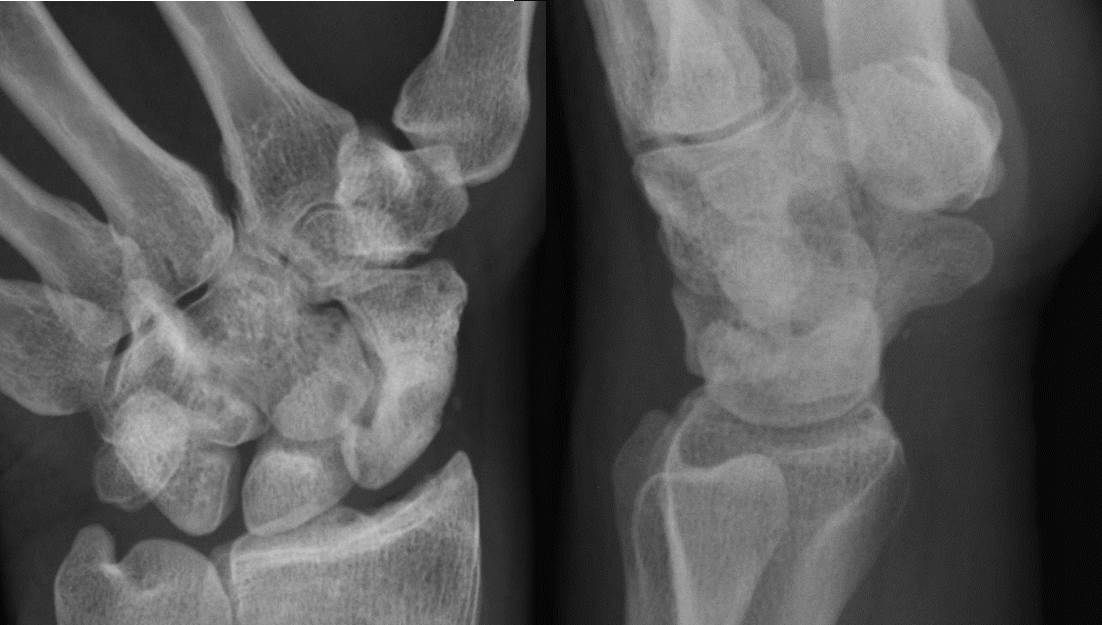
Postoperatively, in case of stable nonunion without SL instability, the wrist is immobilized with a below elbow thumb spica splint. In contrast, in case of unstable nonunion, the wrist is immobilized with an above elbow thumb spica splint for 2 weeks for the protection of RL pinning which was removed after 2 weeks, after which the below elbow thumb spica cast is applied for 8 weeks. Plain radiographs are taken every week until bone union is achieved; however, a computed tomography (CT) scan is taken to confirm the bone union because CT scan is a much more reliable tool than plain radiographs in the evaluation of scaphoid union and deformity [33]. Bone union was clinically assessed as the absence of tenderness at the anatomical snuffbox and radiologically assessed as the disappearance of the fracture line with bony trabecular across the original fracture. When radiological union is confirmed, the K-wires are removed (Fig. 11). We achieved bone union at an average of 9.6 weeks (range, 7−14.3 weeks) in 50 cases.
Go to :
RESULTS
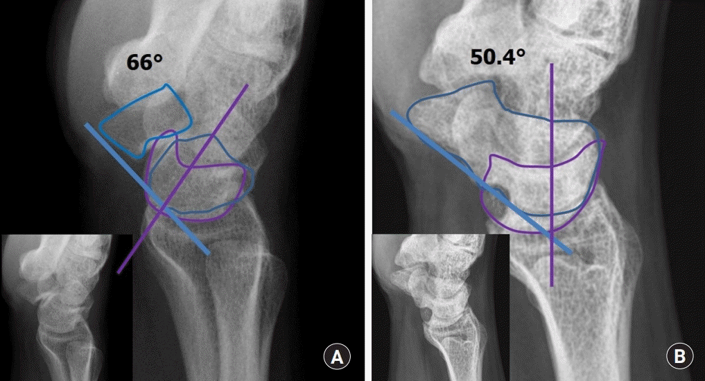
Waist nonunion is the most common site. Except for cases that are diagnosed early, the gaps are usually large and DISI deformity is common. Moreover, SNAC changes can arise early in the waist nonunion. Given the large defect, significant volumes of cancellous bone graft are required for firm and packed bone graft, and such large volumes can increase the operation time. However, since the capsules and ligament are usually intact in the waist nonunion, the grafted bone is less likely to escape. Bone union was achieved in 35 of 36 nonunion patients. In fifteen patients with SNAC stage I, satisfactory outcomes were achieved both clinically and radiologically with the mean SL angle significant improved from an average 66°±7.9° preoperatively to 50.4°±7.5° at the final follow-up (p=0.001) (Fig. 12).
Slade and Gillon [20] extended the range of indications of arthroscopy to a humpback deformity, which was correctable by using arthroscopy, and a case of AVN was identified on the magnetic resonance imaging. They achieved a 96% union rate in 108 patients treated with arthroscopic assisted reduction and dorsal percutaneous rigid headless screw fixation and selective bone grafting. Nevertheless, arthroscopy was not used for osteoarthritis or pseudarthrosis and an uncorrectable deformity.
Kim et al. [34] reviewed 36 unstable scaphoid nonunion patients who underwent arthroscopically assisted osteosynthesis with or without bone grafting and cannulated headless screw fixation. Bone union was achieved in 86% of patients at a mean of 11±2.7 weeks. Radiological parameters such as scaphoid axial length, lateral intrascaphoid angle (LISA), SL angle and reversed carpal height ratio were significantly improved after surgery. The range of wrist motion was unchanged after surgery, but the grip strength, mean Disabilities of the Arm, Shoulder, and Hand (DASH) scores and Patient Related Wrist Evaluation (PRWE) scores improved significantly after surgery. The radiological parameters of the scaphoid and carpal alignment in patients who achieved bone union did not correlate with clinical wrist function. The authors concluded that arthroscopic reduction and osteosynthesis of chronic unstable scaphoid nonunion is limited for restoration of normal carpal alignment but has positive effects on the recovery of clinical wrist function. They did not use arthroscopy for the patients with SNAC.
Kang et al. [35] reviewed 33 scaphoid nonunion patients who underwent arthroscopic bone grafting and screw fixation. The mean follow-up time was 33 months and 97% scaphoid nonunions healed successfully. The mean visual analogue scale (VAS) score, mean active flexion-extension angle, mean grip strength, mean Mayo wrist score (MWS) and mean DASH scores improved significantly after surgery. They found that 17 patients had coexisting intraarticular pathology including TFCC tears, intrinsic ligament tears and mild radioscaphoid degenerative changes. They noted arthroscopic management of scaphoid nonunion was effective. Although intraarticular pathologies commonly coexisted with scaphoid nonunion, the patients generally achieved good results. They did not use arthroscopy for the patients with AVN of the proximal fragments, humpback deformity or DISI deformity and advanced SNAC (stage II or higher).
Oh et al. [36] retrospectively reviewed 62 patients with unstable scaphoid nonunion who underwent arthroscopic (28 patients) or open (34 patients) bone grafting and internal fixation. They achieved a 96.4% union rate in the arthroscopic group and 97.1% in the open group. At the 2-year follow-up, VAS scores, grip strength, ROM, MWS, and DASH scores showed no statistical differences between groups. In radiographic assessment, SL angle, RL angle, and LISA were similar, whereas scaphoid height/length ratio was excellent through open technique. In subgroup analysis of patient with carpal collapse deformities, all radiographic measures in arthroscopic group were significantly higher than those in the open group. This indicates less correction of carpal collapse deformities in the arthroscopic group. They concluded that arthroscopic and open bone grafting and internal fixation in treating unstable scaphoid nonunions did not show any significant differences in clinical and radiologic outcomes at a minimum of 2 years after operation. In scaphoid nonunion with carpal collapse deformities, open bone grafting restored better carpal alignment than arthroscopic bone grafting.
Go to :
LIMITATIONS OF ARTHROSCOPIC OSTEOSYNTHESIS TECHNIQUE
Several circumstances precluded the use of arthroscopic bone grafting for scaphoid nonunion. First, a surgeon should not attempt using this technique if he/she lacks experience in wrist arthroscopy and arthroscopic anatomy. When we first attempted this operation, we could not find the nonunion site. We then operated using an open technique. Therefore, sufficient wrist arthroscopy experience is extremely important before attempting this operation. Second, we consider the salvage procedure as more effective for severe SNAC and severely destructed wrist joint combined with avascular nonunion; having non-reconstructable fragmented proximal pole than arthroscopic bone grafting. Third, significant arthrofibrosis of the wrist joint causes difficulty in arthroscopic bone grafting. An arthroscopic bone grafting for scaphoid nonunion and delayed union should be performed, with the exception of the above-mentioned limitations.
Go to :
CONCLUSION
Arthroscopic bone grafting and percutaneous K-wires fixation seem to be an effective treatment method for patients with scaphoid nonunion. It can provide comprehensive assessment of the fracture status and combined injuries at the time of arthroscopic treatment. This technique can preserve the biology of scaphoid fragments which may promote the bone union.
Go to :
 XML Download