Abstract
The soft tissue structure near the medial malleolus is an area where the bone is covered only with thin skin and subcutaneous tissue without structures like ligaments or tendons. In addition to the above anatomical features, wearing normal footwear should be considered when reconstructing the soft tissues in these areas. Therefore, it may be desirable to restore the original thickness of the soft tissue when performing a reconstruction. The authors reconstructed defects on the medial malleolus of a 50-year-old woman, after wide excision of a malignant melanoma, using only a deep fascial flap based on the posterior tibial artery-based perforator. This technique is thought to be a good option for reconstructing soft tissue defects in this area.
The soft tissue of the medial malleolus is an area where the bone is covered only with thin skin and subcutaneous tissue without structures like ligaments or tendons. Therefore, considering the anatomical characteristics, to use of the thinnest and most durable tissue possible during reconstruction is a great challenge for surgeons [1]. The reconstruction of this area should be performed considering the shape and function of the beneficiary after reconstruction, after gaining an accurate understanding of the location and size of the defect and its relationship with important structures such as bone, muscle, and nerves.
This includes (1) the anatomical characteristics of the protruded shape, (2) functional elements, such as wearing or ambulation of normal footwear, and (3) the fact that this area is exposed and needs to be considered from an aesthetic point of view.
Considering these factors, we present a case of reconstruction of the soft tissue defect in the medial malleolar area after wide excision of malignant melanoma using a deep fascial flap based on the perforator of the posterior tibial artery.
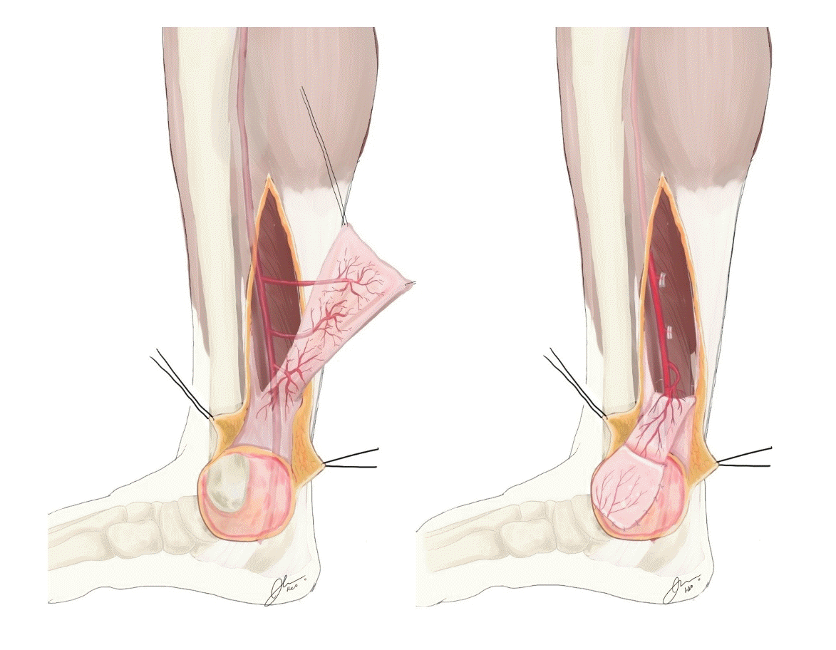
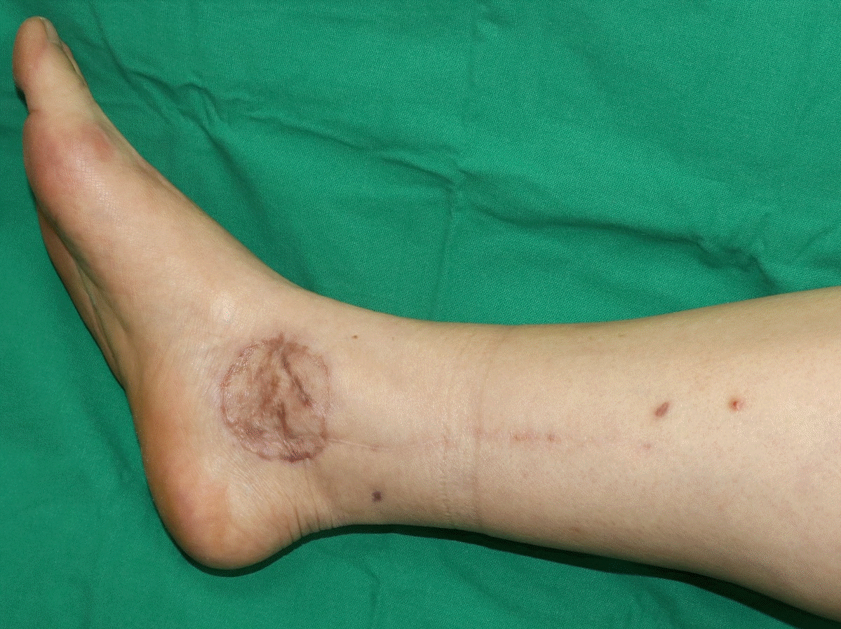
A 50-year-old female patient with no specific disease and family history visited our clinic with a 0.8 cm×0.7 cm sized dark pigmented mole on her right ankle. With gross view, the entire range of the pigmented lesion was 2.3 cm×2.0 cm sized. A preoperative tissue biopsy revealed a 0.3-mm deep (according to Breslow depth) superficial spreading-type malignant melanoma. Metastasis to Lymph node or distant organ was not found in oncologic evaluation, which corresponds to stage IA with T1aN0M0. Therefore we planned for surgical resection of tumor. We also planned a reconstruction using a posterior tibial artery-based adipofascial perforator flap after wide excision (Fig. 1). The resection range included the 2-cm horizontal range from the border of the lesion and the vertical range that included the entire skin layer and the periosteum. Immediately after extensive excision, frozen biopsy was performed at the four edges and two bases of the defect to confirm the absence of cancer cells. The defect measured 4.5 cm×4.2 cm and the location of the posterior tibial artery was determined using Doppler. A longitudinal incision was made in the anterior portion of the posterior tibial artery, on the posterior medial side of the lower extremity, to expose the deep fascia layer. At this time, the skin and subcutaneous fat layer on the deep fascia layer was considered to be too thick to cover the medial malleolar bone and we decided to use the deep fascia alone. A 2-cm proximal point from the medial malleolus was set as the pivot point, and the fascia was cut at the proximal 7 cm from the pivot point and then dissected toward the subfascial plane (Fig. 2). After confirming the distribution pattern on branches derived from the perforator in the fascia, the 7.0 cm×4.0 cm flap encircled around them was elevated. The flap was designed to be narrower toward the distal part to help turn over the flap based on the pivot point, and pinpoint bleeding at the flap tip confirming that the blood flow was intact. After the flap was turned over to the bare bone area, it was folded once into two layers for reinforcement, fixed on the exposed bone of the medial malleolus and covered with split-thickness skin graft (Figs. 3, 4). The donor site was repaired after insertion of a silicon drain. A splint for ankle fixation was used for 2 weeks after surgery. Postoperative monitoring was performed using Doppler in the predominant perforator region, and the first dressing and drainage tube removal was performed 3 days after surgery. Two weeks after the operation, the skin grafts were confirmed to be well taken, and after discharge, the patient was followed up by outpatient examination 11 months after the operation (Fig. 5). No further disease progression or metastasis was observed on oncologic evaluation.
Written informed consents were obtained.
The soft tissue of the medial malleolus is an area where the bone is covered only with thin skin and subcutaneous tissue without structures like ligaments or tendons. Therefore, there is less soft tissue available at the periphery, and reconstruction using a local flap is limited. For this reason, free fasciocutaneous flaps as well as free muscle flaps or fasciocutaneous reverse sural flaps [2] have been used. The rationale for using posterior tibial artery-based deep fascial flaps compared with this previous reconstruction method is explained below.
In soft tissue reconstruction of the medial malleolar area, the thickness of the thin flap is a very important factor considering the functional and aesthetic aspects of the ankle after recovery. The fasciocutaneous flap is too thick to replace the normal skin and fascia of the medial malleolar area. The deep fascial flap we used here is an extremely thin and pliable tissue, with an average thickness of 0.9 mm, which can be easily elevated. It is a dynamic tissue with complex vasculature and split-thickness skin graft performed on this fascia-only flap can easily take. Postoperative photographs of this case show that the thickness is almost the same as the original [3]. It is suitable for areas requiring very thin flaps. Areas such as ankles may cause contour deformity even if a very small amount of fat tissue is included, which interferes with wearing normal footwear. Therefore, considering the shortcomings of the conventional fasciocutaneous flap or free flap, such as limitation to the footwears which can be worn or additional debulking procedures for bulky flaps, this is an outstanding point of the deep fascia-only flap.
The posterior tibial artery has many segmental perforators in the posteromedial aspect of the lower leg. According to Drimouras et al. [4], five perforators are produced on average, which tend to gather in the lower two-thirds. The direction of the perforators varies widely and mostly emerges as a septum between the soleus muscle and the flexor digitorum longus. Each perforator has a diameter of more than 1 mm and is accompanied by 2 venae comitantes, which are considered stable.
The deep fascia supplied by the posterior tibial artery has a sufficient subfascial vascular network and can have sufficient viability when used alone, if preserved during dissection.
Deep fascia is a tissue that is well suited for engraftment, even as an allograft, and it is sufficient to use the fascia lata as a biological mesh in abdominal hernia. Stability is expected to double when performed using the perforator of the posterior tibial artery. In addition, it exhibits strong resistance to traction and has a histologic characteristic that can withstand the movement of the leg itself or shearing caused by footwear [3]. Therefore, a deep fascia-only flap using the perforator is particularly suitable for the medial malleolus.
First, the flap elevation technique is relatively simple and the operation time is short. Second, there is no need to sacrifice important blood vessels for micro-anastomosis. Third, the donor site morbidity is very low compared to a fasciocutaneous free flap. Because the dissection plane is a relatively shallow layer of the lower extremity, there is little chance of damage to major structures such as nerves, blood vessels, muscles, etc. Fourth, there is no need for a secondary debulking procedure due to excessive thickness of the flap after placement of a fasciocutaneous free flap [5].
The only consideration in the intraoperative situation is a definite understanding of the anatomical structure. The posterior tibial artery is a vessel that supplies all the superficial compartments, except the medial head of the gastrocnemius muscle, and delivers a perforator that feeds this compartment through the deep fascia [6]. This segmental perforator produces a good anastomotic arcade, supplying the deep fascia [4]. Surgeons must practice careful dissection during surgery to avoid injury to these structures.
Adipofascial flaps and fasciocutaneous flaps using the perforator of the posterior tibial artery have been used in the past [5,7,8]. Attempts have also been made to reconstruct the lower limbs using free fascial flaps, including the fascia-only anterolateral thigh free flap [9,10]. However, the use of perforator based deep fascia-only flaps is the first concept to the best of my knowledge.
Of course, the deep fascia-only flap with split-thickness skin graft have disadvantages in skin texture, and the possibility of contracture, compared to conventional fasciocutaneous flaps or free flaps. However, the authors would like to emphasize that, given the functional characteristics of the ankle, which requires the wearing normal footwear, it has much more functional advantages than conventional methods with bulky flaps.
Considering the above, therefore, this deep-fascia only posterior tibial artery perforator flap may be a good option for medial malleolar region reconstruction.
REFERENCES
1. Schaverien MV, Hamilton SA, Fairburn N, Rao P, Quaba AA. Lower limb reconstruction using the islanded posterior tibial artery perforator flap. Plast Reconstr Surg. 2010; 125:1735–43.

2. Ciofu RN, Zamfirescu DG, Popescu SA, Lascar I. Reverse sural flap for ankle and heel soft tissues reconstruction. J Med Life. 2017; 10:94–8.
3. Stecco C, Tiengo C, Stecco A, et al. Fascia redefined: anatomical features and technical relevance in fascial flap surgery. Surg Radiol Anat. 2013; 35:369–76.

4. Drimouras G, Kostopoulos E, Agiannidis C, et al. Redefining vascular anatomy of posterior tibial artery perforators: a cadaveric study and review of the literature. Ann Plast Surg. 2016; 76:705–12.
5. Heymans O, Verhelle N, Peters S. The medial adiposofascial flap of the leg: anatomical basis and clinical applications. Plast Reconstr Surg. 2005; 115:793–801.

6. Ward KL, Romano A, Rodriguez-Collazo ER. Cadaveric atlas for orthoplastic lower limb and foot reconstruction of soft tissue defects. Clin Surg. 2018; 3:2001.
7. Galumbeck M, Colen LB. Soft tissue reconstruction: coverage of lower leg: rotational flap. Orthop Clin North Am. 1993; 24:473–80.
8. Kyung H, Choi JI, Song SH, Oh SH. Posterior tibial artery perforator-based adipofascial flap for skin necrosis and exposed extensor tendon after fixation of distal tibiofibular fracture. J Wound Manag Res. 2018; 14:54–7.

Fig. 1.
A 0.8 cm×0.7 cm sized dark pigmented mole was noted on right ankle. Reconstruction using a posterior tibial artery-based perforator flap was planned. The running of the posterior tibial artery on the skin was marked as photo with Doppler.
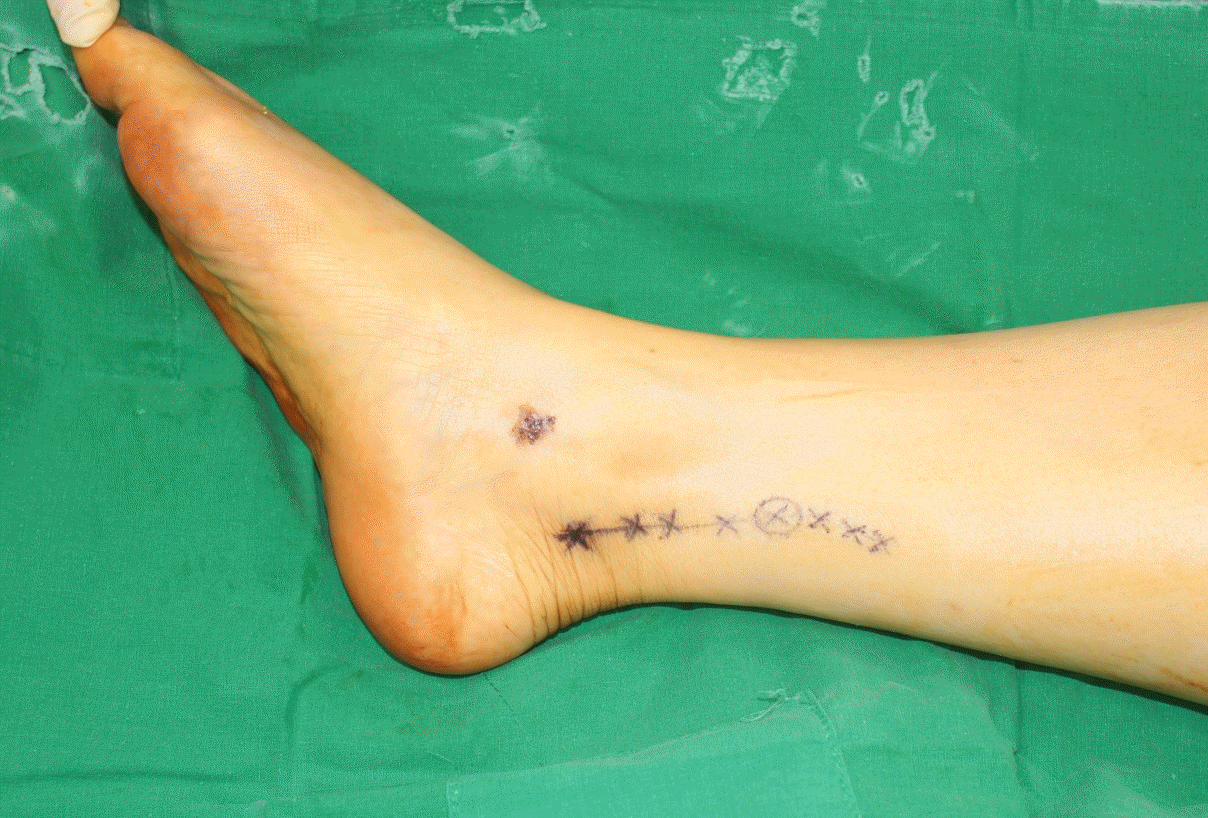
Fig. 2.
After wide excision, a 2-cm proximal point from the medial malleolus was set as the pivot point, and the fascia was cut at the proximal 7 cm from the pivot point and then dissected toward the subfascial plane.
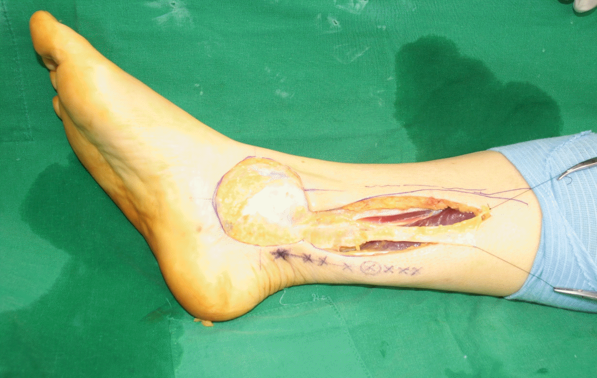
Fig. 3.
After the flap was turned over to the defect, it was folded once into two layers for reinforcement, fixed on the exposed bone of the medial malleolus.
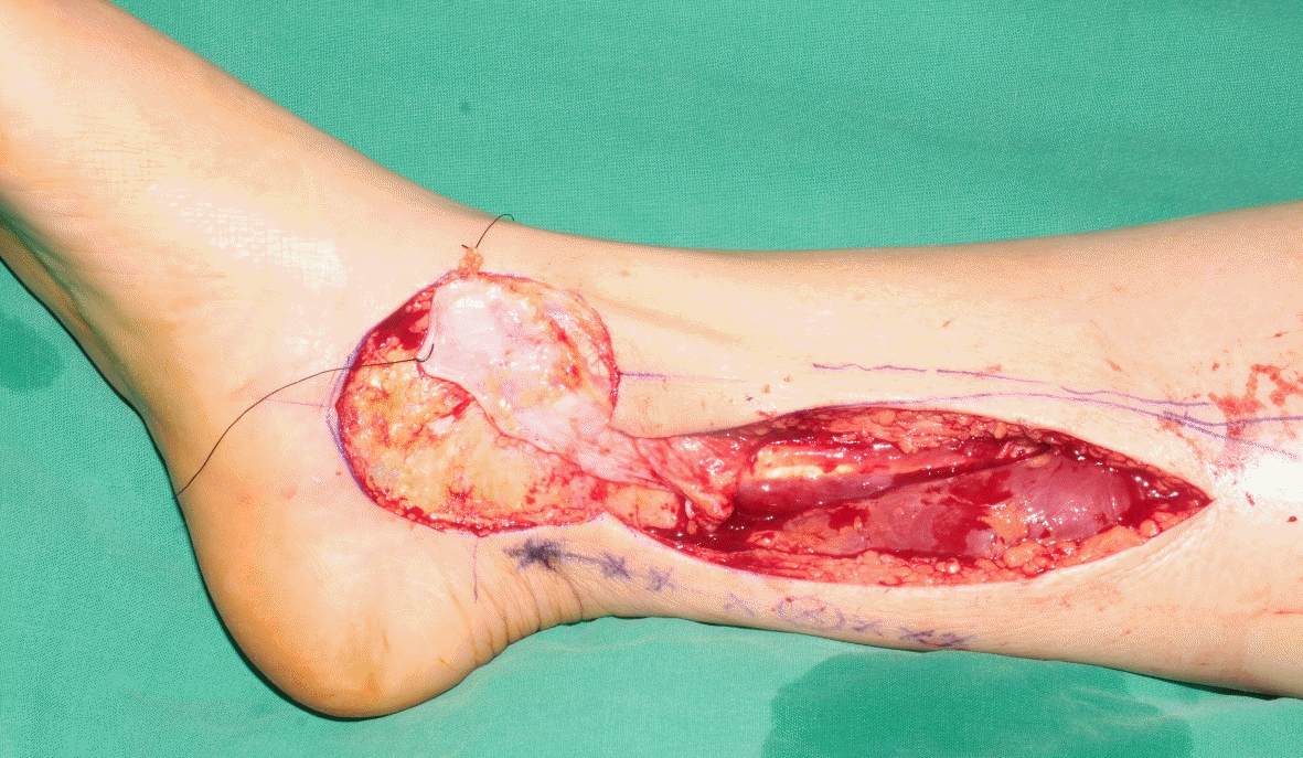
Fig. 4.
Schematic illustration of the deep fascial flap. It is shown that the perforator vessels based on posterior tibial artery are segmented and branch out to supply deep fascia (left). The deep fascial flap is turned over based on the pivot point, and the distal perforator is feeding the whole flap (right).


 PDF
PDF Citation
 Print
Print


 XML Download