

 PDF
PDF Citation
 Print
Print


I. Introduction
Implant failure is the term used for dental implants that require removal or are lost. Early implant failure (EIF) is defined as implant failure before being put into use, i.e., pre-prosthetic phase1.
Despite the progressive increase in the success rate of dental implants, a certain percentage of implant failures remain with unknown etiologies. Clinicians should be aware of any potential risk that may affect implant osseointegration and failure2,3.
Among the local causes of EIF is poor bone quality, a lack of primary stability due to surgical trauma, and infection that disturbs primary bone healing3. Other systemic conditions that influence the initial healing process of bone include uncontrolled diabetes mellitus, osteoporosis, corticosteroids, bisphosphonate therapy, and collagen disorders3-5.
Implant site is among the biological factors that may affect implant survival. There has been considerable debate regarding implant site and failure6-8. Several studies have tried to compare implant failure rates regarding implant insertion region of the jaw. Studies have reported that implants fail in the maxilla more than the mandible9-13. Furthermore, the maxillary anterior region exhibited the highest rate of implant failure. Factors contributing to higher implant failure in the maxillary arch compared to mandibular arch are not yet understood14,15. The high implant failure rate observed in the maxilla suggests that low bone density may be a contributing factor responsible for EIF15. Smoking may modulate implant failure by influencing bacterial infections in the maxillary and mandibular arches16.
Many clinical trials have evaluated the locations of implants and concluded that implants inserted in the anterior maxilla failed more than those placed in the posterior maxilla. Moreover, failures in the anterior mandible are more common than posterior failures, with the posterior maxilla recording the highest rate of failure15. A recent study reported that the two jaw bones exhibited the same rate of implant failure and stated that implantation site is not a modulating factor influencing EIF rate17.
EIF is among the worst problems in implantology. Inappropriate surgical protocols, surgical difficulties, decrease in bone volume or quality at the recipient site, and systemic conditions are all confounding factors14.
Implantation sites could play an influential role in the occurrence of EIF. Therefore, the purpose of this systematic review is to analyze high-quality studies to determine if there is a correlation between early failure and implantation site.
II. Materials and Methods
The recommendations of the PRISMA (Preferred Reporting Items for Systematic Reviews and Meta-Analyses) statement are as follows18.
1. Research question
Is there is any correlation between site of implantation (maxillary anterior, maxillary posterior, mandibular anterior, or mandibular posterior) and EIF?
2. Search strategy
MEDLINE, EMBASE, Web of Science, Cochrane Library, and Egyptian Knowledge Bank (EKB) were individually searched for studies in English from the year 1998 to the present. Manual searches were performed to identify references from primary studies and systematic reviews for relevant data.
The search used a combination of controlled vocabulary “Dental implants”[MeSH Terms] OR (“oral implants”[All Fields] AND “failure”[All Fields] AND “early”[All Fields]) OR “titanium dental implants”[All Fields] OR “early implant failure”[All Fields] OR “dental implant site”[All Fields].
3. Study selection
All abstracts and full articles were screened, and the following inclusion and exclusion criteria were applied.
4. Inclusion criteria
The chosen studies met the criteria of retrospective, prospective, and randomized clinical trials; all age ranges; both sexes; all types of implants either inserted into the drilled site or after use of an osteotome; all patients regardless of smoking status and use of antibiotics.
5. Exclusion criteria
Studies that were excluded from the analysis were those on cadavers, animals, patients with systemic problems affecting bone density or mineralization, patients under treatment with corticosteroids or bisphosphonate therapy, reported failures after prosthetic fixation, implantation in grafted sites, or after sinus lift procedures.
6. Data extraction
The extracted data are shown in a table and comprise authors, publication year, study type, site of implantation, number of implants, number of failed implants, and study power.
7. Quality assessment
Most of the included studies were retrospective. Randomized or blinded studies were impossible, and studies with small sample size or improper methodology were rejected.
8. Assessment of heterogeneity
The chi-square test for heterogeneity was performed, and the extent of the inconsistency of treatment effects (I2) across trials was measured.
We considered heterogeneity as substantial if I2 was greater than 30% or if there was a low P-value (less than 0.10) in the chi-square test for heterogeneity.
We used a fixed-effect meta-analysis for combining data to assume that the studies were estimating the same underlying treatment effect. If there was clinical heterogeneity sufficient to expect that the underlying treatment effects differed between trials or if substantial statistical heterogeneity was detected, we explored this followed by random-effects and subgroup analyses.
9. Assessment of reporting biases
Publication bias was appraised using the visual assessment of symmetry of funnel plots. If asymmetry was detected, we performed exploratory analyses to investigate it.
10. Methodological quality assessment
Data synthesis and meta-analysis were performed according to the rules adopted by the Cochrane Collaboration reviewer’s handbook19. All statistical analyses were conducted using the Review Manager software (RevMan ver. 5.3).
11. Meta-analysis
The dichotomous analysis in the form of number of failed implants in proportion to total implants inserted was analyzed with a risk ratio (RR) (relative risk) and 95% confidence interval (CI) for each study. Random effects were selected in the comparison of maxillary and mandibular implant failures because of heterogeneity. Subgroup analysis of anterior and posterior sites in both bones was performed with a fixed effect and 95% CI for accurate results. Risk difference (RD) (absolute risk) was used to declare the results and extraction of the conclusions.
III. Results
1. Study selection
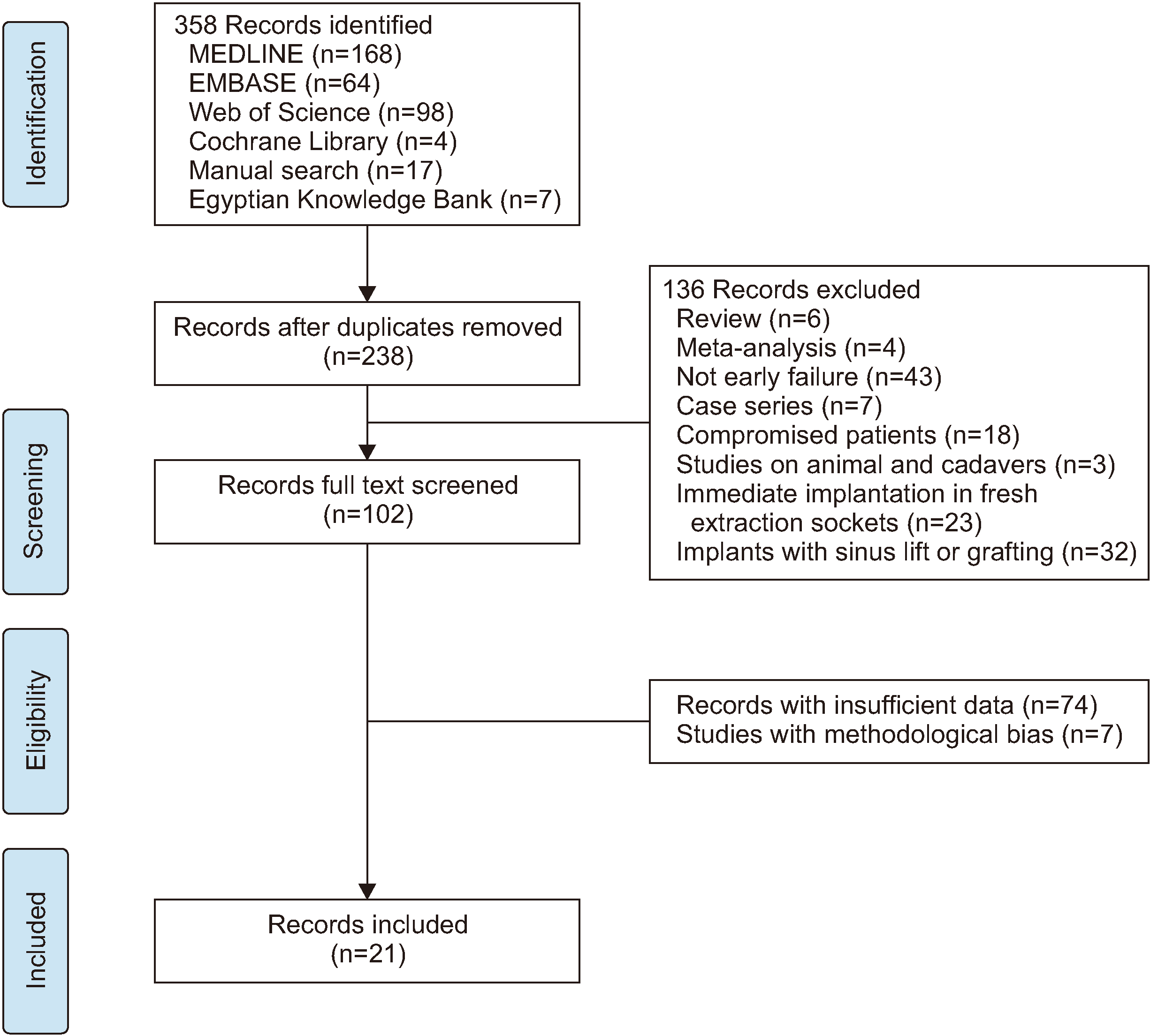
After searching the electronic databases, 341 publications were identified; 17 more articles were added after a manual search (total of 358). Removal of duplicates (120 studies) left 238 studies, studies not meeting the inclusion criteria were rejected (136 studies), and the remaining 102 studies were subjected to full-text reading. Among them, 74 studies were excluded because of insufficient data, 7 studies had methodology bias, and a final total of 21 studies4,10,14,15,20-36 were included in this review.(Fig. 1)
3. Implant failure: Comparison of implant failures in the maxilla and mandible
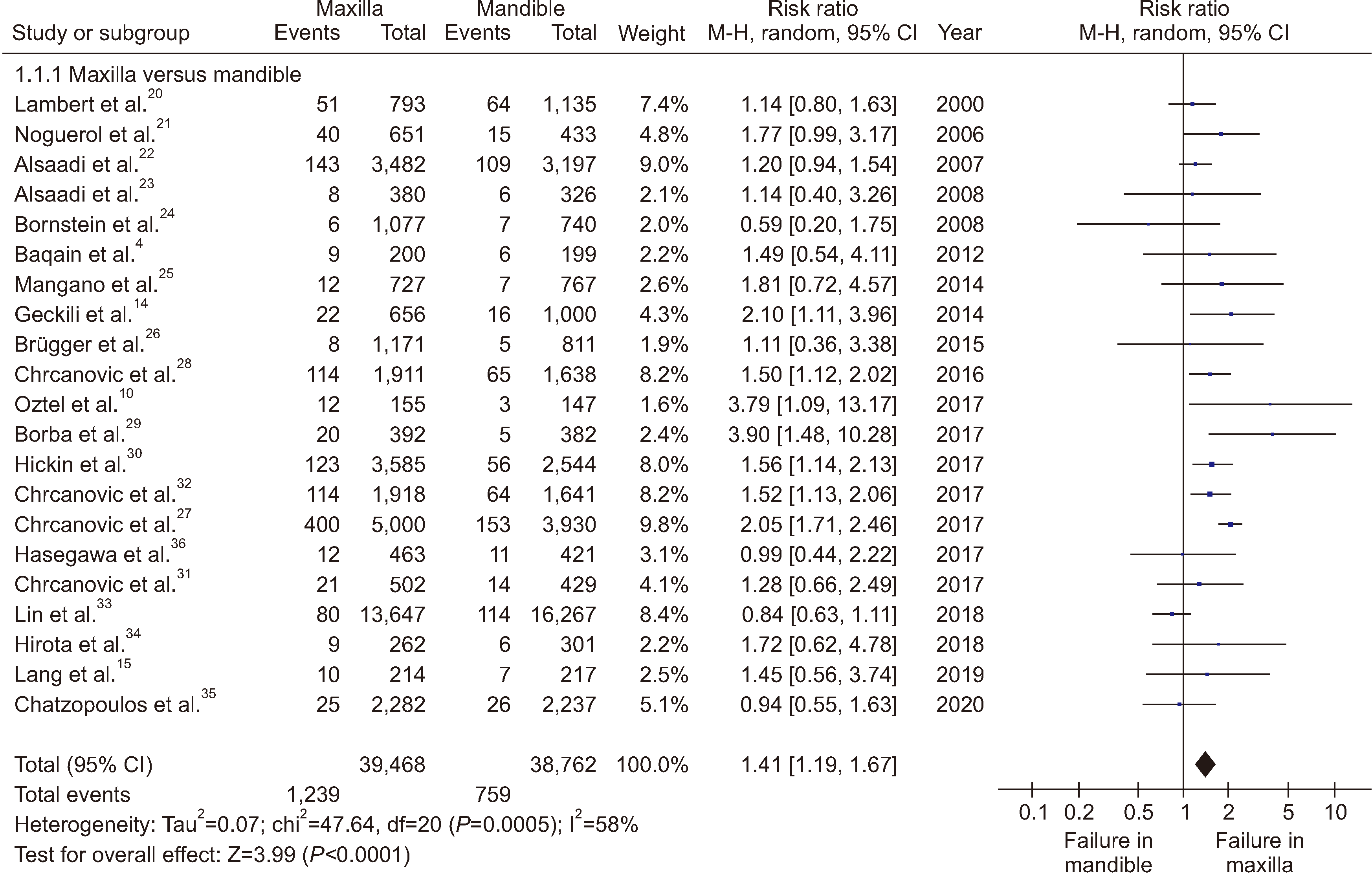
Analysis of the included 21 studies involved a total of 78,230 implants, among them 39,468 implants in the upper jaw bone and 38,762 implants placed in the lower jaw bone. A total of 1,239 implants failed in the upper jaw (3.14%) and 759 implants failed in the lower jaw (1.96%).
Total subgroup analysis of the 21 identified studies4,10,14,15,20-36 applying a random effect, RR, and CI of 95% revealed higher failure in the maxilla compared to the mandible (RR 1.41; 95% CI [1.19, 1.67]; P<0.0001; I2=58%).(Fig. 2) The RD analysis showed a 1% increase in the maxilla compared to the mandible.
4. Subgroup analysis
1) Anterior maxillary region compared with the anterior mandibular region
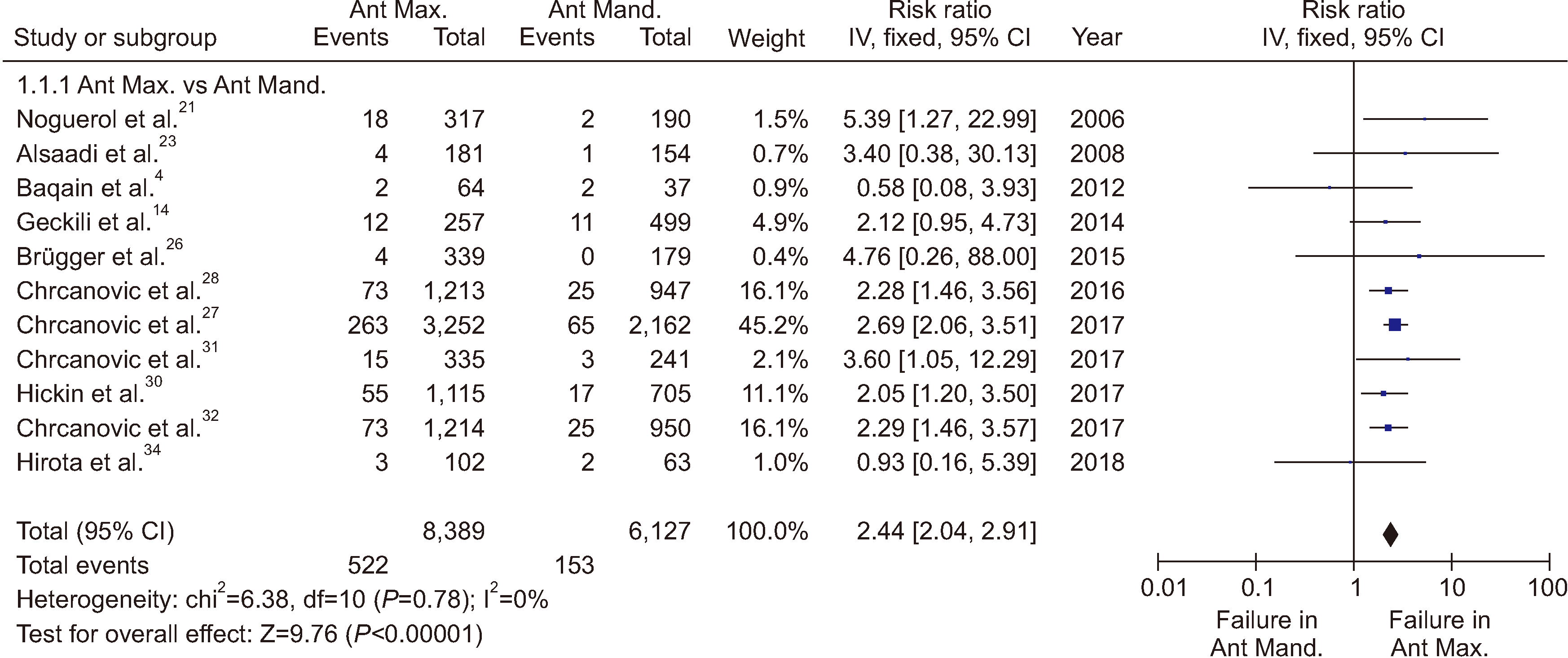
Data were extracted from 11 studies4,14,21,23,26-28,30-32,34 with a total of 14,516 implants. Among those implants, 8,389 were placed in the maxillary esthetic zone and 6,127 implants in the lower anterior region of the mandible. There was a total of 522 failed implants within the anterior maxillary region (6.2%) compared to 153 failed implants within the anterior mandible region (2.5%).
Total subgroup analysis of the 11 identified studies applying a fixed effect, RR, and CI of 95% revealed significant failure in the anterior maxilla compared to the anterior mandible (RR 2.44; 95% CI [2.04, 2.91]; P<0.00001; I2=0%).(Fig. 3)
The RD analysis reported a 3% increase in risk failure in the anterior maxillary region compared to the anterior mandibular region.
2) Posterior maxillary region compared with the posterior mandibular region
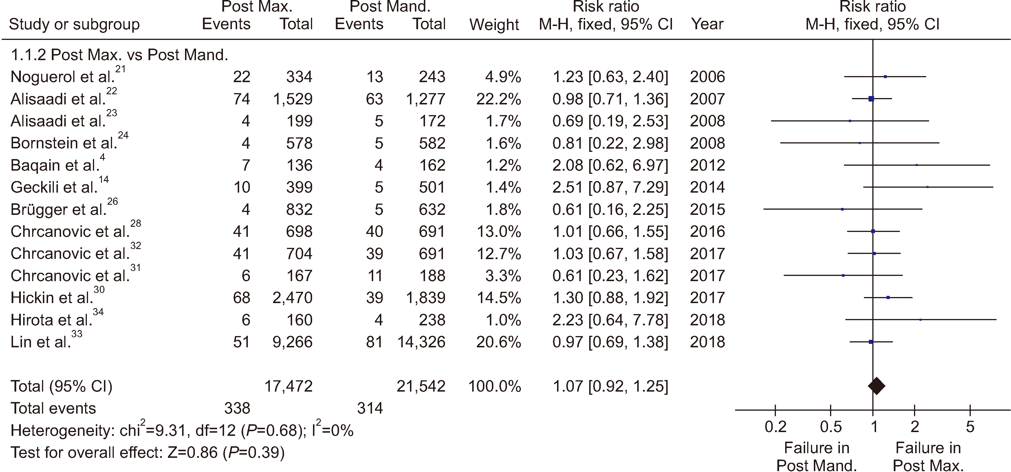
Data were extracted from 13 studies4,14,21-24,26,28,30-34 with a total of 39,014 implants; among them, 17,472 were inserted into the maxillary posterior region and 21,542 in the lower posterior region of the mandible.
There was a total of 338 failed implants in the maxillary posterior region (1.9%) compared to 314 failed implants within the mandibular posterior region (1.46%).
Total subgroup analysis of the 13 identified studies applying a fixed effect, RR, and CI of 95% revealed a non-significant difference in failure between the posterior maxilla and the posterior mandible (RR 1.07; 95% CI [0.92, 1.25]; P=0.68; I2=0%).(Fig. 4)
The RD analysis reported the same risk in the posterior maxillary region as in the posterior mandibular region.
3)
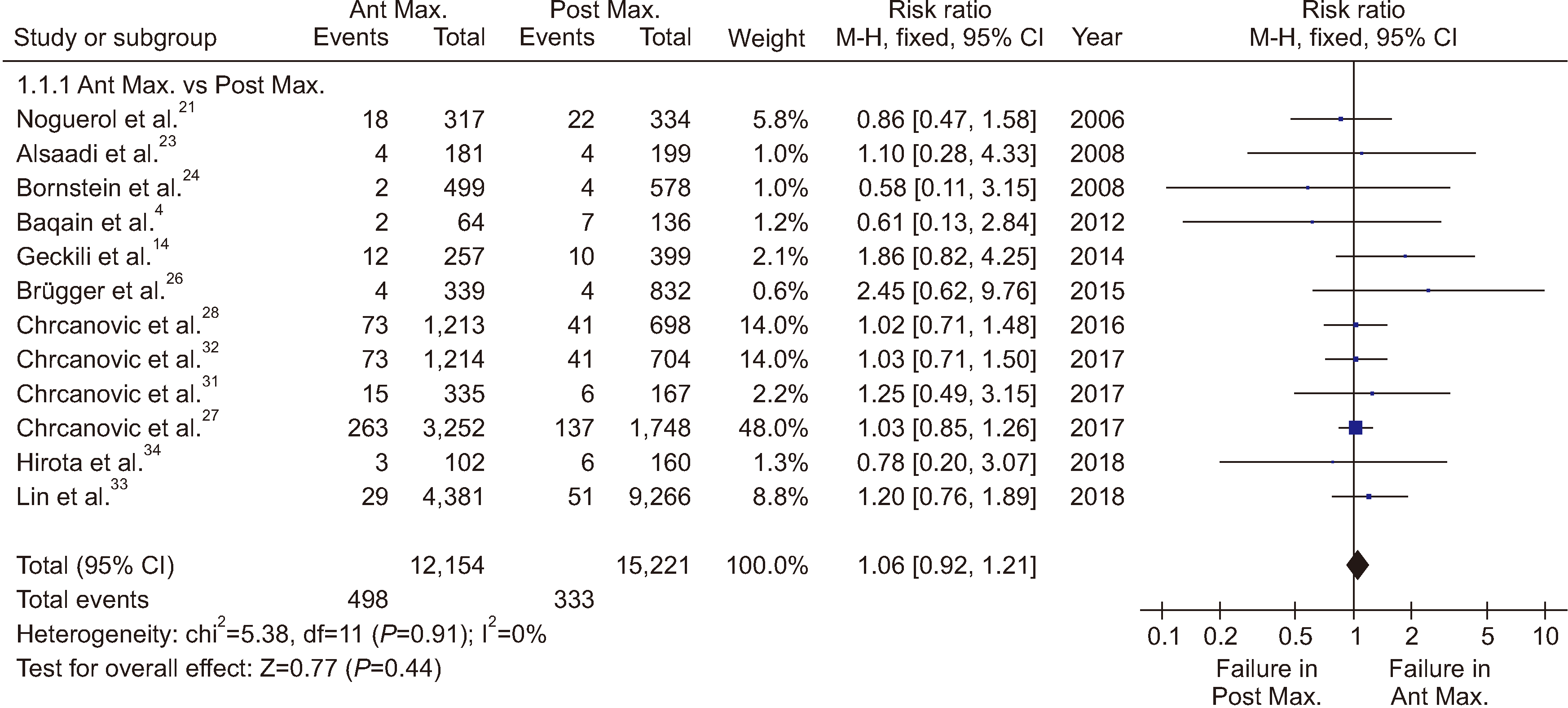
Anterior maxillary region compared with the posterior maxillary region
Data were extracted from 12 studies4,14,21,23,24,26-28,31-34 including a total of 27,375 implants: 12,154 inserted within the maxillary anterior region and 15,221 placed in the posterior region.
A total of 498 implants failed within the anterior region (4%) compared to 333 in the posterior region (2.19%).
Total subgroup analysis of the 12 studies applying a fixed effect, RR, and CI of 95% revealed a non-significant difference in failure between anterior and posterior maxillary regions (RR 1.06; 95% CI [0.92, 1.21]; P=0.44; I2=0%).(Fig. 5)
The RD analysis reported the same risk of failure in both the anterior and posterior maxillary regions.
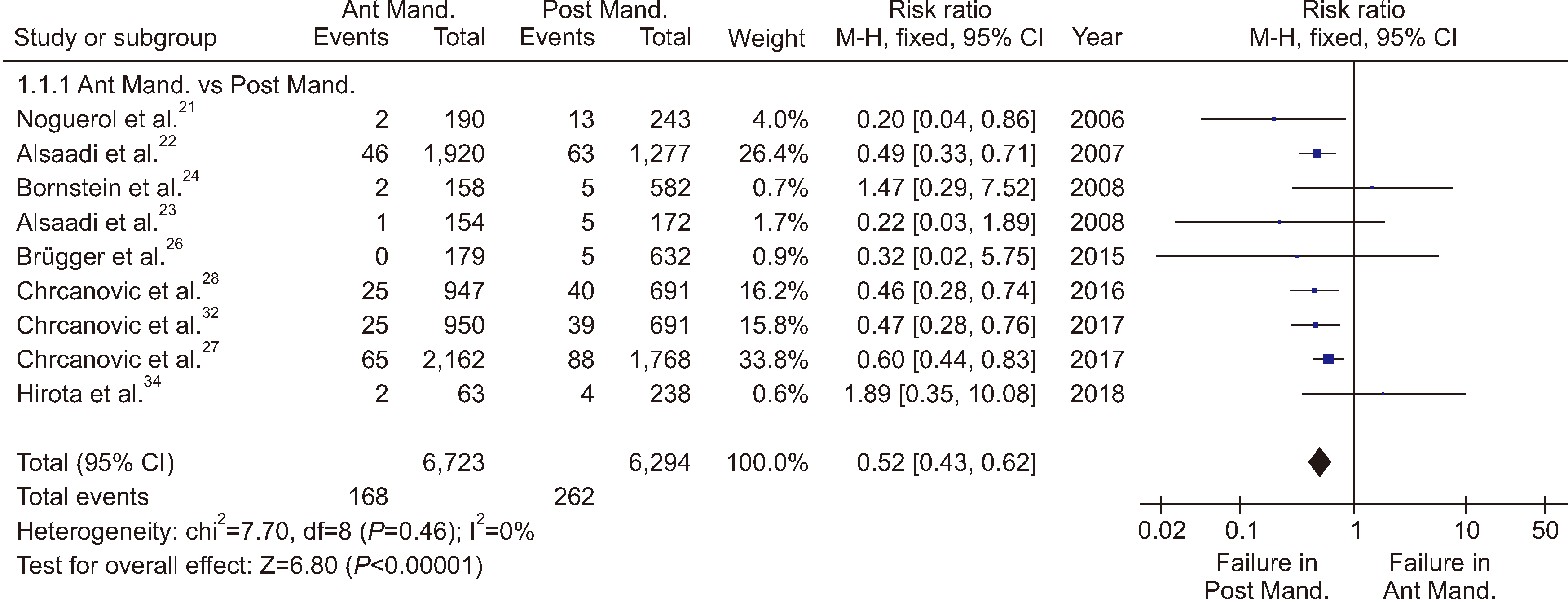
4) Anterior mandibular region compared with the posterior mandibular region
Data were extracted from nine studies21-24,26-28,32,34 with a total of 13,017 implants: 6,723 placed in the mandibular anterior region and 6,294 placed in the posterior region.
A total of 168 implants failed within the anterior region (2.5%) compared to the 262 failed implants in the posterior region (4.2%).
Total subgroup analysis of the nine identified studies applying a fixed effect, RR, and a CI of 95% revealed a significant difference between the anterior and posterior mandibular regions, with more failures in the posterior mandible (RR 0.52; 95% CI [0.43, 0.62]; P=0.46; I2=0%).(Fig. 6)
The analysis of RD reported 2% greater risk failure within the posterior mandibular region compared to the anterior region.
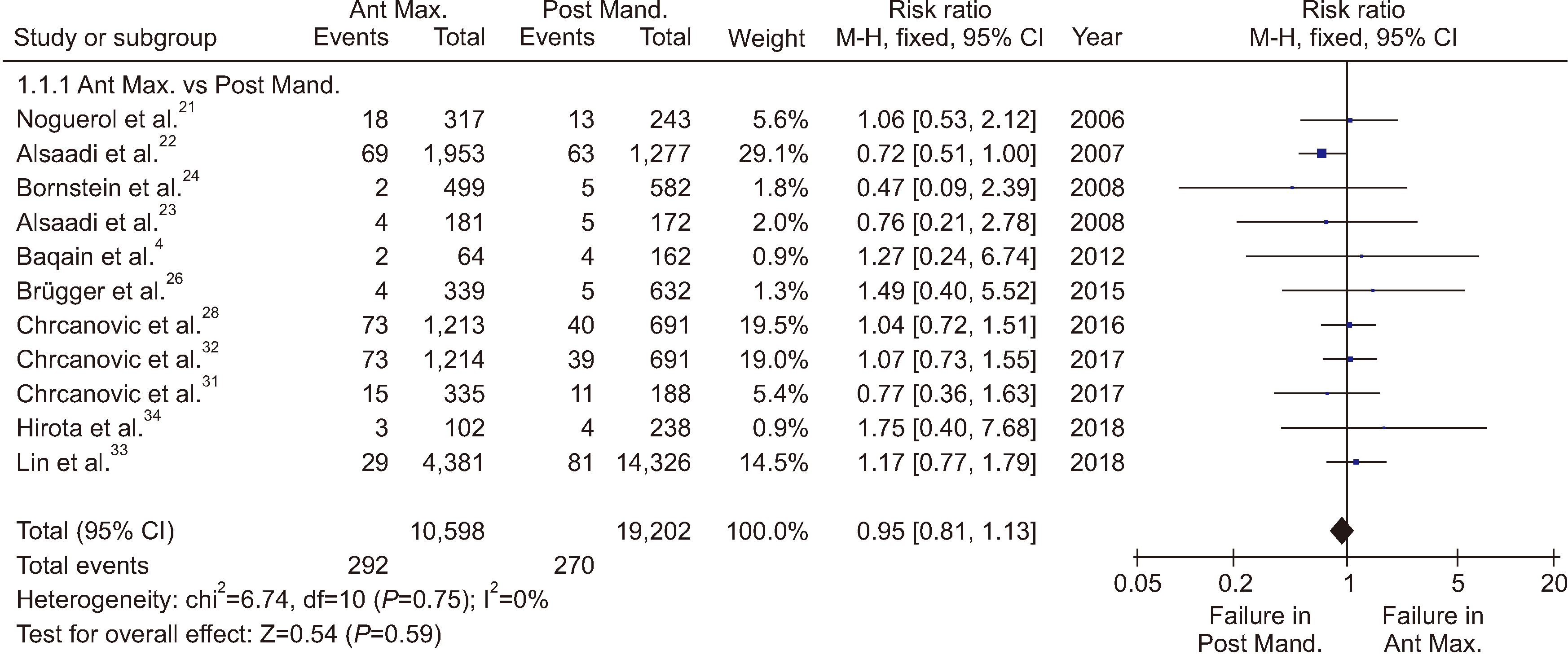
5) Anterior maxillary region compared with the posterior mandibular region
Data were extracted from 11 studies4,21-24,26,28,31-34 with a total of 29,800 implants, among which 10,598 were inserted in the maxillary anterior region and 19,202 placed in the posterior mandibular region.
A total of 292 implants failed in the anterior maxillary region (2.8%) compared to 270 failed implants in the posterior mandibular region (1.4%).
Total subgroup analysis of the 11 identified studies applying a fixed effect, RR, and a CI of 95% revealed a non-significant difference in failure between anterior maxilla and posterior mandibular regions (RR 0.95; 95% CI [0.81, 1.13]; P=0.59; I2=0%).(Fig. 7)
The RD analysis reported the same risk in the anterior maxillary and posterior mandibular regions.
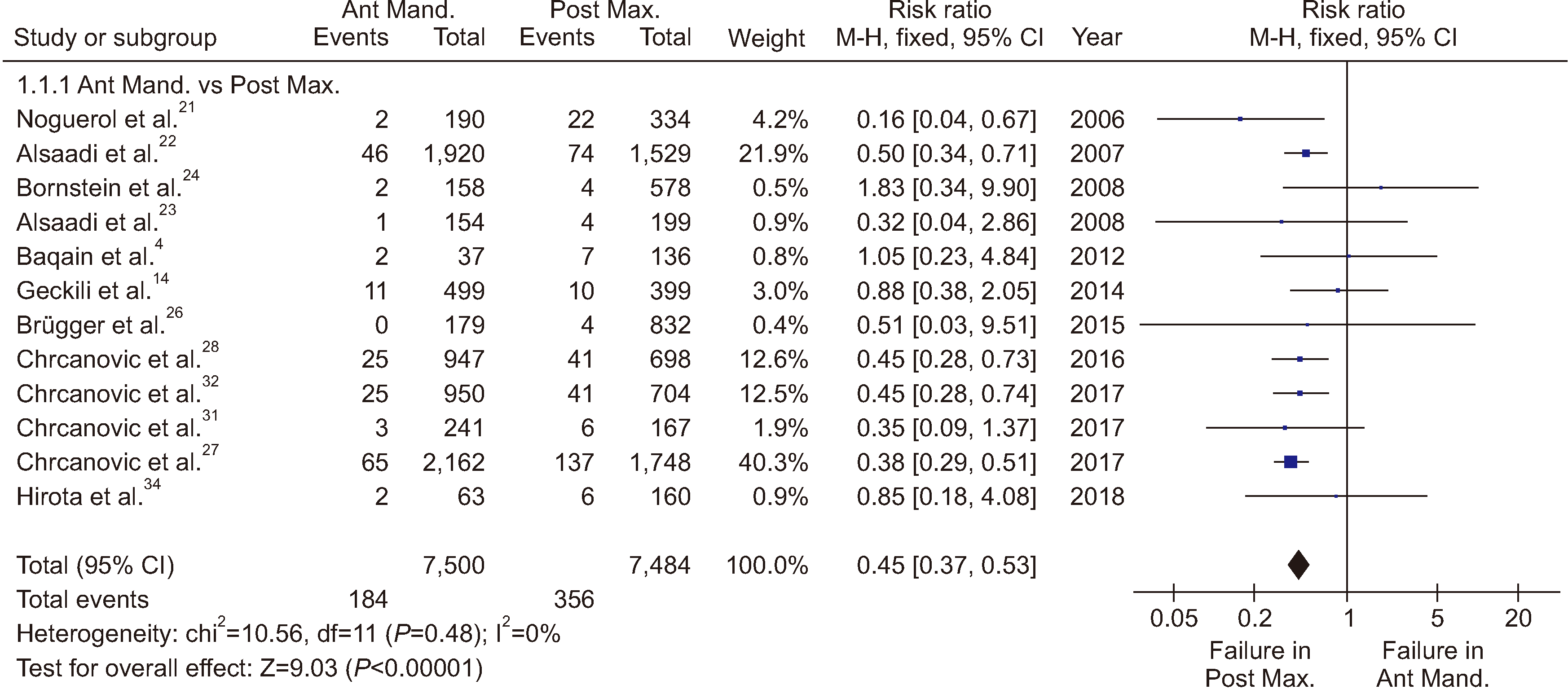
6) Anterior mandibular region compared with the posterior maxillary region
Data were extracted from 12 studies4,14,21-24,26-28,31,32,34 with a total of 14,984 implants, among which 7,500 were inserted into the anterior mandibular region and 7,484 placed in the posterior maxillary region.
A total of 184 implants failed in the anterior region of the mandible (2.5%) compared to 356 failed implants in the maxillary posterior region (4.8%).
Total subgroup analysis of the 12 studies applying a fixed effect, RR, and CI of 95% revealed significant failure in the posterior maxilla compared to the anterior mandible (RR 0.45; 95% CI [0.37, 0.53]; P<0.00001; I2=0%).(Fig. 8)
The RD analysis reported a 3% higher risk failure in the posterior maxillary region compared to the anterior mandibular region.
IV. Discussion
EIF depends on the surgeon’s skill, the type and site of implant, the condition of the bone, and the circumstances during the healing process.
The primary aim of this review was to explore any association between the site of implantation and EIF by comparing studies with implants inserted in different regions in either the upper and lower jaws to determine the most influential site on implant early failure and to change the practice of using a single implant design for all anatomical regions.
EIFs occur due to formation of fibrous tissue before osseointegration or micro-movements of the implants during healing. EIFs are linked to the quality of bone, implant design, bacterial invasion, or improper site preparation that impede the osseointegration process.
In the current review, studies of a total of 78,230 dental implants with recorded failures after insertion in different anatomical regions were analyzed using a fixed effects model. Studies could not ascertain all the confounding factors that could lead to EIFs.
In this review, we attempted to exclude studies with implants placed in immediate post-extraction sites according to the results of Quirynen et al.37 who linked pathology of the extracted tooth to early failure of the inserted implant. Furthermore, studies that included implants inserted after sinus elevation or bone expansion were excluded from analysis because of the presence of multiple confounding factors.
The percentage of EIFs in this review was 2.55%, and the published failure incidence varied from 2.5% to 3.8%22,38-42.
In agreement with Kim and Lee43, the maxilla has a thinner cortical plate and lower bone density than the mandible. Sites with poor bone quality may be at risk of inability to establish primary stability of the implants resulting in EIFs. High failure rates in the mandibular posterior region have been determined to be caused by overheating of the bone44.
Bone type affects EIF rates, yet some studies included implants inserted in poor quality bone without impacting the implant success rate23,45. A study conducted by Baqain et al.4 reported no difference in early failures for implants inserted in different bone types. Alsaadi et al.22,23 concluded that the implant site did not exhibit a significant effect on implant failures in their prospective study. Esposito et al.6 found that the success rate in the posterior maxillary region was the highest of the anterior maxillary region, posterior mandible, and anterior mandible. The results of the subgroup analysis in the current review agree with them which revealed a non-significant difference in failure between the posterior maxilla and posterior mandible (RR 1.07; 95% CI [0.92, 1.25]; P=0.68; I2=0%) rejecting the hypothesis of bone density.
The results of the current review confirmed that the failure rate of maxillary implants was significantly higher than that of mandibular implants, and implants placed in the maxilla result in two times more failures than those in the mandible.
This review examined the role of implant site as a risk factor responsible for early implant loss in both jaw bones. A RD of 1% was detected between the upper and lower jaws.
Subgroup analysis of the anterior maxillary region compared with the anterior mandibular region revealed more frequent EIF within the upper anterior region (6.2%) compared to the lower anterior region (2.5%). Applying a fixed effect and RR, a significant failure in the anterior maxilla was exhibited compared to the anterior mandible (RR 2.44; 95% CI [2.04, 2.91]; P<0.00001; I2=0%). A 3% RD was detected between the mandibular and maxillary anterior regions. This could be explained by the larger number of risk factors possible for the anterior maxillary region compared to any other site, in the form of lower bone density, undesirable forces due to angulation of the implants, and thin cortical plates. The anterior maxillary region exhibited the lowest success rate compared to the posterior maxillary, posterior mandible, and anterior mandible regions.
Posterior implants in both arches have also shown significant failures compared to anterior mandibular implants.
Restriction of the risk of failure to the insertion site is unfair, but it helps in improving implant designs and surgical techniques to be modified according to insertion site.
Retrospective studies may have the risk of missing data and inaccurate interpretation of the results. Narrowing of the inclusion criteria increases the homogeneity between studies but could result in exclusion of some trials with valuable data.
The percentage of EIF is high compared to failures after loading. This review identified a single primary factor for such failures, and the author recommends further evaluations of implantation site is consideration of other risk factors.
Construction of variable implant designs according to the bone quality of each anatomical site is recommended. Implants designed for high-risk regions should differ in design and surface characteristics compared to those inserted in low failure rate regions.
Implants that depend on bone compression to increase primary stability are not suitable in areas with dense bone. Finally, recently introduced modified implants in the form of nano-topographic surface treatments, high surface wettability, and growth factors are suggested to be placed in high-risk areas such as the anterior maxillary region based on cost benefits.
V. Conclusion
The anterior maxilla is a critical site for EIFs compared to other alveolar sites. Implants inserted in the anterior mandible exhibited the best success rate compared with other alveolar bone sites.
The maxilla is riskier than the mandible regarding EIFs, yet the difference between the posterior maxillary and the posterior mandibular regions is not significant. The RD between the maxilla and mandible is related to the high-risk anterior maxillary region.
 XML Download