Journal List > Obstet Gynecol Sci > v.63(4) > 1144677

Amirian, Mahani, and Abdi: Role of interleukin-6 (IL-6) in predicting gestational diabetes mellitus

Abstract

Gestational diabetes mellitus (GDM) is the most common pregnancy-associated metabolic disorder that is steadily increasing worldwide. Early diagnosis of pregnant women susceptible to GDM is the first step for deploying effective preventive treatment to reduce maternal, fetal, and neonatal complications. The diagnostic process of GDM is still controversial and interleukin-6 (IL-6) is one of the most recent markers used for the diagnosis of GDM. In this study, we aimed to systematically review the role of IL-6 in the diagnosis of GDM. In this systematic review, Google Scholar, Scopus, PubMed, ISI Web of Science, ProQuest, and MEDLINE databases were searched using the following keywords: GDM, screening, and IL-6, with the time interval 2009–2020. The quality of articles was assessed using the Strengthening the Reporting of Observational Studies in Epidemiology checklist. Twenty-four articles with desired quality that met the inclusion criteria were selected and reviewed further. Sixteen studies showed a statistically significant association, while 8 studies did not report any relationship between IL-6 levels and GDM. Based on the results of these studies, assessing the serum IL-6 levels can be investigated a newly established diagnostic biomarker for GDM. Therefore, through early diagnosis of susceptible women, effective measures can be implemented to reduce its complications.

Introduction

Diabetes is a metabolic disorder with steadily increasing prevalence. By 2014, 422 million adults were reported to have diabetes and at least 629 million people will be affected by 2045 if appropriate measures are not taken to reduce it. It has also been reported that high blood sugar causes 4 million deaths each year. The effects of diabetes go beyond the individual level, as it also affects the family and society and have wide-ranging socio-economic consequences [1]. Gestational diabetes mellitus (GDM) is a type of Diabetes, which is defined as diabetes diagnosed during the second or third trimester of pregnancy without prior detection [2]. GDM is a heterogeneous disorder resulting from the interactions between environmental and genetic factors [3]. Obesity and advanced maternal age are associated with the increasing prevalence of GDM worldwide. GDM heightens the potential risk of type 2 diabetes onset in the mother and her offspring [4]. The prevalence of GDM worldwide is estimated to be 17%, varying across different regions, with an estimate of 10% in North America and 25% in Southeast Asia [5].
GDM is associated with adverse pregnancy outcomes, including preeclampsia, polyhydramnios, fetal macrosomia, stillbirth, and neonatal complications such as hypoglycemia, hyperbilirubinemia, hypocalcemia, polycythemia, and respiratory distress [5]. Consequences of GDM extend beyond infancy and pregnancy, increasing the risks of metabolic syndrome, impaired glucose tolerance, and obesity in the offspring of affected mothers; it is a robust marker for the diagnosis of type 2 diabetes and diabetes-associated vascular diseases for the mother [6]. Various studies have shown that the lifestyle changes during pregnancy, especially in the early stages of pregnancy, can help in reducing the risk of GDM and also improve the adverse consequences associated with it [7].
Pregnancy represents a complex metabolic and physiological state in women. Insulin resistance plays a crucial role in the pathophysiology of GDM; in normal pregnancy, it can occur due to the increased secretion of diabetogenic placental hormones [8]. Despite over five decades of research, a common consensus on an internationally accepted screening method for GDM is yet to be achieved. Disagreements include the optimal time for screening, appropriate screening test, and general or selective screening methods [9]. According to World Health Organization (WHO), GDM can now be diagnosed with gestational glucose tolerance test using 75 grams of glucose at 24–28 gestational weeks [10]. Oral glucose tolerance test (OGTT) is an unpleasant test requiring consumption of 75 grams of glucose and delaying gastric discharge, which can cause nausea and vomiting. It is also a time-consuming method that requires overnight fasting before collection of 3 blood samples [11]. Moreover, an increase in the maternal blood glucose and fetal growth pathway occurs before 24 weeks of gestation, which has not been diagnosed in GDM. Early diagnosis of pregnant women with GDM and timely treatment can reduce the short- and long-term complications associated with it [12]. Researchers are currently investigating various markers to diagnose GDM, including interleukin-6 (IL-6) [13]. IL-6 is a cytokine produced by immune, adipose, and endothelial cells, and can have significant effects on glucose metabolism. IL-6 also affects pancreatic islet beta cells and enhances insulin secretion [14]. Additionally, inflammatory markers such as IL-6 have also been implicated in the pathogenesis of type 2 diabetes [15].
Numerous studies have been conducted on the association of IL-6 with GDM. Some studies have shown a statistically significant association between elevated IL-6 levels and GDM [4,13,16], while others did not report such relationships [17,18]. To this end, considering the contradictory results regarding the association between IL-6 and GDM, and based on the literature, there is no systematic review discussing the association between IL-6 and GDM. Therefore, in this systematic review, we aimed to investigate the association between IL-6 and GDM.

Methods

1. Search strategy

This study was conducted based on the guidelines of Preferred Reporting Items for Systematic Reviews and Meta-Analyses. In order to collect data in a systematic manner, reliable databases such as MEDLINE, ISI Web of Science, PubMed, Scopus, Google Scholar, and ProQuest were used (Table 1).

2. Inclusion and exclusion criteria

Inclusion criteria included all the observational articles published in English and Persian from January, 2009 to February, 2020, in which the healthy pregnant women were in the age group of 18–40 years and screened for gestational diabetes during 24–28 gestational weeks.
Lack of access to the full text of articles, protocol studies, case studies, brief reports, all non-Persian and non-English articles, as well as studies on high-risk pregnant women (over 40 years of age, with body mass index (BMI) over 30, family history of type 2 diabetes, history of polycystic ovary syndrome, thyroid problems, hypertension, diabetes, and tobacco use, and any other disorders affecting the maternal and neonatal health) were excluded.

3. Study selection

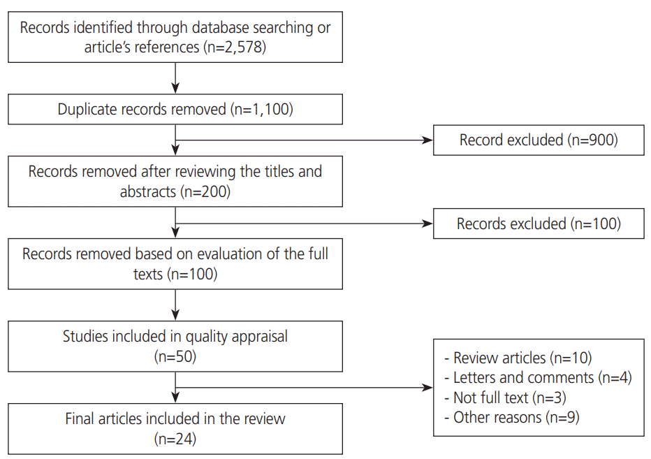
In the initial search, 2,578 articles were fetched. Two different researchers reviewed these articles and disagreements were resolved by a third one. Subsequently, 1,478 duplicated articles were removed. After reviewing the titles and abstracts, 900 more articles were excluded. After reviewing the full text of the articles in the next step, 100 articles were removed due to incompetence. Finally, 24 articles were considered sufficiently qualified and eligible for further reviewing (Fig. 1).

4. Quality assessment

Strengthening the Reporting of Observational Studies in Epidemiology (STROBE) statements were used to assess the quality of the studies. The STROBE statement as a valid tool consists of a checklist of 22 items to assess the quality of different parts of the observational studies [19,20].

5. Data extraction

Initially, the selection and evaluation of studies were performed independently by the 2 researchers, and the disagreements were resolved by a third one. Information on the first author’s name, year of publication, geographic region, study design, participants, BMI, sample, test time, test analysis method, diagnostic criteria of GDM, and levels of IL-6 were extracted and considered for the analysis.

Results

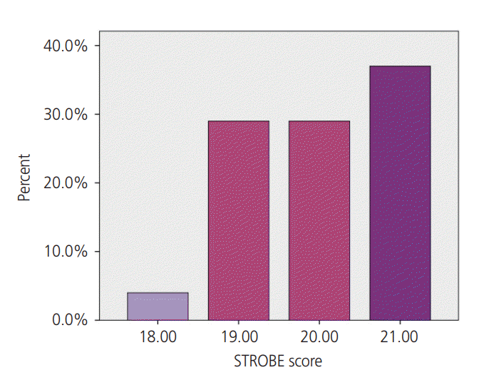
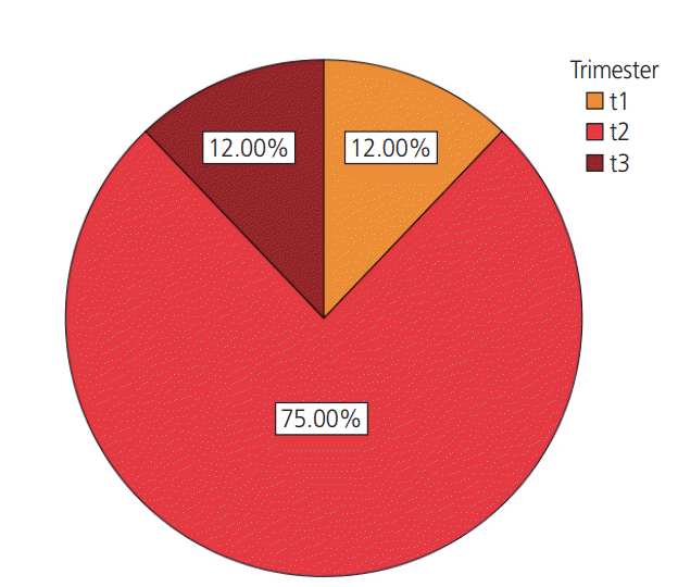
According to the process of search for articles, flowchart of which is presented in Fig. 1, 24 relevant high quality articles were selected and considered in this study after thoroughly reviewing the selected articles. Quality of the selected articles was assessed on the basis of the STROBE checklist (Fig. 2). These studies included articles published from 2009 to 2020, which were mainly case-control (n=17), cross-sectional (n=2), observational (n=2), cohort (n=2), and longitudinal (n=1) studies. A total of 2,806 pregnant women participated in these studies and blood samples were collected for measuring IL-6 levels in 3 studies during the first trimester (12%), in 18 studies during the second trimester (75%), and in 3 studies during the third trimester (12%) (Fig. 3).
The characteristics of the selected studies are listed in Table 2. These studies were conducted in different countries, including China (6), Turkey (4), US (2), Poland (2), India (2), Australia (1), Brazil (1), Prague (1), Tunisia (1), Saudi Arabia (1), Canada (1), Finland (1), and Greece (1). Moreover, for the diagnosis of GDM, 5 studies used the guidelines of Carpenter and Coustan, 1 study used the Indian criteria, 2 studies used the guidelines of International Association of Diabetes and Pregnancy Study Groups (IADPSG), 5 studies used the American Diabetes Association (ADA) guidelines, 4 studies used the National Diabetes Data Group guidelines, 2 studies used the China Diabetes Association Diabetes Branch guidelines, 1 study used the Australasian Diabetes in Pregnancy Society guidelines, 1 study used the Polish Diabetes Association guidelines, 1 study used the American College of Obstetricians and Gynecologists (ACOG) guidelines, 1 study used the Canadian Diabetes Association guidelines, 1 study used the WHO guidelines, 1 study used the Endocrine Society Clinical Practice Guideline/ACOG guidelines, 1 study used the ACOG/ADA guidelines, and 1 study used the Carpenter and Coustan/ADA guidelines. The majority of the studies measured IL-6 levels using the enzyme-linked immunosorbent assay (18), while the others employed multiplex immunoassay (4) or chemiluminescent immunoassay (2).
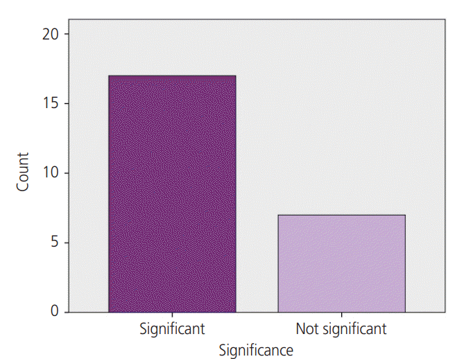
Fig. 4 shows the significance level of the selected studies. As shown in this diagram, 16 studies exhibited a significant relationship between the serum level of IL-6 and GDM. Therefore, we can conclude that the serum IL-6 level may act as a suitable diagnostic marker for GDM.

Discussion

In the present systematic review, 24 articles were reviewed, most of which demonstrated a significant relationship between IL-6 levels and GDM. Thus, we concluded that IL-6 can be used as a marker to predict the occurrence of gestational diabetes.
Accurate and early diagnosis of women at a high risk of developing GDM provides an opportunity to manage the prenatal care models and apply future interventions to reduce the progression of gestational diabetes, and thereby its associated health care expenses and side effects [40]. However, the diagnostic criteria for GDM is still debatable. A previous study reported a linear relationship between maternal blood glucose levels and adverse perinatal outcomes [41]. The IADPSG recommends that the studies aimed to diagnose gestational diabetes must develop simpler, more cost-effective methods that do not require OGTT [42]. In recent years, there has been a great interest in identifying the role of inflammation during the development of GDM. Inflammatory factors can act as insulin antagonists and cause insulin resistance [43]. IL-6, as a cytokine, plays a crucial role in the pathophysiology of glucose intolerance and serves as a potential serum marker for early screening of glucose intolerance [43].
In type 2 diabetes, inflammatory cytokines may induce insulin resistance by suppressing multiple pathways in target tissues that are responsible for proper insulin signaling [44]. Insulin resistance is associated with abnormal secretion of proinflammatory cytokines, such as IL-6 [4]. In non-pregnant women, BMI and high body fat mass have been found to be associated with elevated levels of serum IL-6 [13]. IL-6 is also secreted by the placenta during pregnancy, which can lead to a chronic inflammatory process in adipose tissue and further assist the development of pregnancy-induced insulin resistance [45]. In GDM, similar to type 2 diabetes, insulin resistance is implicated in the pathophysiology [46]. Type 2 diabetes is considered a chronic inflammatory disease and IL-6 is a risk factor for the development of type 2 diabetes. Therefore, due to similar mechanisms underlying the onset of GDM and type 2 diabetes, IL-6 might act as an effective marker in predicting GDM.
In this systematic review, quantitative analysis of available data on the relationship between serum IL-6 levels and GDM was performed based on 24 selected articles, of which 16 articles showed an association between elevated serum IL-6 levels in women and GDM. For example, the results of a case-control study conducted by Siddiqui et al. [13] that aimed to investigate the association of IL-6 and C-reactive protein with GDM in Indian women demonstrated that the serum levels of IL-6 in women with GDM was significantly higher than in control women. Moreover, IL-6 levels were also associated with pre-pregnancy BMI, fasting blood sugar, and postprandial blood sugar . A study conducted by Yu et al. [22], which aimed at investigating the changes in gut flora and various inflammatory factors in patients with GDM, showed that the serum levels of inflammatory factors, including IL-6, were significantly higher in the case group than that in the control group; moreover, patients with GDM were highly susceptible to intestinal flora imbalances with elevated inflammatory factors, which affected the immune function in these patients and may play an important role in the development of diabetes. Furthermore, the findings of the study conducted by Zhao et al. [23], which aimed to examine the possible association of inflammatory markers with glucose intolerance and GDM in Chinese women, indicated that IL-6 levels were significantly higher in pregnant women with GDM or glucose intolerance compared to those in the healthy control group, and that there was a positive relationship between inflammatory cytokines, BMI, and HbA1c . In the study conducted by Zhang et al. [25], aimed at investigating the association between inflammatory and metabolic biomarkers in women with GDM in Mongolia, showed that the levels of inflammatory and placental biomarkers, including IL-6, in both serum and placenta showed a significant difference between women with GDM and those with healthy pregnancies.
The present review also included studies that did not show a correlation between the serum levels of IL-6 and GDM, such as the study conducted by Abell et al. [24], which aimed at evaluating the relationship between GDM risk using fasting glucose and serum biomarkers at the early pregnancy, and indicated that the serum IL-6 levels did not improve the ability to predict the risk of GDM. The results of another study conducted by Gümüş et al. [29] aimed to evaluate the possible association between clinical and biochemical parameters with GDM and gingivitis; this study showed that there was no association between the serum IL-6 levels and GDM. Similar results were obtained in an another study conducted by Özyer et al. [34] that aimed at investigating the association of inflammatory mediators, including IL-6, with glycemic status in pregnancy; the results of this study indicated that the maternal serum levels of inflammatory mediators are not relevant for assessing GDM during the late second or early trimester. The reason for such differences in the results of these studies can be attributed to the use of different methods and kits for measuring IL-6 levels and dissimilar diagnostic criteria for GDM. Also, the effects of confounding variables on serum IL-6 levels and GDM were not considered in all of the selected articles.
Increased IL-6 secretion during pregnancy has been associated with GDM in several studies. Conversely, OGTT testing with 75 grams of oral glucose, which is currently the gold standard test, is performed almost late in pregnancy and requires overnight fasting. Moreover, consumption of glucose is not very pleasant for a pregnant woman and it requires the collection of blood samples 3 times during the process. However, assessing IL-6 levels does not have the above-mentioned challenges and it is easy, affordable, and tolerable for a pregnant woman. Therefore, IL-6 can be used as a marker for assessing the risk of GDM in pregnant women. Considering the increasing prevalence of gestational diabetes worldwide and the need for timely diagnosis and treatment of GDM to reduce adverse maternal and fetal complications, an acceptable marker such as IL-6 is urgently needed.

Conclusion

The results of this study indicate that serum IL-6 levels are significantly higher in pregnant women with GDM than in healthy pregnant women. Therefore, the evaluation of this marker as an acceptable, inexpensive, and readily available diagnostic criterion for assessing the risk of GDM can be investigated.
One of the limitations of this systematic review is the heterogeneity of the diagnostic criteria for GDM. Different commercial kits and assays have been employed to measure the serum IL-6 levels. Conversely, based on the extensive research and use of multiple studies with different ethnicities, it is difficult to control the effect of this variable. Additionally, all the previous studies that were considered did not offer adequate clinical information for performing a robust meta-analysis.

ACKNOWLEDGMENTS

We would like to thank Alborz University of Medical Sciences.

Notes

Conflict of interest

No potential conflict of interest relevant to this article was reported.

Ethical approval This study has a code of ethics No. IR.ABZUMS.REC.1398.218 approved by Alborz University of Medical Sciences.

Patient consent There is no need for patient consent in this review article.

References

1. World Health Organization. Classification of diabetes mellitus. Geneva: World Health Organization;2019.
2. American Diabetes Association. Classification and diagnosis of diabetes: standards of medical care in diabetes-2019. Diabetes Care. 2019; 42:S13–28.
3. Koivusalo SB, Rönö K, Klemetti MM, Roine RP, Lindström J, Erkkola M, et al. Gestational diabetes mellitus can be prevented by lifestyle intervention: the Finnish Gestational Diabetes Prevention Study (RADIEL): a randomized controlled trial. Diabetes Care. 2016; 39:24–30.
4. Sudharshana Murthy KA, Bhandiwada A, Chandan SL, Gowda SL, Sindhusree G. Evaluation of oxidative stress and proinflammatory cytokines in gestational diabetes mellitus and their correlation with pregnancy outcome. Indian J Endocrinol Metab. 2018; 22:79–84.
crossref
5. Durnwald C. Diabetes mellitus in pregnancy: screening and diagnosis [Internet]. Waltham (MA): UpToDate, Inc;c2019 [cited 2019 Nov 25]. Available from: https://www.uptodate.com/contents/diabetes-mellitus-in-pregnancyscreening-and-diagnosis.
6. Caughey AB. Gestational diabetes mellitus: obstetric issues and management [Internet]. Waltham (MA): UpToDate, Inc;c2019 [cited 2019 Nov 30]. Available from: https://www.uptodate.com/contents/gestationaldiabetes-mellitus-obstetric-issues-and-management.
7. Song C, Li J, Leng J, Ma RC, Yang X. Lifestyle intervention can reduce the risk of gestational diabetes: a meta-analysis of randomized controlled trials. Obes Rev. 2016; 17:960–9.
crossref
8. Mirghani Dirar A, Doupis J. Gestational diabetes from A to Z. World J Diabetes. 2017; 8:489–511.
crossref
9. Cunningham FG, Leveno KJ, Bloom SL, Dashe JS, Hoffman BL, Casey BM, et al. Williams obstetrics. 25th ed. New York (NY): Mcgraw-hill;2018.
10. World Health Organization. Diagnostic criteria and classification of hyperglycaemia first detected in pregnancy. Geneva: World Health Organization;2013.
11. Amreen S, Suneel A, Shetty A, Vasudeva A, Kumar P. Use of glycosylated HbA1c and random blood sugar as a screening tool for gestational diabetes mellitus in first trimester. Int J Reprod Contracept Obstet Gynecol. 2018; 7:524–8.
crossref
12. Huhn EA, Rossi SW, Hoesli I, Göbl CS. Controversies in screening and diagnostic criteria for gestational diabetes in early and late pregnancy. Front Endocrinol (Lausanne). 2018; 9:696.
crossref
13. Siddiqui S, Waghdhare S, Goel C, Panda M, Soneja H, Sundar J, et al. Augmentation of IL-6 production contributes to development of gestational diabetes mellitus: an Indian study. Diabetes Metab Syndr. 2019; 13:895–9.
crossref
14. Suzuki T, Imai J, Yamada T, Ishigaki Y, Kaneko K, Uno K, et al. Interleukin-6 enhances glucose-stimulated insulin secretion from pancreatic β-cells: potential involvement of the PLC-IP3-dependent pathway. Diabetes. 2011; 60:537–47.
crossref
15. Lainampetch J, Panprathip P, Phosat C, Chumpathat N, Prangthip P, Soonthornworasiri N, et al. Association of tumor necrosis factor alpha, interleukin 6, and C-reactive protein with the risk of developing type 2 diabetes: a retrospective cohort study of rural thais. J Diabetes Res. 2019; 2019:9051929.
crossref
16. Yang Y, Liu L, Liu B, Li Q, Wang Z, Fan S, et al. Functional defects of regulatory T cell through interleukin 10 mediated mechanism in the induction of gestational diabetes mellitus. DNA Cell Biol. 2018; 37:278–85.
crossref
17. Braga FO, Negrato CA, Matta MFBD, Carneiro JR, Gomes MB. Relationship between inflammatory markers, glycated hemoglobin and placental weight on fetal outcomes in women with gestational diabetes. Arch Endocrinol Metab. 2019; 63:22–9.
crossref
18. Šimják P, Cinkajzlová A, Anderlová K, Kloučková J, Kratochvílová H, Lacinová Z, et al. Changes in plasma concentrations and mRNA expression of hepatokines fetuin A, fetuin B and FGF21 in physiological pregnancy and gestational diabetes mellitus. Physiol Res. 2018; 67:S531–42.
crossref
19. Abdi F, Ozgoli G, Rahnemaie FS. A systematic review of the role of vitamin D and calcium in premenstrual syndrome. Obstet Gynecol Sci. 2019; 62:73–86.
crossref
20. Rahnemaie FS, Zare E, Zaheri F, Abdi F. Effects of complementary medicine on successful breastfeeding and its associated issues in the postpartum period. Iran J Pediatr. 2019; 29:e80180.
crossref
21. Wang X, Liu J, Wang D, Zhu H, Kang L, Jiang J. Expression and correlation of Chemerin and FABP4 in peripheral blood of gestational diabetes mellitus patients. Exp Ther Med. 2020; 19:710–6.
crossref
22. Yu H, Liu Z, Dong S. Changes in intestinal flora, TNF-α, L-17, and IL-6 levels in patients with gestational diabetes mellitus. Eur J Inflamm;Forthcoming 2018.
23. Zhao X, Liu J, Shen L, Wang A, Wang R. Correlation between inflammatory markers (hs-CRP, TNF-α, IL-1β, IL-6, IL-18), glucose intolerance, and gestational diabetes mellitus in pregnant women. Int J Clin Exp Med. 2018; 11:8310–6.
24. Abell SK, Shorakae S, Harrison CL, Hiam D, MorenoAsso A, Stepto NK, et al. The association between dysregulated adipocytokines in early pregnancy and development of gestational diabetes. Diabetes Metab Res Rev. 2017; 33:e2926.
crossref
25. Zhang J, Chi H, Xiao H, Tian X, Wang Y, Yun X, et al. Interleukin 6 (IL-6) and tumor necrosis factor α (TNF-α) single nucleotide polymorphisms (SNPs), inflammation and metabolism in gestational diabetes mellitus in Inner Mongolia. Med Sci Monit. 2017; 23:4149–57.
crossref
26. Bossick AS, Peters RM, Burmeister C, Kakumanu N, Shill JE, Cassidy-Bushrow AE. Antenatal inflammation and gestational diabetes mellitus risk among pregnant African-American women. J Reprod Immunol. 2016; 115:1–5.
crossref
27. Oztop N, Kusku-Kiraz Z, Dervisoglu E, Dinccag N, Genc S. The association of glycemic markers with plasma adipocytokine levels in women with gestational diabetes. J Diabetes Metab. 2016; 7:1000699.
crossref
28. Qu X, Yu H, Jia B, Yu X, Cui Q, Liu Z, et al. Association of downregulated HDAC 2 with the impaired mitochondrial function and cytokine secretion in the monocytes/macrophages from gestational diabetes mellitus patients. Cell Biol Int. 2016; 40:642–51.
crossref
29. Gümüş P, Özçaka Ö, Ceyhan-Öztürk B, Akcali A, Lappin DF, Buduneli N. Evaluation of biochemical parameters and local and systemic levels of osteoactive and B-cell stimulatory factors in gestational diabetes in the presence or absence of gingivitis. J Periodontol. 2015; 86:387–97.
crossref
30. Hassiakos D, Eleftheriades M, Papastefanou I, Lambrinoudaki I, Kappou D, Lavranos D, et al. Increased maternal serum interleukin-6 concentrations at 11 to 14 weeks of gestation in low risk pregnancies complicated with gestational diabetes mellitus: development of a prediction model. Horm Metab Res. 2016; 48:35–41.
crossref
31. Kim SY, Sy V, Araki T, Babushkin N, Huang D, Tan D, et al. Total adiponectin, but not inflammatory markers Creactive protein, tumor necrosis factor-α, interluekin-6 and monocyte chemoattractant protein-1, correlates with increasing glucose intolerance in pregnant Chinese-Americans. J Diabetes. 2014; 6:360–8.
32. Kuźmicki M, Telejko B, Lipińska D, Pliszka J, Wilk J, Wawrusiewicz-Kurylonek N, et al. The IL-6/IL-6R/sgp130 system and Th17 associated cytokines in patients with gestational diabetes. Endokrynol Pol. 2014; 65:169–75.
33. Nergiz S, Altınkaya ÖS, Küçük M, Yüksel H, Sezer SD, Kurt Ömürlü İ, et al. Circulating galanin and IL-6 concentrations in gestational diabetes mellitus. Gynecol Endocrinol. 2014; 30:236–40.
crossref
34. Özyer Ş, Engin-Üstün Y, Uzunlar Ö, Katar C, Danışman N. Inflammation and glycemic tolerance status in pregnancy: the role of maternal adiposity. Gynecol Obstet Invest. 2014; 78:53–8.
crossref
35. Mrizak I, Arfa A, Fekih M, Debbabi H, Bouslema A, Boumaiza I, et al. Inflammation and impaired endotheliumdependant vasodilatation in non obese women with gestational diabetes mellitus: preliminary results. Lipids Health Dis. 2013; 12:93.
crossref
36. Abdel Gader AG, Khashoggi TY, Habib F, Awadallah SB. Haemostatic and cytokine changes in gestational diabetes mellitus. Gynecol Endocrinol. 2011; 27:356–60.
crossref
37. Morisset AS, Dubé MC, Côté JA, Robitaille J, Weisnagel SJ, Tchernof A. Circulating interleukin‐6 concentrations during and after gestational diabetes mellitus. Acta Obstet Gynecol Scand. 2011; 90:524–30.
crossref
38. Pöyhönen-Alho M, Ebeling P, Saarinen A, Kaaja R. Decreased variation of inflammatory markers in gestational diabetes. Diabetes Metab Res Rev. 2011; 27:269–76.
crossref
39. Kuzmicki M, Telejko B, Szamatowicz J, Zonenberg A, Nikolajuk A, Kretowski A, et al. High resistin and interleukin-6 levels are associated with gestational diabetes mellitus. Gynecol Endocrinol. 2009; 25:258–63.
crossref
40. Harrison CL, Lombard CB, East C, Boyle J, Teede HJ. Risk stratification in early pregnancy for women at increased risk of gestational diabetes. Diabetes Res Clin Pract. 2015; 107:61–8.
crossref
41. Farrar D, Duley L, Dowswell T, Lawlor DA. Different strategies for diagnosing gestational diabetes to improve maternal and infant health. Cochrane Database Syst Rev. 2017; 8:CD007122.
crossref
42. Weinert LS. International Association of Diabetes and Pregnancy Study Groups recommendations on the diagnosis and classification of hyperglycemia in pregnancy: comment to the International Association of Diabetes and Pregnancy Study Groups consensus panel. Diabetes Care. 2010; 33:e97.
crossref
43. Kleiblova P, Dostalova I, Bartlova M, Lacinova Z, Ticha I, Krejci V, et al. Expression of adipokines and estrogen receptors in adipose tissue and placenta of patients with gestational diabetes mellitus. Mol Cell Endocrinol. 2010; 314:150–6.
crossref
44. Khodabandehloo H, Gorgani-Firuzjaee S, Panahi G, Meshkani R. Molecular and cellular mechanisms linking inflammation to insulin resistance and β-cell dysfunction. Transl Res. 2016; 167:228–56.
crossref
45. Sun D, Li F, Zhang Y, Xu X. Associations of the prepregnancy BMI and gestational BMI gain with pregnancy outcomes in Chinese women with gestational diabetes mellitus. Int J Clin Exp Med. 2014; 7:5784–9.
46. Vejrazkova D, Vcelak J, Vankova M, Lukasova P, Bradnova O, Halkova T, et al. Steroids and insulin resistance in pregnancy. J Steroid Biochem Mol Biol. 2014; 139:122–9.
crossref

Fig. 1.
Flow diagram for searching the articles.
ogs-20020f1.tif
Fig. 2.
Strengthening the Reporting of Observational Studies in Epidemiology (STROBE) score of different studies.
ogs-20020f2.tif
Fig. 3.
Trimester in which the interleukin-6 test was performed.
ogs-20020f3.tif
Fig. 4.
Assessing the significance level of different studies.
ogs-20020f4.tif
Table 1.
Search strategy
No. Search term
#1 ‘Gestational diabetes’ [tiab], OR ‘GD’ [tiab], OR ‘Gestational Diabetes Mellitus’ [tiab], OR ‘GDM’ [tiab], OR ‘Diabetes, Pregnancy- Induced’ [tiab], OR ‘Pregnancy-Induced Diabetes ‘ [tiab]
#2 ‘Screening’ [tiab], OR ‘Predicting’[tiab], OR ‘Diagnosis’[tiab]
#3 ‘ Interleukin-6’ [tiab], OR ‘IL-6’ [tiab]
#1 AND #2
#1 AND #3
#1 AND #2 AND #3
Table 2.
Results from a systematic review of studies
Author Geographic region Study design GDM group
Control group
Sample Time of collect test (wk) Method of analysis test Diagnostic criteria of GDM GDM group
Control group
Results (P-value)
Participants BMI Participants BMI IL-6 IL-6
Wang et al. [21] China Case-control 60 NR 50 NR Serum 24–28 ELISA ADA 96.66 (88.33–106.66)a) 83.32 (78.34–88.33)a) Significant (P<0.001)
Braga et al. [17] Brazil Observational 78 27.8 (23.6–32.1)a) 98 22.8 (20.9–27.3)a) Serum 24–28 Multiplex immunoassay Carpenter and Coustan 0.175 (0.120–0.298)a) 0.155 (0.100–0.325)a) Not significant (P=0.77)
Sudharshana Murthy et al. [4] India Observational 30 25.7b) 30 25b) Serum 24–28 ELISA India criteria 2.96±1.37c) 2.88±1.21c) Significant (NR)
Siddiqui et al. [13] India Case-control 53 25.3±4.08c) 50 23.28±2.95c) Serum 24–31 ELISA (disclose) ADA 0.72±0.67d) 0.39±0.70d) Significant (P=0.001)
Šimják et al. [18] Prague Case-control 12 V1: 26.04±3.75d) 12 V1: 27.63±4.59d) Serum V1: 28–32 Multiplex immunoassay IADPSG V1 :4.33±0.81d) V1: 2.78±0.37d) Not significant (NR)
V2: 27.59±3.54d) V2: 28.74±4.63d) V2: 36–38 V2: 5.02±1.13d) V2: 3.69±0.29d)
Yang et al. [16] China Cohort 21 31.60±3.70c) 34 31.70±3.10c) Serum <13 ELISA Carpenter and Coustan 17/5 (12–23)a) 11 (5–7)a) Significant (P=0.001)
Yu et al. [22] China Case-control 80 NR 60 NR Serum 24–29 ELISA CDADB 59.44±3.95 22.81±1.54 Significant (P=0.021)
Zhao et al. [23] China Cross sectional 29 28.10±3.20c) 32 24.10±3.00c) Serum 24–28 ELISA Carpenter and Coustan 5.10±1.20c) 2.96±0.70c) Significant (P<0.001)
Abell et al. [24] Australia Cohort 25 28.0 (26.0–31.0)a) 78 29.5 (25.5–34.0)a) Serum 12–15 ELISA ADIPS 2.03 (1.67–2.90)a) 1.92 (1.57–2.56)a) Not significant (P=0.428)
Zhang et al. [25] China Case-control 60 38.68±9.50c) 60 28.74±7.26c) Serum 24–28 Multiplex immunoassay ESCPG/ACOG 5.85±1.41c) 3.91±1.66c) Significant (P=0.002)
Bossick et al. [26] USA Case-control 18 37.10±7.70c) 167 28.40±7.10c) Serum 13–28 Multiplex immunoassay ACOG/ADA 5.40±2.00c) 3.90±2.10c) Significant (P=0.002)
Oztop et al. [27] Turkey Case-control 30 26.4 (17.3–44.1)a) 20 24 (21–26)a) Serum 24–26 ELISA ADA 3.10±0.90c) 2.70±0.70c) Significant (P=0.04)
Qu et al. [28] China Case-control 53 32.17±4.36 57 23.62±3.51 Serum 22–30 ELISA Carpenter and Coustan 4.43±0.45c) 2.57±0.19c) Significant (P=0.0004)
Gümüş et al. [29] Turkey Case-control 101 27.00±5.00c) 66 22.00±3.00c) Serum 24–28 ELISA ADA 19.21±29.00c) 15.49±19.13c) Not significant (P=0.933)
Hassiakos et al. [30] Greece Case-control 40 25.36e) 94 23.16e) Serum 11–14 Chemiluminescent immunoassay IADPSG 2.0 (1.60)f) 1.5 (1.01)f) Significant (P=0.001)
Kim et al. [31] USA Case-control 32 24.1±0.9d) 227 24.9±0.6d) Serum 24–28 ELISA Carpenter and Coustan 2.6±0.2d) 2.9±0.9d) Not significant (NR)
Kuźmicki et al. [32] Poland Case-control 46 V1: 25.0 (24.2–28.0)a) 45 V1: 23.0 (20.4–26.2)a) Serum V1: 24–28 ELISA PDA V1: 0.99 (0.86–1.27)a) V1: 0.89 (0.75–1.07)a) Significant (P<0.05)
V2: 25.0 (24.2–28.0)a) V2: 23.0 (20.4–26.2)a) V2: 29–32 V2: 1.13 (0.98–1.57)a) V2: 1.02 (0.83–1.22)a)
Nergiz et al. [33] Turkey Case-control 30 30.20±5.60c) 30 29.10±30.00c) Serum >24 ELISA ACOG 32.5 (24.8–48)a) 7.1 (4.2–9.8)a) Significant (P=0.001)
Özyer et al. [34] Turkey Case-control 35 30.40±4.30c) 70 25.30±3.80c) Serum 24–28 Chemiluminescent sequential immunometric assay Carpenter and Coustan/ADA 2.0 (2.0–2.00)a) 2.0 (2.0–2.51)a) Not significant (P=0.08)
Mrizak et al. [35] Tunisia Case-control 28 28.50±1.80c) 24 29.7±2.23c) Serum 24–26 ELISA NDDG 77.84±10.98c) 53.17±1.45c) Significant (P=0.002)
Abdel Gader et al. [36] Saudi Arabia Cross-sectional 150 34.40±5.90c) 100 30.60±4.60c) Serum 35–40 ELISA NDDG 13.70+2.50c) 13.90+15.30c) Not significant (NR)
Morisset et al. [37] Canada Longitudinal 20 28.20±7.50 27 24.20±4.30 Serum 26.1±3.7 ELISA CDA 1.47±0.72 0.90±0.32 Significant (P≤0.05)
Pöyhönen-Alho et al. [38] Finland Case-control 38 30.6 21 26.9 Plasma 24–28 ELISA ADA 4.9±2.21c) 4.91±2.82c) Not significant (P=0.58)
Kuzmicki et al. [39] Poland Case-control 81 22.1 (20.5–24.9)a) 82 23.1 (20.3–25.0)a) Serum 24–31 ELISA WHO 1.0 (0.7–1.5)a) 0.8 (0.5–1.1)a) Significant (P=0.006)

GDM, gestational diabetes mellitus; BMI, body mass index; IL, interleukin; V1, visit 1; V2, visit 2; ELISA, enzyme-linked immunosorbent assay; ADA, American Diabetes Association; IADPSG, International Association of Diabetes and Pregnancy Study Groups; CDADB, China Diabetes Association Diabetes Branch; ADIPS, Australasian Diabetes in Pregnancy Society; ESCPG, Endocrine Society Clinical Practice Guideline; ACOG, American College of Obstetricians and Gynecologists; PDA, Polish Diabetes Association; NDDG, National Diabetes Data Group; CDA, Canadian Diabetes Association; WHO, World Health Organization; NR, not reported; SD, standard deviation.

a) Median (interquartile range);

b) Mean;

c) Mean±SD;

d) Means±standard error of the mean;

e) Median;

f) Median (SD).

TOOLS
Similar articles