INTRODUCTION
Various research and debates have been conducted about whether glycated hemoglobin (HbA1c)—an index for the overall hyperglycemia—is more related to fasting hyperglycemia (FHG) or postprandial hyperglycemia (PPG). In 1997, Avignon et al. [
1] first measured blood glucose at different times of the day to study the relative effect of FHG and PPG on HbA1c. They found that PPG was a better predictor of overall hyperglycemia than FHG. Analyzing the diabetes control and complication trial (DCCT) data, Rohlfing et al. [
2] also showed that PPG had a linear association with HbA1c and had a stronger association with overall hyperglycemia than FHG. In contrast, Bonora et al. [
3] reported that FHG had a stronger association with overall hyperglycemia than PPG.
To re-examine these associations, Monnier et al. [
4] had patients with type 2 diabetes mellitus (T2DM) blood glucose measured at four points of time and calculated the area under the blood glucose curve. In their study, the contribution of PPG to overall hyperglycemia was high in the group with relatively good blood glucose control, while that of FHG increased with poorer blood glucose control.
Following the study by Monnier et al. [
4], several other investigations were conducted using the same method. For example, the study by Riddle et al. [
5] classified targets according to HbA1c in the same way as Monnier et al. [
4] and recognized the contribution of FHG and PPG. The authors also categorized participants by HbA1c into the same groups as in the study by Monnier et al. [
4] and examined the contributions of FHG and PPG. They pointed out that the contribution of FHG in all groups was 70% or greater. Furthermore, despite a slight rise in the contribution of FHG as HbA1c increased, no statistically significant difference was observed.
Previous studies have shown conflicting results despite using the same analysis methods, and only a few studies have been carried out on Asian populations. Therefore, we conducted this study aiming to do the following: (1) examine how the contribution of FHG and PPG changes with HbA1c in Korean T2DM patients and (2) analyze the factors, other than HbA1c, associated with FHG and PPG.
Go to :

METHODS
Participants
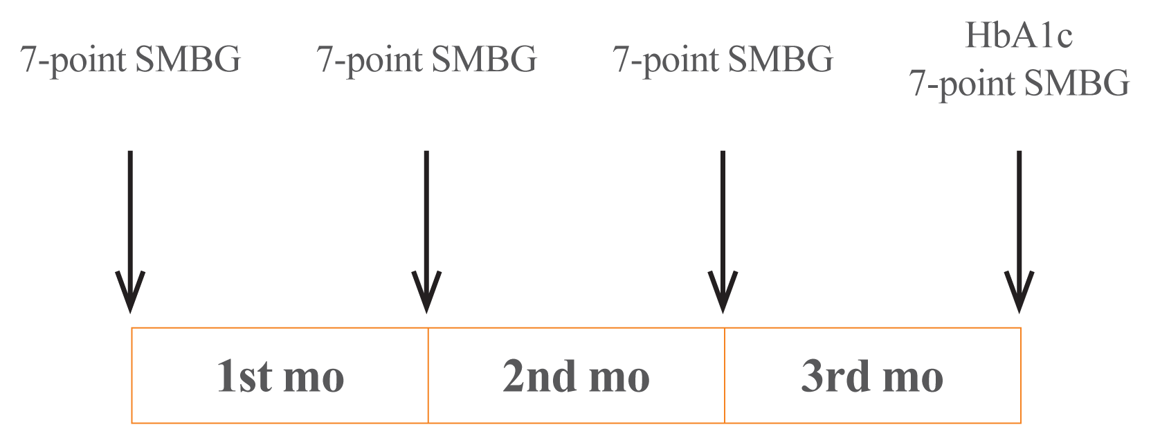
In this study, we retrospectively collected and analyzed the data of patients visiting the outpatient clinic in the Division of Endocrinology and Metabolism, Department of Internal Medicine, Jeju National University Hospital, from August 2009 to October 2011. The patients must meet the inclusion criteria as follows: (1) they suffered from T2DM; (2) their age range must be between 20 and 80 years old; and (3) they had performed 7-point self-monitoring blood glucose (SMBG), i.e., had recorded their own blood glucose levels seven times a day, for 4 days (sessions) as recommended by a medical professional. Days 1, 2, and 3 were in the beginning of the first, second, and third months, respectively, while day 4 was at the end of the third month (
Fig. 1).
 | Fig. 1Measurement of seven-point self-monitoring of blood glucose (SMBG) and glycated hemoglobin (HbA1c). 
|
Patients were excluded from the study if (1) they had type 1 diabetes (serum C-peptide <0.6 ng/mL or increased anti-glutamic acid decarboxylase antibody titer, if they received multiple doses of insulin from the time of diagnosis, or if they had a history of diabetic ketoacidosis); and (2) they were administered insulin or α-glucosidase inhibitors. They were excluded because the medications can have certain effects on PPG. After applying the inclusion and exclusion criteria, we had a final sample of 194 patients.
This study was approved by the Institutional Review Board of the Jeju National University Hospital (IRB File No. JEJUNUH 2015-06-006) and was exempted from informed consent requirements because of its retrospective nature.
Study procedure
All participants performed 7-point SMBG immediately before each meal, 2 hours after each meal, and before sleeping. The recording of SMBG was conducted for 4 days (sessions) selected in the 3 consecutive months. In the last session, HbA1c was also measured (
Fig. 1).
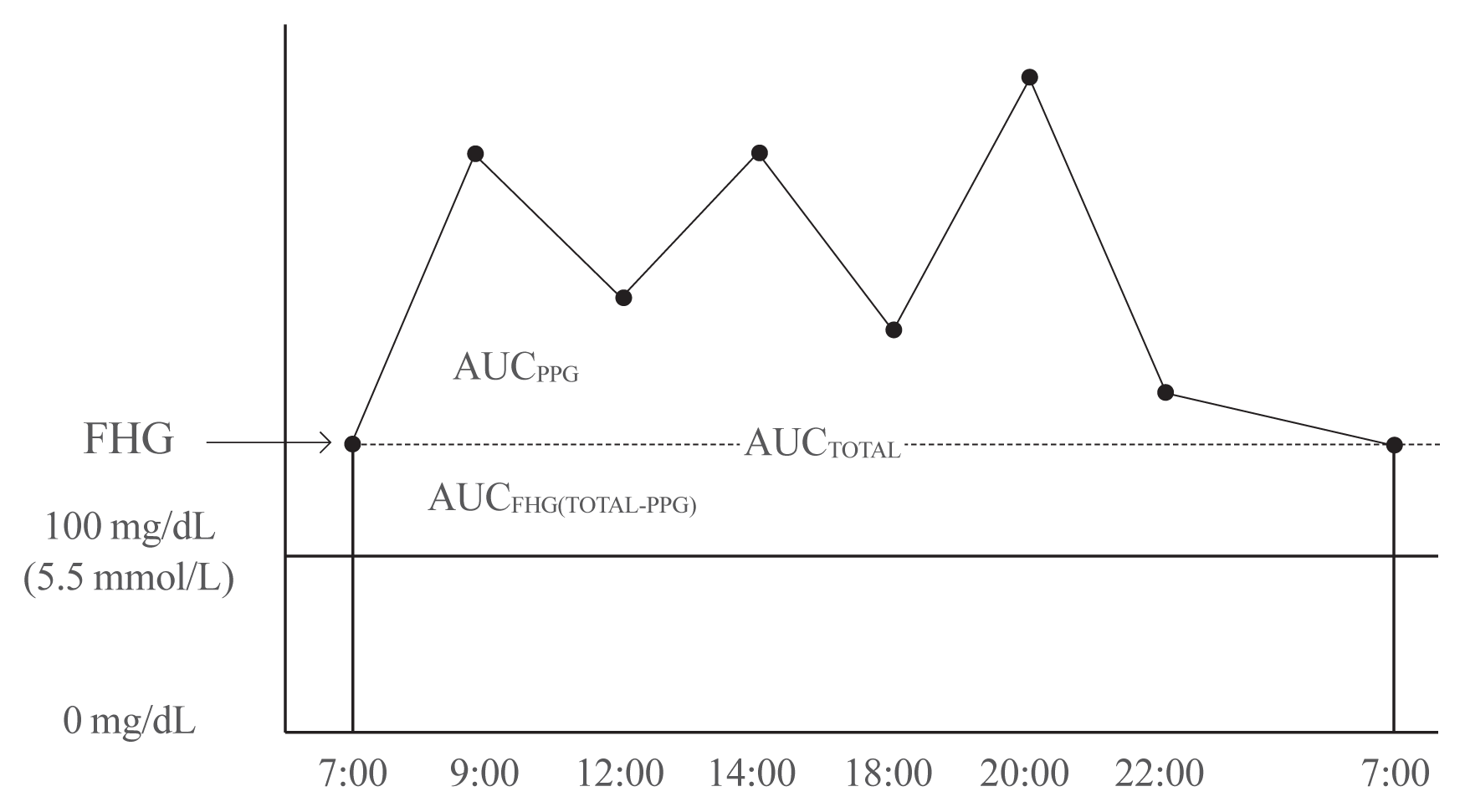
Using 7-point SMBG profiles, we calculated the area under the curve (AUC). The AUC
total was defined as the area that corresponds to 5.6 mmol/L (100 mg/dL) or greater. We used 100 mg/dL as the cut-off point because it is defined as the upper limit of normal fasting glucose by the American Diabetes Association [
6]. AUC
FHG was calculated by AUC
total-AUC
PPG, and the contribution of FHG was AUC
FHG/AUC
total (%). AUC
PPG refers to the area above the fasting glucose level in the graph, and the contribution of PPG on the overall hyperglycemia was calculated by AUC
PPG/AUC
total (%) (
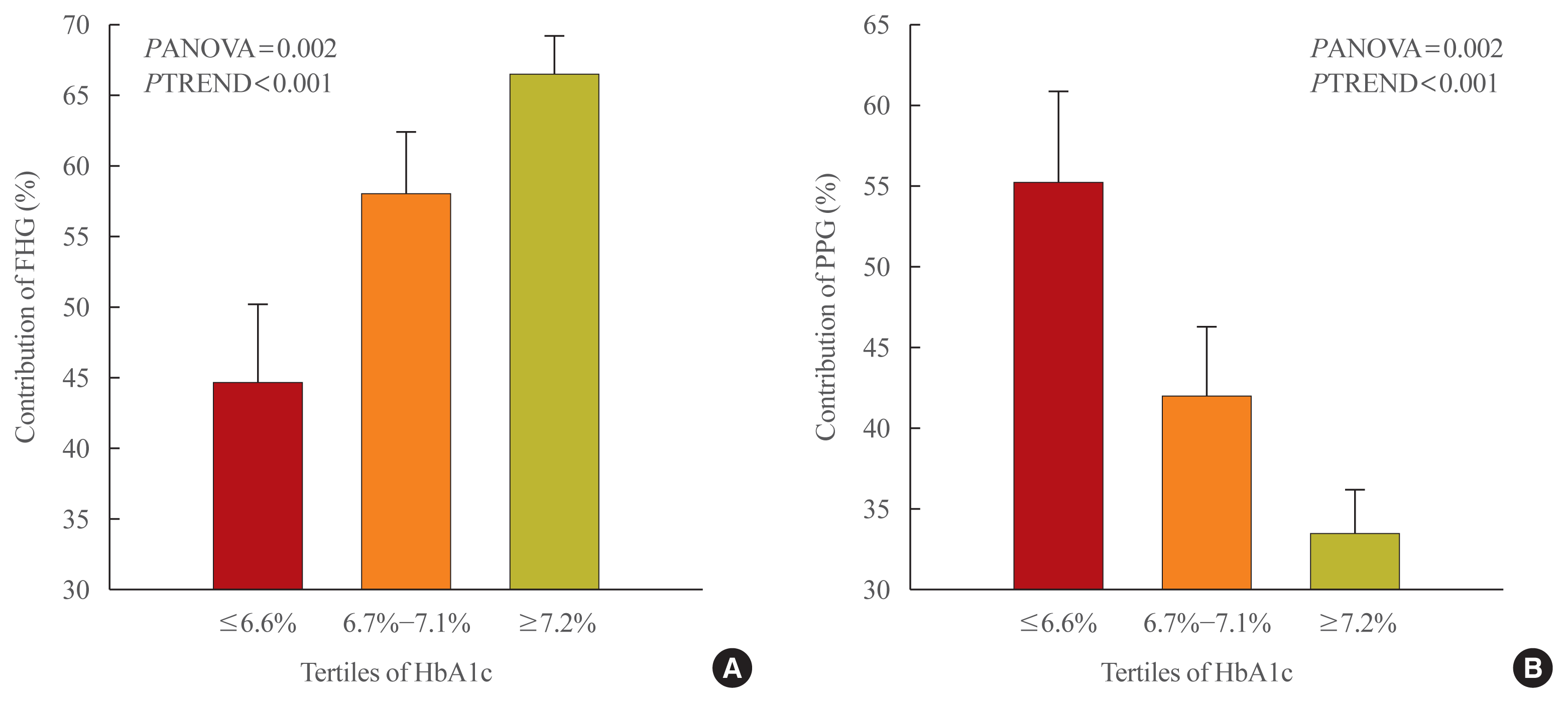
Fig. 2). We used these calculations (i.e., the mean values of contribution) to identify the association between FHG and PPG with HbA1c. We divided the patients into three groups according to HbA1c tertiles: Group 1 (6.6% or below), Group 2 (6.7% to 7.1%), and Group 3 (7.2% or above). The mean contributions of FHG and PPG were then compared among the three groups.
 | Fig. 2The relative contributions of fasting hyperglycemia (FHG) and postprandial hyperglycemia (PPG) to overall hyperglycemia according to the tertiles of glycated hemoglobin (HbA1c). AUC, area under the curve. 
|
Demographic characteristics, medical history, and the history of hypoglycemic agent use were investigated through interviews and electronic medical records, and physical examinations were performed by a physician. Some blood tests were performed after 12 hours of fasting. They included HbA1c, C-peptide, high sensitivity C-reactive protein (hsCRP), total cholesterol, triglyceride (TG), high-density lipoprotein cholesterol, low-density lipoprotein cholesterol, creatinine, aspartate transaminase (AST), and alanine transaminase (ALT).
Statistical analysis
The results are presented as mean±standard deviation (SD) for continuous variables and as percentages for categorical variables. Moreover, graphs were plotted with mean±SD. We used analysis of variance (ANOVA) with the linear trend test to examine the difference between the contribution of FHG and that of PPG to overall hyperglycemia by HbA1c tertile, and the change in the contributions. Additionally, to identify the factors affecting the two types of hyperglycemia, we performed a multivariate linear regression model in which AUCPPG and AUCFHG were treated as dependent variables, while independent variables included those significantly associated with AUCPPG and AUCFHG in the simple correlation analysis and Student’s t test. Non-normally distributed variables were logarithmically transformed and analyzed. In this study, we used SPSS version 18.0 (SPSS Inc., Chicago, IL, USA) to analyze data and set the level of significance at P<0.05.
Go to :

DISCUSSION
This study assessed not only the contribution of FHG and PPG to overall hyperglycemia but also the factors affecting these two types of hyperglycemia.
Many studies have been conducted on the contributions of fasting or PPG to overall blood glucose control; nevertheless, their results were found to be inconsistent. Monnier et al. [
4] reported that the relative contributions of FHG and PPG differed by the progression of diabetes. To assess these contributions, they categorized patients into different groups based on HbA1c tertiles and calculated the AUC. We based on their methods to analyze data of Korean patients; however, our study differed from theirs in several respects [
4]. First, the number of patients in our study was small (
n=194); therefore, we divided patients into three groups according to HbA1c tertiles instead of five groups as categorized by Monnier et al. [
4]. Second, to have more accurate calculation of the areas and contributions, we selected patients who measured their own blood glucose at 7 points of time during the day (i.e., immediately before each meal, 2 hours after each meal, and before sleeping), compared to 4 points of time as mentioned in the study by Monnier et al. [
4]. Third, Monnier et al. [
4] calculated AUC
total with the cut-off point of 6.1 mmol/L (110 mg/dL), compared to 5.5 mmol/L (100 mg/dL) in our present study (To use this cut-off point, we referred to the American Diabetes Association’s upper limit of the normal fasting glucose) [
6]. Finally, patients in our present study had better blood glucose control than those in the study by Monnier et al. [
4], as the mean HbA1c value in our study was lower (7.0% vs. 8.8%). Despite these differences, both studies shared the same result that the contribution of FHG increased and that of PPG decreased as HbA1c increased. This result was consistent with that of a study by Kikuchi et al. [
7] and that of another study by Wang et al. [
8]. Kikuchi et al. [
7] conducted a study to assess the correlation between AUC and HbA1c in Japanese T2DM patients, but not the contributions of FHG and PPG. Their study results, however, pointed out that postprandial and fasting glucose were significantly associated with HbA1c in groups with better and poorer blood glucose control, respectively. In 2011, Wang et al. [
8] categorized participants into five groups by HbA1c in a similar manner as the present study to assess the contributions of FHG and PPG among Asian T2DM patients. Like our study results, theirs also showed that the contribution of PPG tended to increase in the group with low HbA1c and FHG in the group with high HbA1c.
Unlike previous research, this study also analyzed factors other than HbA1c that might affect FHG and PPG. When controlling for HbA1c and other factors, FHG showed a significant correlation with TG and waist circumference. It has been suggested in previous studies that TG and waist circumference increased fasting glucose by affecting insulin resistance. In 2002, Kametani et al. [
9] followed-up patients for 9 years and found that obesity, hypertension, hypertriglyceridemia, and family history of diabetes were significant risk factors of impaired fasting glucose. Furthermore, in 2013, Lin et al. [
10] reported that the risk of impaired fasting glucose increased with heightened levels of TG. However, most previous studies showed that obesity and hypertriglyceridemia were factors related to impaired fasting glucose. Unlike our study, they did not thoroughly assess the effects of TG or waist circumference on fasting glucose in patients with diabetes.
Apart from HbA1c, age was the only factor affecting PPG. The correlation between hyperglycemia and age has been previously reported [
11]. In 2013, Munshi et al. [
12] analyzed the fasting and postprandial contribution to overall hyperglycemia using the AUC method. They found that in higher age groups, the contribution of postprandial glucose was greater than that of fasting glucose. Chiu et al. [
13] explained that as age increases, the functions of beta-cells decrease, resulting in the relative rise in the contribution of postprandial glucose.
Based on the results of this study, we provided the following recommendations for the treatment of patients with diabetes. First, the contribution of PPG is generally higher in well-controlled patients; therefore, it is difficult to reach the target HbA1c by reducing only fasting glucose levels. In a previous study by Woerle et al. [
14], patients who reached the target HbA1c of below 7.0% had a significantly lower postprandial glucose level than that of those who did not. However, the study also showed no differences in the fasting glucose level. As the relative contribution changes with the degree of glycemic control, medications should be prescribed accordingly. Second, medication must be selected considering the age even if the HbA1c is the same, as the relative contribution of PPG increases with age which is typically accompanied by the decreased secretion of insulin. Third, the contribution of FHG is high in patients with obesity or high levels of TG. The use of drug that usually reduces fasting glucose level, such as metformin and thiazolidinedione, should therefore be considered with priority.
This study encountered some limitations that should be addressed. The first limitation was the retrospective nature of this study. Second, the number of patients (
n=194) was relatively small compared to that of the previous study conducted by Monnier et al. [
4]. Third, the patients in this present study had relatively good control of their blood glucose, which was reflected in their relatively low HbA1c levels. When using tertiles to categorize the study sample into different groups, the group with the poorest control of blood glucose showed a mean HbA1c of 8%. Therefore, it remains unclear whether the results of our study apply to patients with poor blood glucose control. However, our results of high contribution of fasting glucose in higher HbA1c levels and high contribution of postprandial glucose in lower HbA1c levels are consistent with the results of studies outside Korea that include patients with various levels of blood glucose control [
4,
5,
7,
8,
14]. Thus, we can predict similar results from Korean patients with poor glucose control. Fourth, our study calculated the contribution of FHG and PPG using 7-point SMBG instead of continuous glucose monitoring (CGM). Most international studies that have shown proportional relationship between contribution of FHG and HbA1c used 4-point or 7-point SMBG [
4,
5,
7,
12,
14–
16]. Thus, we also used the same method and validated that the contribution of FHG is high at higher levels of HbA1c in Korean diabetes patients. However, as measuring blood glucose using CGM can more accurately differentiate fasting and postprandial glucose than using SMBG, detailed studies assessing the contribution of FHG and PPG in Korean patients using CGM are further required. None the less, this present study made certain contributions to the current literature, as it, to our best knowledge, is the first to use 7-point SMBG in Korean T2DM patients to examine the research topic of interest.
This study assessed the contributions of FHG and PPG in 194 Korean T2DM patients and analyzed data of three HbA1c groups. The contribution of PPG was high in the group with relatively good control of blood glucose, whereas that of FHG gradually increased with elevated HbA1c. Two other factors, namely waist circumference and TG, were also significantly associated with FHG. However, apart from HbA1c, age was the only factor that had a statistically significant association with PPG.
Go to :


 PDF
PDF Citation
 Print
Print


 XML Download