

 PDF
PDF ePub
ePub Citation
 Print
Print


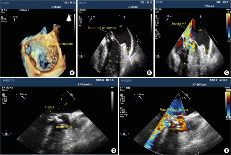
A 74-year-old male patient was admitted to our intensive care unit with a diagnosis of pneumonia. During follow-up period, the patient was connected to mechanical ventilation due to acute respiratory failure. On medical history, he was diagnosed with lung cancer and treated with the combination of chemotherapy and radiotherapy, which was applied to the right lobe of the lung. The heart was left outside the radiation exposure. The patient was consulted to our cardiology clinic because of persistent tachycardia and fever. At the time of examination, a grade IV pansystolic murmur at the cardiac apex radiating to the axilla was noted. Laboratory analysis revealed a leukocytosis (26,240/mm3) and elevated levels of C-reactive protein (135.2 mg/dL). Transthoracic echocardiography was performed providing a normal left ventricle systolic function, vegetation on the anterolateral mitral valve, and severe mitral regurgitation. On transesophageal echocardiography (TEE) examination, there was large vegetation on the anterolateral mitral valve along with aneurysm formation and rupture (Figure 1A-B-C, Movie 1-2-3). A large fistula originating from non-coronary cusp of the aortic valve to the left atrium was present on TEE examination (Figure 1-D-E, Movie 4-5). Results of urine and feces cultures that were taken at different times were negative, however; two sets of blood culture yielded a growth of Escherichia coli (E. coli). The patient was consulted to cardiovascular surgery for operation and pre-operative coronary angiography was planned. However, he died on the 4th day of hospitalization in the intensive care unit. E. coli infective endocarditis of the native valves is a very rare clinical finding.1) Moreover, to the best of our knowledge, this is the first case of native infective endocarditis of E. coli presented with a large fistula formation from aorta to the left atrium along with mitral valve aneurysm formation and rupture, which are rarely seen together.
Tufan Cinar, MD , Mert İlker Hayiroğlu, MD, Vedat Çiçek, MD, Mehmet Uzun, MD, Ahmet Lütfullah Orhan, MD
, Mert İlker Hayiroğlu, MD, Vedat Çiçek, MD, Mehmet Uzun, MD, Ahmet Lütfullah Orhan, MD
References
1. Rangarajan D, Ramakrishnan S, Patro KC, et al. Native valve Escherichia coli endocarditis following urosepsis. Indian J Nephrol. 2013; 23:232–234. PMID: 23814428.
SUPPLEMENTARY MATERIALS
Movie 2
Transesophageal echocardiography showing the large vegetation on the anterolateral mitral valve along with aneurysm formation and rupture.

Movie 3
Transesophageal echocardiography showing the large vegetation on the anterolateral mitral valve along with ruptured aneurysm causing severe mitral regurgitation with color Doppler imaging.
Figure 1
(A) Ruptured mitral valve aneurysm on 3D echocardiography. (B) The large aneurysm and vegetation on the anterolateral mitral valve. (C) The severe MR to the LA due to aneurysm rupture. (D) The fistula formation of aorta to LA. (E) The color Doppler flow through the fistula between the aorta and LA. LA: left atrium, LAA: left atrium appendage, LV: left ventricle, MR: mitral regurgitation.
 XML Download