Journal List > J Cardiovasc Imaging > v.28(2) > 1144598

Garg, Agrawal, and Sharma: Non-Obstructive Accessory Mitral Valve Tissue in the Left Ventricular Outflow Tract with PerimembranousVentricular Septal Defect: A Rare Entity
A 19-year-old male was referred to us for evaluation of heart murmur. The patient was asymptomatic and in good physical condition. On examination, his blood pressure was 126/80 mmHg, and pulse rate was 80 beats/min. Grade IV/VI systolic murmur was heard at the left sternal border.
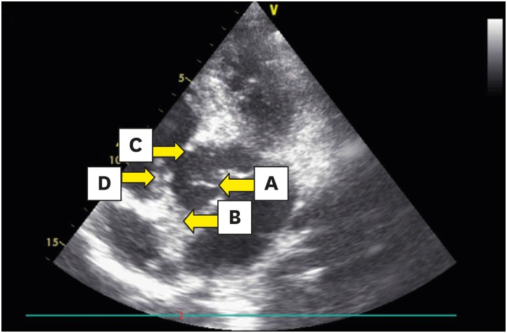
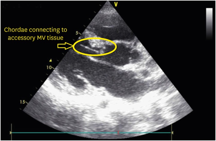
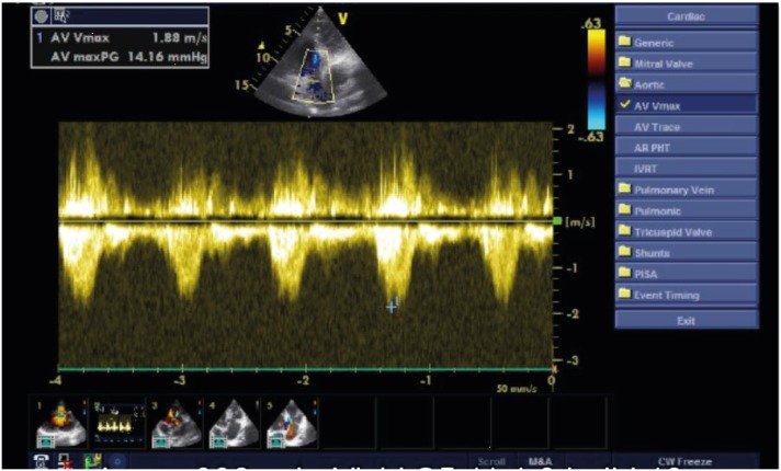
His electrocardiogram (ECG) showed a normal sinus rhythm. A two-dimensional transthoracic echocardiogram was performed in the usual manner with a Vivid S5 General Electric (Milwaukee, WI, USA) ultrasound system and a 3 MHz transducer. The result showed situs solitus and normal arrangement of atria and ventricles with no atrioventricular or ventricular-arterial discordance. Transthoracic echocardiography showed a large perimembranous ventricular septal defect (VSD) partially obstructed by a septal leaflet of the tricuspid valve (Figure 1, Movies 1, 2, 3). A mobile filamentous structure was also seen attached to the ventricular side of the base of the anterior mitral valve leaflet with chordal attachment from anterior papillary muscles. This structure was moving in systole into the left ventricular outflow tract (LVOT) (Figure 1, 2, Movies 1, 4, 5). No relevant subaortic obstruction was demonstrated with a 14.16 mmHg maximum gradient across the LVOT (Figure 3).
The patient underwent elective surgery for a diagnosis of perimembranous VSD and non-obstructive accessory mitral valve tissue. The operation showed successfully closure of the VSD and excision of accessory mitral tissue in the LVOT.

Notes

Conflict of Interest: The authors have no financial conflicts of interest.

SUPPLEMENTARY MATERIALS

Movie 1

Apical five-chamber view showing filamentous AMVT moving in the LV outflow tract with a perimembranous VSD. AMVT: accessory mitral valve tissue, Ao: aorta, LA: left atrium, LV: left ventricle, RA: right atrium, RV: right ventricle, VSD: ventricular septal defect.

Movie 2

Parasternal long-axis view showing colored flow through the VSD. Ao: aorta, LA: left atrium, LV: left ventricle, RV: right ventricle, VSD: ventricular septal defect.

Movie 3

Apical five-chamber view showing turbulent colored flow through the perimembranous VSD partially obstructed by a septal leaflet of the tricuspid valve. Ao: aorta, LA: left atrium, LV: left ventricle, RA: right atrium, RV: right ventricle, VSD: ventricular septal defect.

Movie 4

Parasternal long-axis view showing filamentous AMVT moving in the LV outflow tract. AMVT: accessory mitral valve tissue, Ao: aorta, LA: left atrium, LV: left ventricle, RV: right ventricle.

Movie 5

Parasternal long-axis view showing filamentous accessory mitral valve tissue attached to the chordae.
Figure 1

Apical five-chamber view; arrows point to filamentous accessory mitral valve tissue (A), aortic valve (B), perimembranous ventricular septal defect (C), and septal leaflet of the tricuspid valve (D).

jcvi-28-158-g001
Figure 2

Parasternal long-axis view showing accessory mitral valve tissue attached to the chordae.

jcvi-28-158-g002
Figure 3

Apical five-chamber view with continuous width Doppler showing a maximum pressure gradient of 14.16 mmHg through the left ventricular outflow tract.

jcvi-28-158-g003
TOOLS
Similar articles