

 PDF
PDF ePub
ePub Citation
 Print
Print


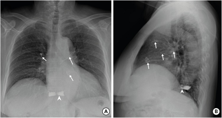
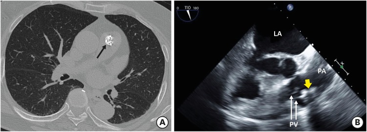
A 72-year-old woman presented with mild dyspnea on exertion 2 months ago. She had a history of myocardial infarction 8 years ago. She had undergone vertebroplasty by injecting polymethylmethacrylate cement in the 10th thoracic body due to a compression fracture 2 years ago (Figure 1). For evaluation of dyspnea, transthoracic echocardiography was undertaken. Echocardiography revealed a lobulated mass attached to the pulmonary valve and a stick at the bifurcation of the pulmonary artery (Movie 1). Simple chest X-ray showed a round radio-opaque mass made of coiled lines in the pulmonary artery and linear materials in the left and right pulmonary arteries (Figure 1). Computed tomography confirmed a mass with high attenuation (Figure 2A). Additionally, transesophageal echocardiography revealed the lobulated mass attached to the pulmonary valve (Figure 2B). The foreign bodies were bone cements that had leaked into the paravertebral vasculature and moved to the pulmonary artery during vertebroplasty. The mass did not lead to pulmonary hypertension and a progressive thromboembolic phenomenon was not found. We decided to monitor the cement emboli regularly with the patient's consent.
Pulmonary bone cement embolism is not a rare complication. The incidence of pulmonary embolism is 2.1% to 26%.1)2) Although most cases are detected incidentally and are asymptomatic, clinical presentation is varied and can include dyspnea, chest pain, cardiac rupture, and/or death.2) In this patient, the mass in the pulmonary artery was detected incidentally, and it was confused with a cardiac mass, vegetation, and pulmonary thromboembolism. We were able to confirm bone cement emboli through a multimodality imaging technique.

 XML Download