

 PDF
PDF ePub
ePub Citation
 Print
Print


Abstract
Purpose:
To investigate normal location of the peroneus longus tendon (PL) in the cuboid groove by evaluating it between ankles with no significant abnormality (asymptomatic group) and those with retromalleolar PL dislocation (dislocation group) using three-dimensional isotropic fast spin-echo (3D-FSE) magnetic resonance imaging (MRI) of the ankle.
Materials and Methods:
Thirty-six and 32 3D-FSE ankle MRI were assigned to the asymptomatic group and the dislocation group, respectively. Using multiplanar reformatted 3D-FSE, qualitative PL location (i.e., outside, overlying, and inside in relation to the cuboid groove), quantitative PL location (i.e., distance between the proximal margins of PL and cuboid groove), and cuboid groove size were measured in lateral, middle, and medial levels of the cuboid groove.
Results:
In the asymptomatic group, 64%, 42%, and 11%, respectively, had the outside or overlying-located PL in lateral, middle, and medial levels of the cuboid groove and the quantitative location gradually decreased from lateral to medial level. Qualitative and quantitative PL locations were not significantly different between the asymptomatic group and dislocation group. Cuboid groove size showed significant negative correlation with quantitative PL location in both groups.
REFERENCES
1.Dombek MF., Lamm BM., Saltrick K., Mendicino RW., Catanzariti AR. Peroneal tendon tears: a retrospective review. J Foot Ankle Surg. 2003. 42:250–258.

2.Heckman DS., Gluck GS., Parekh SG. Tendon disorders of the foot and ankle, part 1: peroneal tendon disorders. Am J Sports Med. 2009. 37:614–625.
3.Philbin TM., Landis GS., Smith B. Peroneal tendon injuries. J Am Acad Orthop Surg. 2009. 17:306–317.
4.Stone TJ., Rosenberg ZS., Velez ZR., Ciavarra G., Prost R., Bencardino JT. Subluxation of the peroneus long tendon in the cuboid tunnel: is it normal or pathologic? An ultrasound and magnetic resonance imaging study. Skeletal Radiol. 2016. 45:357–365.
5.Choo HJ., Lee SJ., Huang BK., Resnick DL. The location of the peroneus longus tendon in the cuboid groove: sonographic study in various positions of the ankle-foot in asymptomatic volunteers. Skeletal Radiol. 2018. 47:1277–1284.
6.Choo HJ., Lee SJ., Kim DW., Jeong HW., Gwak H. Multibanded anterior talofibular ligaments in normal ankles and sprained ankles using 3D isotropic proton density-weighted fast spin-echo MRI sequence. AJR Am J Roentgenol. 2014. 202:W87–94.
7.Stevens KJ., Busse RF., Han E, et al. Ankle: isotropic MR imaging with 3D-FSE-cube–initial experience in healthy volunteers. Radiology. 2008. 249:1026–1033.
8.Ulbrich EJ., Zubler V., Sutter R., Espinosa N., Pfirrmann CW., Zanetti M. Ligaments of the Lisfranc joint in MRI: 3D-SPACE (sampling perfection with application optimized contrasts using different flip-angle evolution) sequence compared to three orthogonal proton-density fat-saturated (PD fs) sequences. Skeletal Radiol. 2013. 42:399–409.
Fig. 1.
Differentiation of the three levels of the cuboid groove. The peroneus longus tendon (PL) location and cuboid groove size are evaluated on the oblique sagittal images of the lateral, middle, and medial levels of the cuboid groove. On the oblique axial image, the three yellow-dotted lines indicate the lateral, middle, and medial levels of the cuboid groove. 1st MT = 1st metatarsal; LAT = lateral; MED = medial; MID = middle
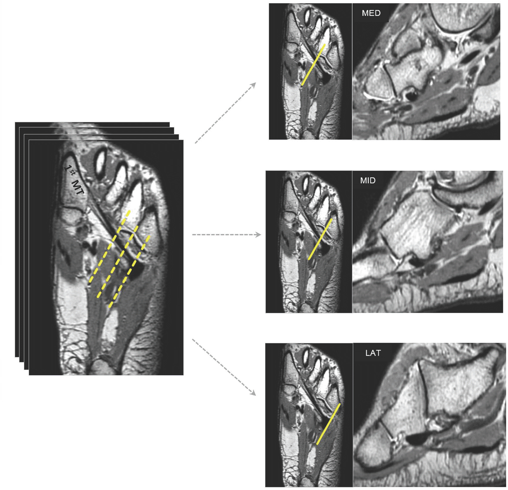
Fig. 2.
Mimetic diagram of the categorization for the qualitative location of the PL . (a) The images show an inside-located type, wherein the whole cross-sectional area of the PL is inside the cuboid groove. (b) The images show an overlying-located type, wherein some part of cross-sectional area of the PL is outside the groove. (c) The images show an outside-located type, wherein the entire cross-sectional area of the PL is outside the groove.
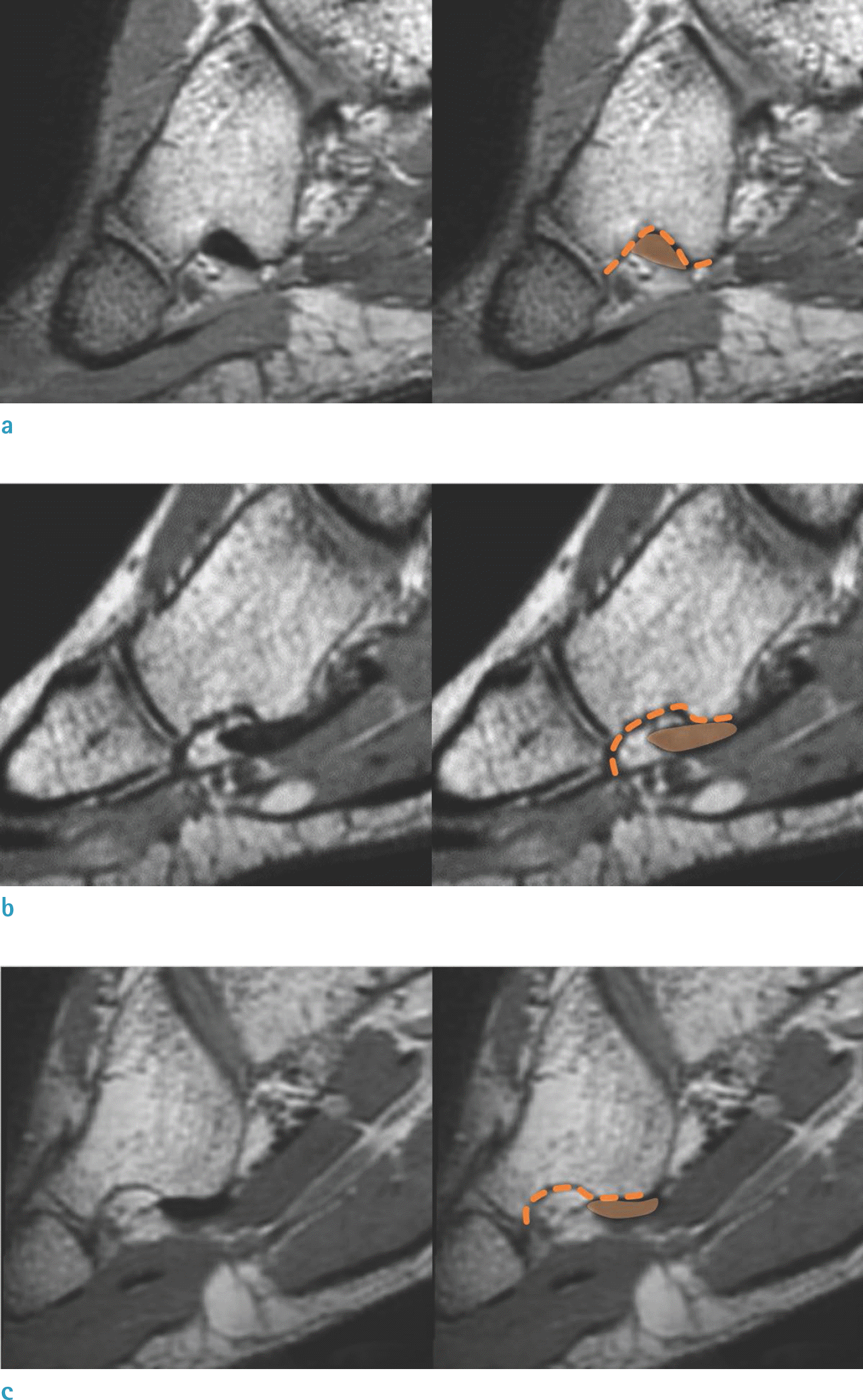
Fig. 3.
Measurement for the quantitative location of the PL. On the oblique sagittal plane, the distance (double-headed arrow) between the proximal margin of cross-sectional area of the PL (orange solid line) and the proximal border of the cuboid groove (orange dotted line) is measured.
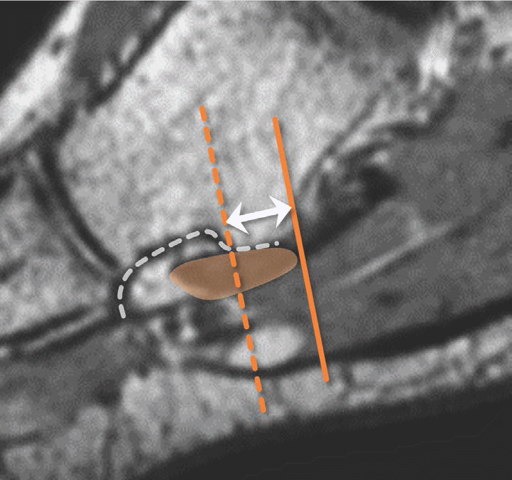
Fig. 4.
Measurement for the width and depth of the cuboid groove. (a) The cuboid groove width (double-headed arrow) indicates the distance between the most inferior contour of the cuboid tuberosity and the anterior edge of the cuboid. (b) Its depth indicates the distance between the deepest point of the cuboid groove and the imaginary line, crossing the most inferior contour of the cuboid tuberosity and the anterior edge of the cuboid.
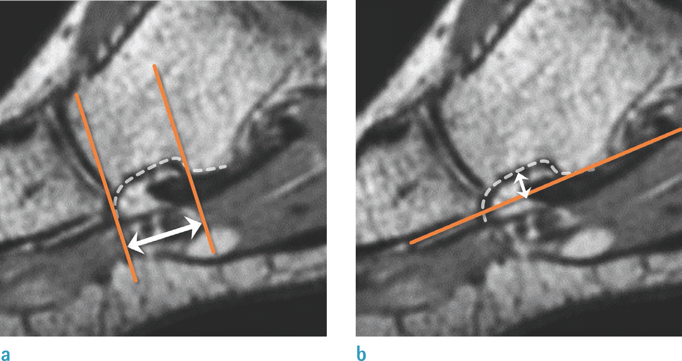
Fig. 5.
Bar graph showing PL location in the asymptomatic group and the dislocation group. The proportions of ankles with the inside-located PL increase as the PL goes through the cuboid groove from the lateral to medial level in the asymptomatic group and the dislocation group, but there is no statistically significant difference.
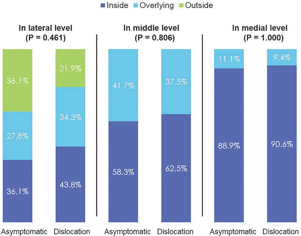
Fig. 6.
An age 19 male with PL dislocation from the retromalleolar fossa. (a) On the axial T2-weighted spin-echo image, the PL (arrow) is anterolateral aspect of the lateral malleolus (arrow). (b-d) On the oblique sagittal reformatted 3D-FSE, the PL (arrow) is ‘inside-located’ in the lateral (b), middle (c) and medial levels of the cuboid groove (d).
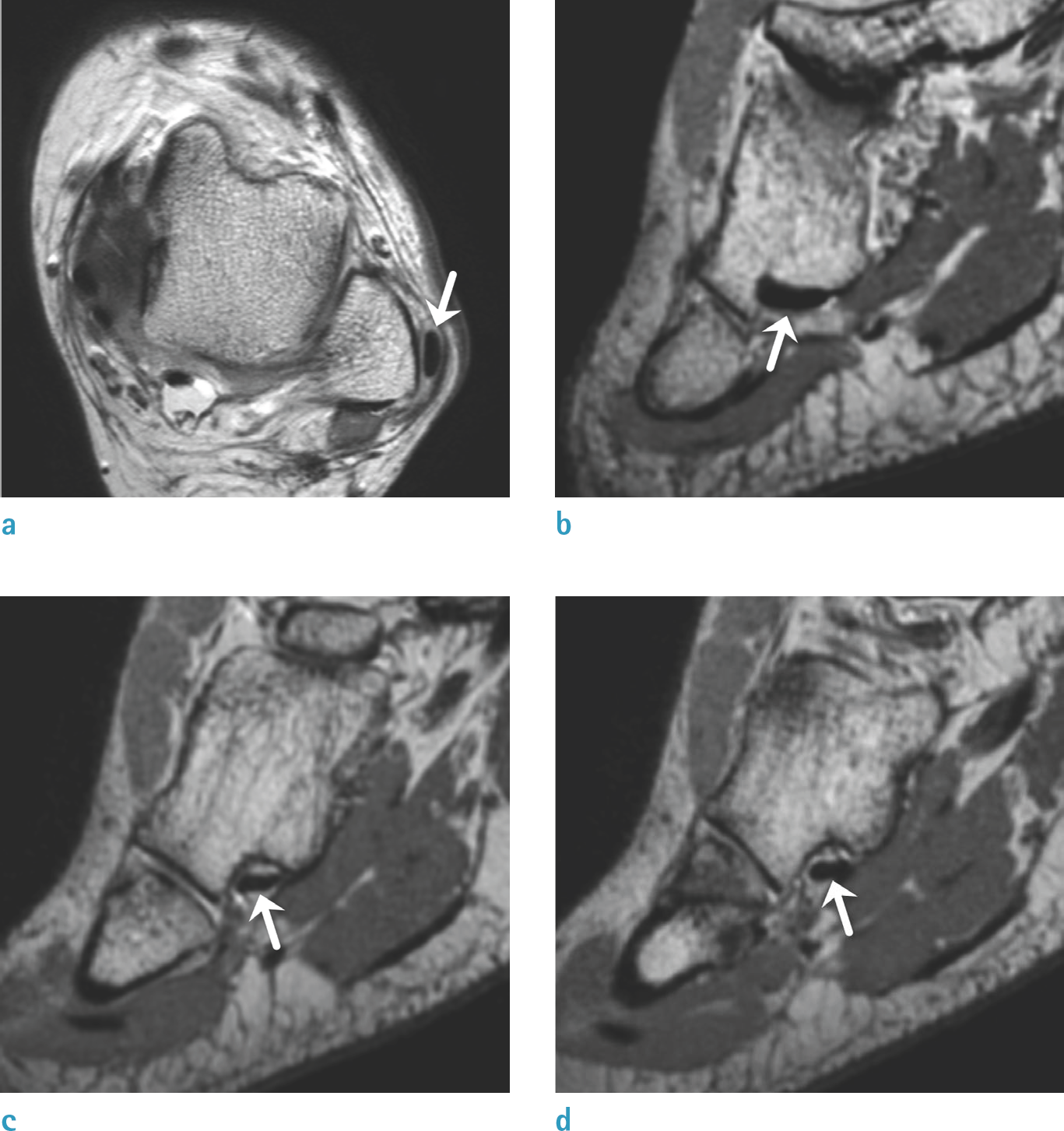
Fig. 7.
An age 19 female with PL dislocation from the retromalleolar fossa. (a) On the axial T2-weighted spin-echo image, the PL (arrow) is dislocated from the retromalleolar fossa (arrow). (b-d) The oblique sagittal reformatted 3D-FSE demonstrates the outside-located PL (arrows) at the lateral level of cuboid groove (b), which is glided entirely into the cuboid groove in its middle and medial levels (c, d).
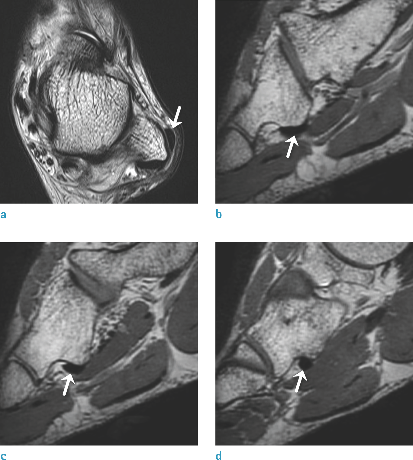
Table 1.
Parameters of Three-Dimensional Fast Spin Echo MRI of the Ankle
Table 2.
Quantitative Peroneus Longus Tendon Location
Table 3.
Width and Depth of Cuboid Groove
Table 4.
Relationship between Peroneus Longus Tendon Location and Cuboid Groove Size
 XML Download