

 PDF
PDF ePub
ePub Citation
 Print
Print


Ju Yup Lee, Kyung Sik Park
References
1. Oh SY, Cunningham J, Saif MW. Colonic metastasis from gastric cancer. Clin Colorectal Cancer. 2014; 13:255–256.

2. Riihimäki M, Hemminki A, Sundquist K, Sundquist J, Hemminki K. Metastatic spread in patients with gastric cancer. Oncotarget. 2016; 7:52307–52316.
3. Duarte I, Llanos O. Patterns of metastases in intestinal and diffuse types of carcinoma of the stomach. Hum Pathol. 1981; 12:237–242.
4. Balthazar EJ, Rosenberg HD, Davidian MM. Primary and metastatic scirrrhous carcinoma of the rectum. AJR Am J Roentgenol. 1979; 132:711–715.
5. Duarte I, Llanos O. Patterns of metastases in intestinal and diffuse types of carcinoma of the stomach. Hum Pathol. 1981; 12:237–242.
6. Jang HJ, Lim HK, Kim HS, et al. Intestinal metastases from gastric adenocarcinoma: helical CT findings. J Comput Assist Tomogr. 2001; 25:61–67.
7. Ogiwara H, Konno H, Kitayama Y, Kino I, Baba S. Metastases from gastric adenocarcinoma presenting as multiple colonic polyps: report of a case. Surg Today. 1994; 24:473–475.
8. Pace U, Contino G, Chiappa A, et al. Metachronous colon metastases from gastric adenocarcinoma: a case report. Case Rep Oncol. 2009; 2:92–96.
9. Lee HC, Yang MT, Lin KY, Tu HY, Zhang TA, Chen PH. Metastases from gastric carcinoma to colon in the form of multiple flat elevated lesions: a case report. Kaohsiung J Med Sci. 2004; 20:552–557.
10. Metayer P, Antonietti M, Oumrani M, Hemet J, Lemoine F, Basuyau J. Metastases of a gastric adenocarcinoma presenting as colonic polyposis. Report of a case. Dis Colon Rectum. 1991; 34:622–623.
11. Katon RM, Brendler SJ, Ireland K. Gastric linitis plastica with metastases to the colon: a mimic of Crohn's disease. J Clin Gastroenterol. 1989; 11:555–560.
Fig. 1.
Abdominal computed tomography demonstrated multiple strictures with proximal bowel dilatation in the (A) ascending, (B) transverse, and (C, D) sigmoid colon (arrows).
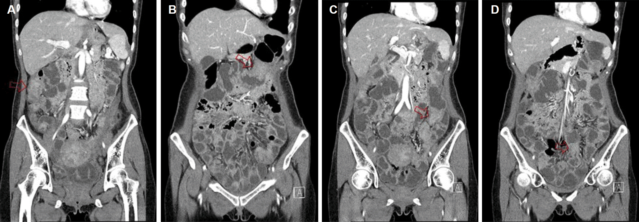
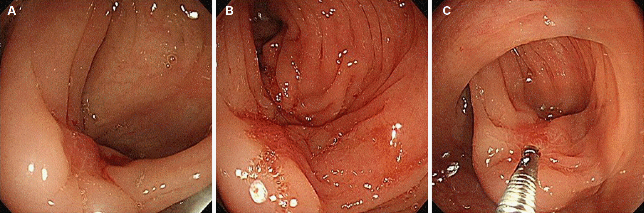
Fig. 2.
Colonoscopy findings. (A, B) Mass-like lesion with surface reddish nodularity and luminal stenosis is noted at the sigmoid colon, 30 cm from the anal verge. The 9.2 mm scope could not pass through the stenotic lesion. (C) Multiple endoscopic biopsy was done at reddish nodular mucosa.
 XML Download