

 PDF
PDF ePub
ePub Citation
 Print
Print


Tae Young Park , Soo Hyung Ryu, Jeong Seop Moon
, Soo Hyung Ryu, Jeong Seop Moon
Abstract
A visceral artery pseudoaneurysm after ERCP is a rare adverse event that is potentially life-threatening. Most cases reported pre-viously originated from the peripancreatic arteries, including the splenic artery, gastroduodenal artery, or pancreaticoduodenal artery. The mechanism of the occurrence of visceral artery pseudoaneurysms after ERCP has not been elucidated until now. Recently, a pseudoaneurysm rupture originating from the superior mesenteric artery after ERCP was observed in a patient without a history of pancreatitis. This paper reports this case with a review of the relevant literature.
References
1. Bergert H, Hinterseher I, Kersting S, Leonhardt J, Bloomenthal A, Saeger HD. Management and outcome of hemorrhage due to arterial pseudoaneurysms in pancreatitis. Surgery. 2005; 137:323–328.

2. de Perrot M, Berney T, Bühler L, Delgadillo X, Mentha G, Morel P. Management of bleeding pseudoaneurysms in patients with pancreatitis. Br J Surg. 1999; 86:29–32.
3. Udd M, Leppäniemi AK, Bidel S, Keto P, Roth WD, Haapiainen RK. Treatment of bleeding pseudoaneurysms in patients with chronic pancreatitis. World J Surg. 2007; 31:504–510.
4. Gaduputi V, Tariq H, Dev A. Visceral arterial aneurysms complicating endoscopic retrograde cholangiopancreatography. Case Rep Gastrointest Med. 2013; 2013:515201.
5. Kurita A, Ito T, Kudo Y, Yazumi S. Rupture of a pseudoaneurysm caused by endoscopic papillary large-balloon dilation. Endoscopy. 2015; 47(Suppl 1):UCTN:E532-E533.
6. Priya P, Bhattacharyya A, Mohammed S, Gulati S, Ghatak S. Angiographic management of pseudoaneurysms of gastroduodenal artery following endoscopic sphincterotomy. Indian J Gastroenterol. 2016; 35:242–244.
7. Flati G, Salvatori F, Porowska B, et al. Severe hemorrhagic complications in pancreatitis. Ann Ital Chir. 1995; 66:233–237.
8. Al-Jeroudi A, Belli AM, Shorvon PJ. False aneurysm of the pancreaticoduodenal artery complicating therapeutic endoscopic retrograde cholangiopancreatography. Br J Radiol. 2001; 74:375–377.
9. Ding Z, Tang XL, Lin R, Han C, Liu J. ERCP-related complication is not the only cause of GI bleeding in post-liver transplantation patients: a case report. Medicine (Baltimore). 2017; 96:e7716.
10. El Hajj II, Sherman S, Pyko M, Lehman GA. Life threatening hemo-bilia after endoscopic retrograde cholangiopancreatography (ERCP). Dig Liver Dis. 2017; 49:1336–1337.
11. Mohapatra S, Moradi D, Broder A. A rare cause of post-sphincterotomy bleeding. Gastroenterology. 2017; 153:1193–1194.
Fig. 1.
Diffuse GB wall edema with pericholecystic fluid collection and subtle calcification in the CBD were noted in the abdominal CT scan. GB, gallbladder; CBD, common bile duct; CT, computed tomography.
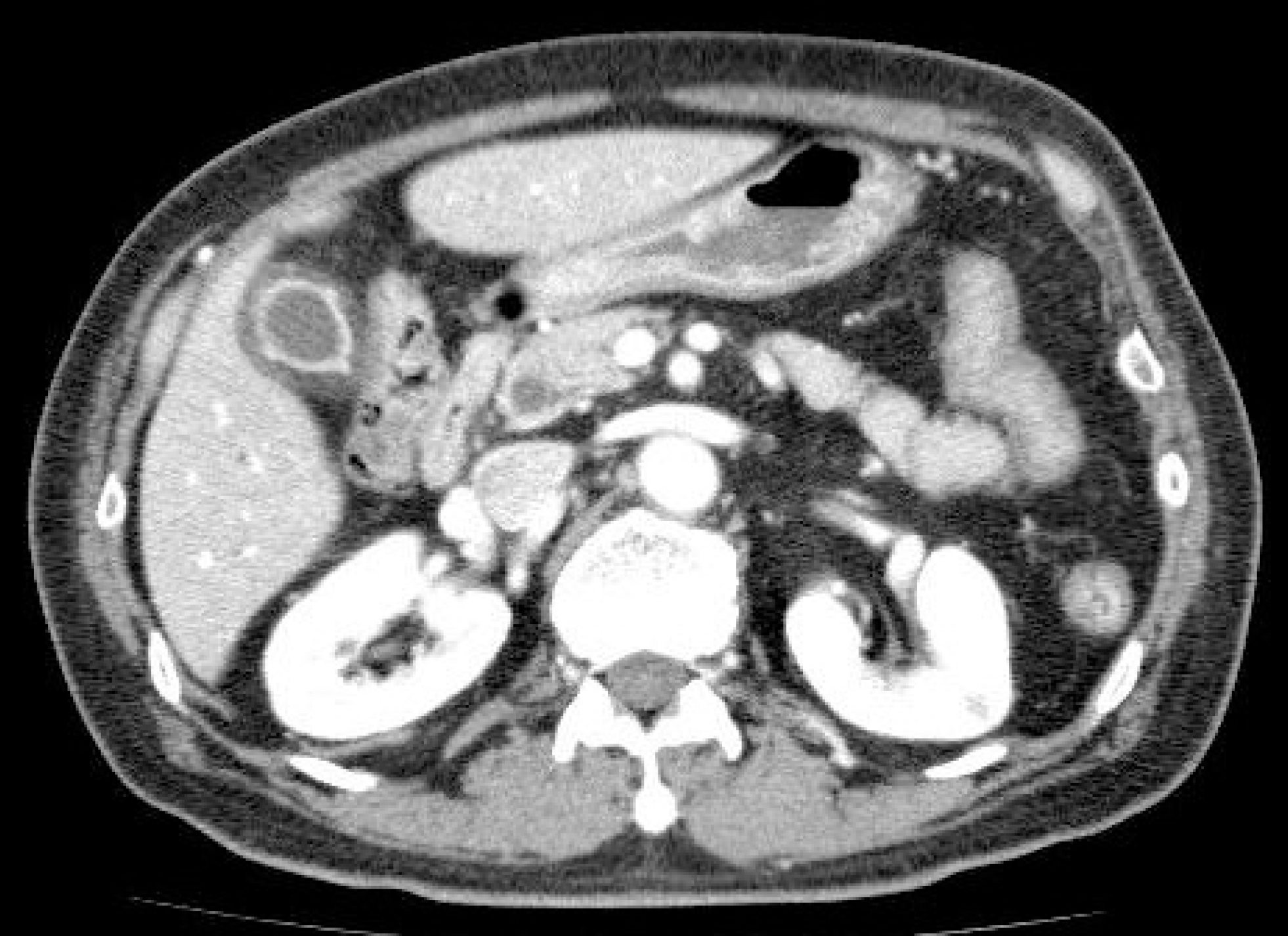
Fig. 2.
Endoscopic retrograde cholangiopancreatography. (A) A filling defect was noted in the distal CBD during cholangiography. (B) After EST, a round stone was removed using a retrieval balloon catheter. CBD, common bile duct; EST, endoscopic sphincterotomy.
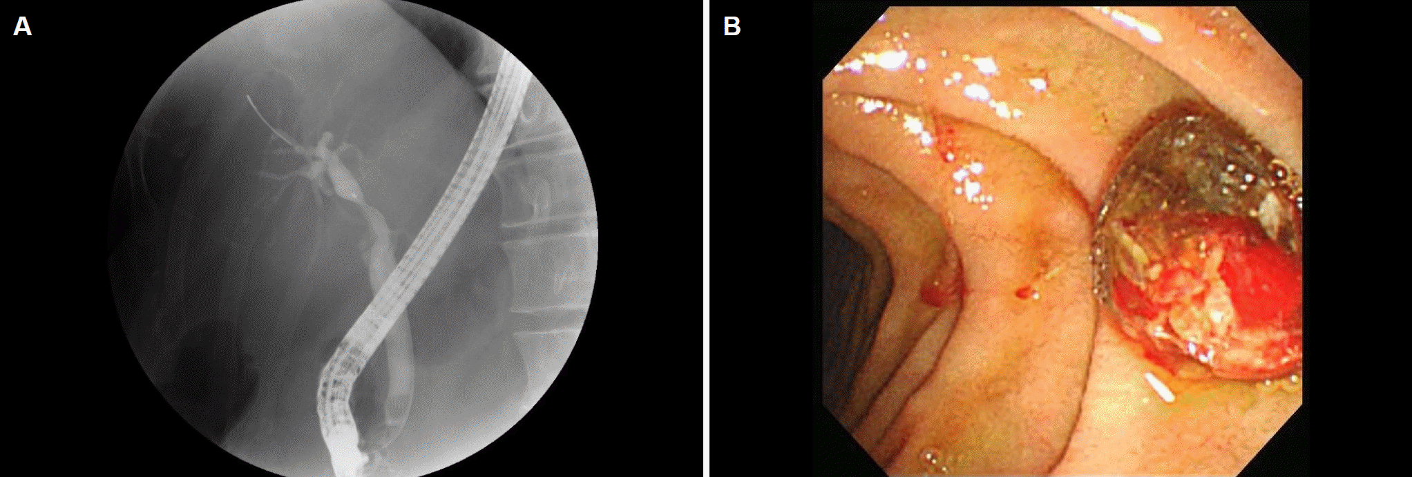
Fig. 3.
2.5 cm sized heart-shaped contrast leakage was noted inside the hematoma on enhanced abdominal CT. CT, computed tomography.
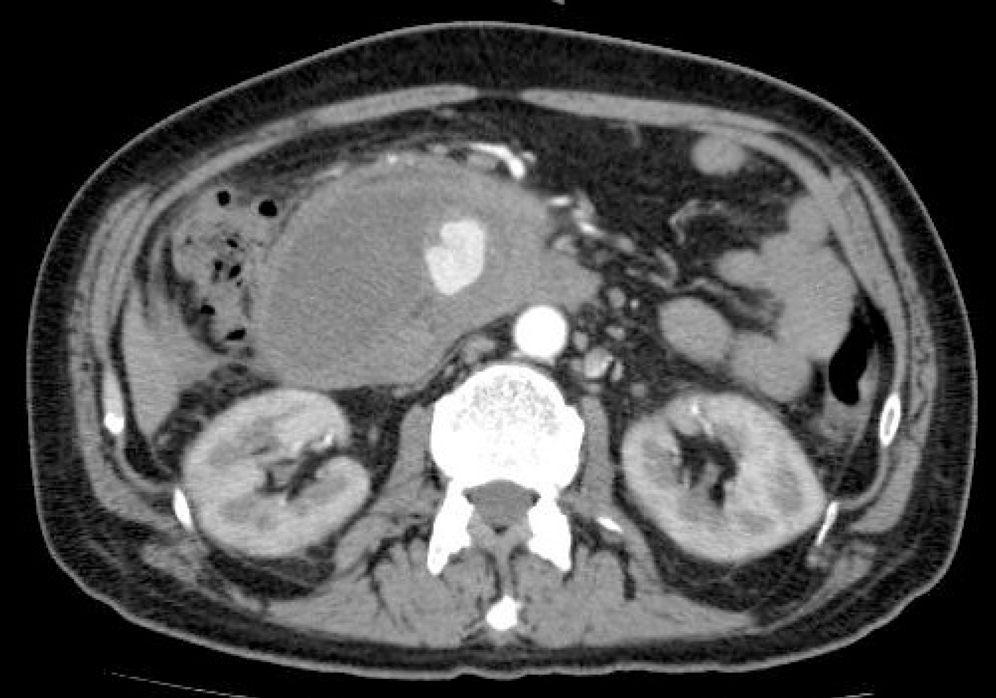
Fig. 4.
Contrast leakage (arrowhead) from the right side wall of the first jejunal branch originating from the SMA was noted on angiography. SMA, superior mesenteric artery.
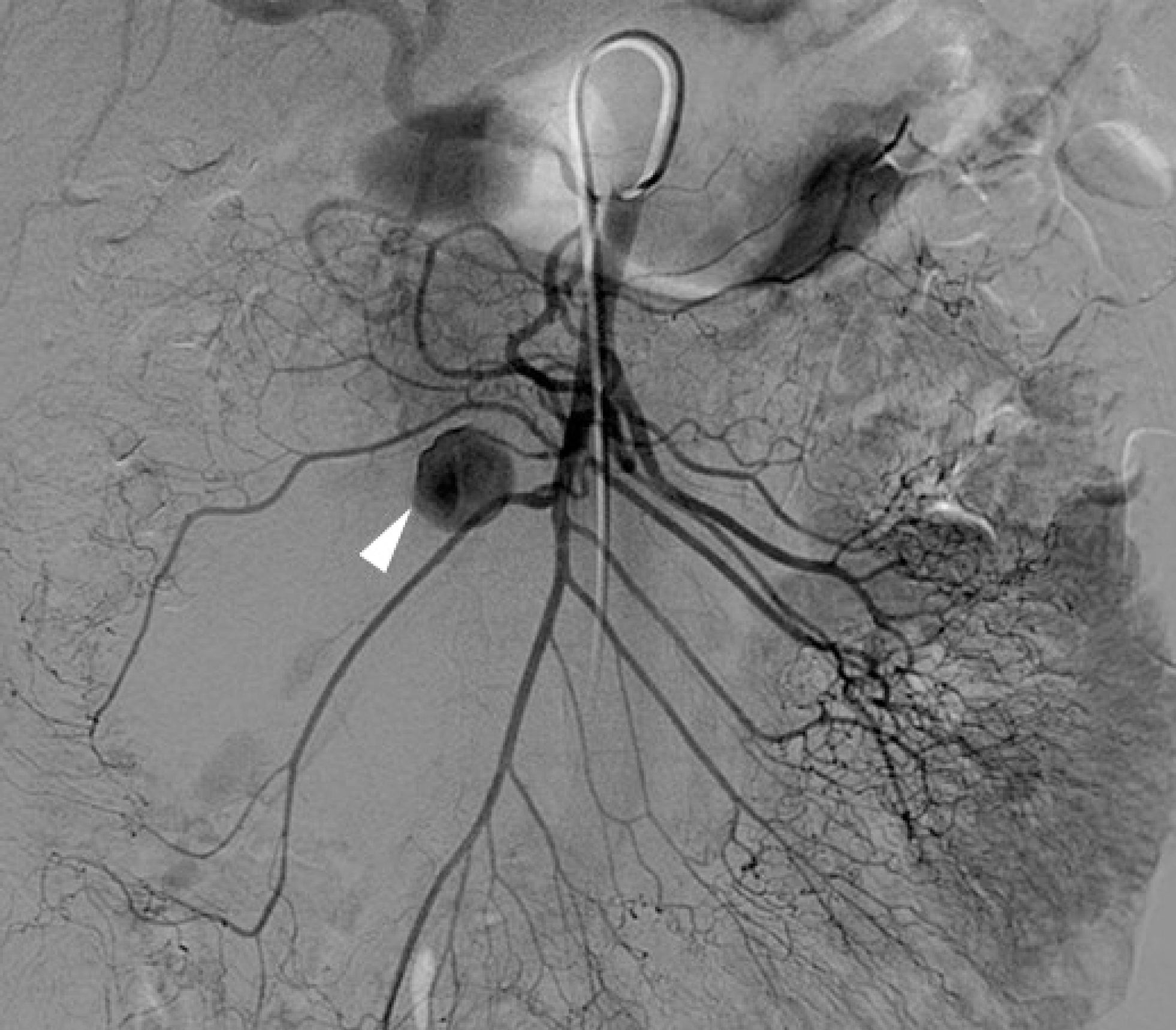
Fig. 5.
No contrast leakage from the embolized first jejunal branch of the SMA was observed on enhanced abdominal CT. SMA, superior mesenteric artery; CT, computed tomography.
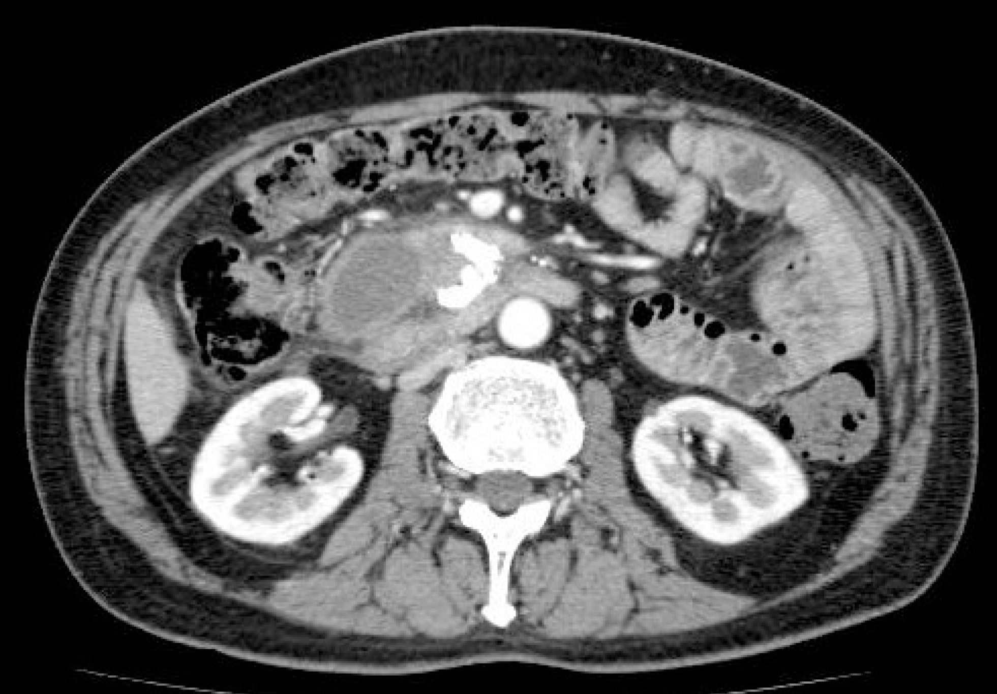
Table 1.
Clinical Features of Visceral Artery Pseudoaneurysm Rupture after ERCP
 XML Download