

 PDF
PDF ePub
ePub Citation
 Print
Print


INTRODUCTION
Osteoarthritis (OA) is a chronic inflammatory disease that progressively debilitates joints and the surrounding tissue. OA limits movement, distorts joint shape, and causes joint pain. OA of the temporomandibular joint (TMJ-OA) poses difficulties with chewing, limits mouth opening, and causes facial asymmetry,1 and nutritional imbalances due to pain and masticatory dysfunction may impede growth and development.23
One study investigating the demographics of patients visiting an orofacial pain clinic in South Korea noted an increase in young TMD patients with OA from 9% in 2000 to 39% in 2008.4 TMJ-OA that starts at a young age inevitably leads to facial deformity and malocclusion.5 To solve these problems, orthodontic treatment with orthognathic surgery is generally required. However, sudden changes in occlusion can aggravate TMJ-OA and worsen the problem, thus treatment cannot be started until TMJ-OA is completely cured.6 Therefore, early treatment before irreversible bony changes occur is needed.
Human growth hormone (hGH) stimulates growth by triggering active chondrocyte growth at the postnatal epiphyseal growth plates of the long bone.7 Exerting growth promoting effect on chondrocytes, hGH has been suggested as a treatment strategy for OA.8 hGH showed a positive effect in the very early healing phase of an osteochondral defect in an animal model.9 Reinecke, et al.10 concluded that hGH could be used as a potential treatment option for OA because of its anabolic effect on joints. Additionally, hGH could be used with the purpose of increasing cartilage metabolism and chondrocyte proliferation.11 Therefore, use of hGH to treat OA could be optimized by avoiding systemic administration and using it locally in only problematic TMJ-OA sites.
Growth hormone (GH) has been shown to be involved in the proliferation of cartilage by stimulating insulin-like growth factor 1 (IGF-1) production in chondrocytes and modulating the growth metabolism of cartilage.1213 Accordingly, IGF-1 could modify the progression of OA via cartilage growth.14 GH regulates cell growth using IGF-1 as a mediator. Immunolocalized studies have confirmed that when GH is directly injected into the growth plate, IGF-1 levels in the growth plate are increased.15 This mechanism was reflected as increases in the mRNA and protein levels of IGF-1 when GH was systemically administered.16 Therefore, IGF-1 and hGH were confirmed to be capable of being utilized as monitoring markers of OA.17
This study sought to document changes that occurred after the local application of GH in TMJ-OA chemically induced in rats by monosodium iodoacetate (MIA) and to evaluate intraarticular injections of hGH as a suitable treatment option for TMJ-OA patients in the future.
MATERIALS AND METHODS
Animals
Seventeen 4-week-old male Sprague-Dawley rats (Koatech, Pyeongtaeck, Korea), weighing 90–100 g, were used, providing a total of 34 TMJs. Animals were housed one rat per cage at 21–22℃ with a 12-h light/12-h dark cycle. All experiments were approved by the Animal Ethics Committee of Pusan National University (PNU-2014-0518) and performed according to Guidelines for the Care and Use of Laboratory Animals.
Study design
TMJ-OA was induced in 4-week-old experimental animals as described in a previous paper.18 Briefly, 50 mL of solution [monosodium iodoacetate (MIA) 1 mg] was injected into the TMJ bilaterally, and 2 weeks later, typical OA stages were observed. After the animals were anesthetized with inhaled isoflurane, a 50-µL intra-articular injection of MIA 1 mg (Sigma, St. Louis, MO, USA) was administered in both condyles. After 2 weeks, 50 µL of phosphate buffered saline (PBS) was injected into the articular space of the right condyle and 50 µL of a solution of 500 µg of recombinant human growth hormone (rhGH) (Pfizer, New York, NY, USA) in PBS was injected into the articular space of the left condyle. The procedures were repeated twice a week at 3-day intervals for a total of four injections. The animals were euthanized after 2 or 4 rhGH injections (Fig. 1). OA was induced in all experimental animals using MIA except in two healthy controls. Ten randomly selected rats were injected with GH (OA+GH injected) and PBS (OA+PBS injected) in each TMJ according to the experimental protocol, and the remaining five were left to self-heal after OA induction (OA-induced only). The bilateral condyles were collected for histological observation, and serum and synovial fluid of the TMJ were collected. Synovial fluid was collected from both condyles by a lavage of 50 µL of sterile saline infused into the joint and withdrawn by syringe.
Micro-computed tomography
Under intraperitoneal anesthesia with Zoletil 0.6 mL/kg (Virbac Korea, Seoul, Korea) and Rompun 0.4 mL/kg (Bayer Korea, Seoul, Korea), the rats were placed on a volumetric computed tomography (CT) scanner (NFR-MXSCAN-G90; NanoFocus-Ray, Iksan, Korea), a micro-CT system designed to minimize motion artifacts and to maintain respiratory anesthesia during scanning to obtain CT images of a live rat. The system was set to 70 kV and 60 mA. A micro-CT scan was performed three times to measure condylar length and to check for MIA-induced arthritis. The CT images were obtained at the following time points: just before starting GH injection (CT1, 6-weekold) and after two (CT2, 7-week-old) or four (CT3, 8-week-old) GH treatments (Fig. 1).
For the measurement of condylar length, a straight line was drawn between two points anatomically situated in the lowest position on two-dimensional reconstructed images. Mandibular head length was measured by drawing a perpendicular line from the highest point of the condylar process toward the former line.19
Subchondral bone OA score
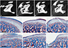
For the scoring of radiographic signs of OA, two individuals participated in this experiment independently. The lateral, central, and medial parts of the condyle were surveyed and graded as previously described.20 In this study, only erosive changes of the condyle were recorded before and after GH treatment. Subchondral bone OA scores were classified as follows: None, 0; Minor, 1; Moderate, 2; or Severe, 3 (Fig. 2A–D).
Histological observation
For histological observation, the bilateral condyles of each rat were dissected, immersed in 10% formaldehyde, and decalcified in Plank-Rychlo solution. Each trimmed specimen was processed following a standard procedure for paraffin preparation: hydration in a graded series of ethanol, clearance by xylene, and embedding in paraffin. The 4-mm-thick frontal sections were deparaffined and visualized by Masson's trichrome staining.
Cartilaginous OA score
OA cartilage histopathology was assessed using Masson's trichrome stained samples. Histological OA grading of cartilage degradation was performed as described previously21: 0, normal; 1, surface irregularities; 2, pannus and surface irregularities; 3, clefts to transitional zones; 4, clefts to radial zones; 5, clefts to calcified zones; and 6, complete disorganization. A pathologist who was blinded to the experiment graded all cartilage sections.
Blood screening for growth hormone and insulin-like growth factor-1
Enzyme-linked immunohistochemistry (ELISA) tests (R&D Systems, Inc., Minneapolis, MN, USA) were used to measure GH and IGF-1 levels in serum and synovial fluid. Whole-blood samples were collected into tubes and centrifuged at 13000 rpm for 10 min at 4℃. Aliquots of serum and synovial fluid were stored at −80℃ until ready for analysis. The assay was conducted according to the manufacturer's recommended protocols. Standard serum diluents and synovial fluid were then added in duplicate to individual wells and incubated for 2 h at 37℃. After washing the plates five times with wash buffer, 100 µL of conjugate was added to each well and incubated for 2 h at room temperature. After incubation, the plates were washed another five times with wash buffer. Substrate solution was then added to the wells and incubated for 30 min at room temperature in the dark. The reaction was stopped with 100 µL of H2SO4 solution (Merck, Darmstadt, Germany). The absorbance was determined using a spectrophotometric plate reader (VersaMax Microplate Reader; Molecular Reader, Toronto, Canada) at a 40-nm wavelength.
Statistical analysis
All statistical analyses were performed using IBM SPSS version 22.0 (IBM Corp., Armonk, NY, USA). The Mann-Whitney test was used to determine significant differences in mean values of two independent groups. The Kruskal-Wallis test was used to compare mean values or scores for changes over three independent groups. The Wilcoxon signed-rank test was used to confirm statistically significant changes in the OA scores of respective samples before and after treatment. Differences with p<0.05 were considered statistically significant.
RESULTS
MIA successfully induces OA in the TMJ of rats
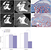
As shown in Fig. 2, the normal controls exhibited sound cortical lining, as well as normal trabecular patterns on micro-CT images (Fig. 2A). The intactness of the structures, including fibrous covering, fibrocartilage, and osteochondral tissue, were histologically confirmed in the tissue sections (Masson's trichrome stain). Normal tissue consisted of five layers: fibrous covering, proliferative region, fibrocartilage region, calcified cartilage region, and subchondral bony region (Fig. 2E). When micro-CT images were taken at 2 weeks after a 50-µL intra-articular injection of 1 mg of MIA (CT1), erosive OA stages could be observed (Fig. 2B–D). Fig. 2 depicts the degrees of increasing TMJ-OA and relative OA scores: the micro-CT images show bony OA scores, while the histologic views represent cartilaginous OA scores.
Fig. 2J denotes a representative histological view showing a cartilaginous OA score of 5. Extensive loss of fibrous covering with only small parts remaining were seen. The underlying cartilage was lost, and subchondral bone was exposed. High-power microscopy revealed bone formation in the areas below the destructed joint, although it was not calcified; rather, it was in an immature state, with the central part of the joint showing extensive trabecular destruction replaced with fibrous tissue. Furthermore, the remaining fibrocartilage and underlying cartilage were thin and irregular in shape, compared to the control group. In the areas of bone exposure, the fibrous covering, fibrocartilage, and underlying cartilage were all destroyed and showed increased numbers of fibroblasts, osteoclasts, and inflammatory cells, with less trabecular calcification (Fig. 2J; cartilaginous OA score=5).
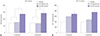
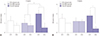
Local GH injections increase IGF-1 levels in synovial fluid, but not throughout the length of the condyle
Changes in GH and IGF-1 levels were observed on ELISA (Figs. 3 and 4). Significant changes in GH occurred in the blood (p=0.003) (Fig. 3A), where IGF-1 levels remained unaffected (Fig. 3B). There were no changes in condylar length among the healthy, OA-induced only, and GH-injected groups (data not shown). When we injected GH directly into the TMJs, we found that IGF-1 levels responded positively in the synovial fluid (p=0.016) (Fig. 4B); however, there were no statistically significant changes in GH levels (Fig. 4A).
Locally injected GH improves OA scores for subchondral bone and cartilage of the TMJ
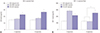
To determine the effect of GH in bony remodeling of arthritic TMJ, OA bone scores were analyzed using micro-CT (Fig. 2). Once MIA-induced OA was confirmed (CT1), images were taken after two GH injections (CT2) and after four GH injections (CT3).
Although scores varied in the degree of induced OA between subjects, the average MIA-induced OA score was 1.55 before starting GH injection. In rats with PBS-injected TMJs, only a small change in average OA scores was observed. On the individual level, most of the subjects showed a slight reduction in OA scores. However, two subjects actually showed an increase in OA scores. On the other hand, GH-injected TMJs showed marked reductions in average OA scores at both after two (Fig. 5A) and four (Fig. 5B) injections, and OA scores in the subchondral bone were significantly better for the TMJs from the GH-injected group, compared to those from the PBS-injected group (Wilcoxon signed-rank test, p=0.010).
Histological OA grading of cartilage degradation via Masson's trichrome staining was performed as described previously.21 As changes in cartilage were difficult to verify on the CT images, confirmation could only be made after the subjects were euthanized at the end of the experiment (Fig. 2E–J). There was only a small difference in cartilaginous OA scores from PBS-injected and GH-injected TMJs after two injections. However, there was a significant difference in OA scores for the PBSinjected and GH-injected TMJ groups after four injections (p<0.01) (Fig. 6B).
Fig. 6 shows CT and Masson's trichrome stained tissue images of test subject number 3, which was treated with four injections of GH in the left TMJ (Fig 6A-a, b, c) and PBS in the right TMJ (Fig 6A-d, e, f). Generalized condylar head OA (score 3) is visible in Fig. 6A-a; this score was much higher than that shown in Fig. 6A-d (score 1), indicating that the defect was much larger at the initial stage. However, images taken after four GH injections revealed that the score had decreased to 1 (Fig. 6A-b). As seen in Fig. 6A-c, the repaired condyle after the GH injection exhibited the maintenance of all cellular layers and ossification of about 10% of the cartilage tissue. On the other hand, the OA score of the TMJ treated with PBS was recorded as 1 before the experiment (Fig. 6A-d), and after four PBS injections, the score rose to 2, showing that the OA had worsened (Fig. 6A-e). The histological findings showed that approximately 50% of the TMJ was composed of bone and reparative tissue, and the remaining 50% showed all cellular layers, although thinning of the hypertrophic layer was observed (Fig. 6A-f).
DISCUSSION
Although several hypotheses for OA progression have been proposed, in general, changes are thought to initiate from cartilage, followed by changes in subchondral bone through cartilage and bone interaction.14 The primary aim of TMJ-OA treatment is reducing TMJ overloading and pain management by eliminating inflammatory responses in the capsular space. OA in middle-aged to older adults appears to be accompanied by degenerative joint changes, whereas OA in adolescents does not display a degenerative etiology.4 Therefore, many attempts have been made to determine the etiology of OA in accordance with etiologic factors, such as genetic factors, parafunctional habits, occlusal disturbance, and sudden dental treatment,122 and to develop treatments to eliminate these etiologic factors. Treatment options include physical therapy, medication, stabilization splint therapy, and behavioral therapy. These treatments are dependent on recovery of the condyle through the physiologic remodeling process, and their results depend on individual's cell activity and OA severity.23 There have been numerous attempts to assist with the regeneration of the cells that form the condyle. Among them, the most common for direct intra-articular injection include hyaluronic acid,24 steroids,25 and IGF-1.26
GH is known to play a definite role in bone growth, and its efficacy has been proven on many occasions. Moreover, it was long ago suggested that GH directly stimulates pre-chondrocyte generation in the germinal cell layer of the growth plate, leading to proliferation of chondrocytes. GH can also activate IGF-1 indirectly to further stimulate chondrocyte proliferation.27 One study involving hypophysectomized rats showed that systemic administration or local injection of GH could stimulate chondrocyte growth in the epiphyseal growth plate.28 Another found that GH improved chondrocyte generation and proliferation independent of IGF-1 when it was injected into experimental animals that could not produce IGF-1.29
Normal condylar tissue consists of five layers: fibrous covering, proliferative region, fibrocartilage region, calcified cartilage region, and subchondral bony region (Fig. 2E). The MIA used in this study is a representative substance that inhibits chondrocyte metabolism, thereby inducing apoptosis and reducing cartilage matrix. When injected intra-articularly, irregular surfaces, loss of superficial cell layers, and generalized growth inhibition can be induced.30 The histological findings of this experiment, such as the loss of superficial cell layers, irregularity in the fibrocartilage region, destruction of trabeculae, and fibrous tissue proliferation into the destructed trabeculae space, were consistent with the results of previous studies. Extensive loss of the fibrous covering with only small parts remaining was seen in induced TMJ-OA. The underlying cartilage was lost, and subchondral bone was exposed (Fig. 2I and J). Furthermore, MIA-induced TMJ-OA showed irregular surfaces, erosions which are consistent findings with those of the CT images of highly advanced OA patients.18 Thus, it was deemed a good method to model human TMJ-OA (Fig. 2D).
According to Wang, et al.,18 starting 3 days after MIA injection, chondrocytes show nuclear condensation and enter apoptosis. Also, TUNEL-positive chondrocytes nearly disappear by 1 week after the injection; after 2 weeks, the shape of subchondral bone is somewhat maintained and erosion becomes visible on CT images. In this study, GH was injected 2 weeks after MIA administration, because this was the point at which the focal erosion is seen most frequently in adolescents.
In the OA+PBS group, IGF-1 levels in synovial fluid were higher after two injections than after four. We assumed that IGF-1 was secreted temporarily into the damaged areas for GH-independent healing in the body. This may explain why there was transient expression of IGF-1 in joints that received only PBS after the induction of OA (Fig. 4B). This same result was observed in a previous study, which demonstrated that IGF-1 in the synovial fluid can be generated in response to damage.31 When OA is progressing, a temporary increase in IGF-1 levels in synovial fluid is assumed due to release from chondrocytes independent from serum GH.
In this study, after GH injection, the amount of GH in the blood increased (Fig. 3A). This increase in serum GH could be due to leakage of injected GH from the articular space. The increase in IGF-1 levels in the blood was not significant after the GH injection (Fig. 3B). However, a distinctive increase was observed in the synovial fluid, and this was higher in the second week after four injections (Fig. 4B). IGF-1 released from the damaged joint surface could induce tissue healing, although in situations in which continuous secretion did not occur, the GH injections maintained IGF-1 concentrations. This supports the notion that using GH could be an effective treatment option for OA.
In this study, local GH injection improved subchondral bone OA scores in damaged TMJs (Fig. 5), and a marked improvement in cartilaginous OA scores was observed after four GH injections (Fig. 6B). From this, a hypothesis can be made to suggest that GH either directly acts on chondrocytes for regeneration or improves the inflammatory response, therefore providing the right environment for cartilage regeneration. GH is known to stimulate cells directly and indirectly by controlling IGF-1 locally or systemically to achieve a major role in linear bone growth.7 Visnapuu, et al.32 reported that GH receptors were located in the mineralized area of the rat TMJ and that IGF-1 receptors were detected in the cartilage layer of the condyle. In this study, locally injected GH was able to bind to receptors in the exposed subchondral bone. After binding to its chondrocyte membrane receptor, GH activates a number of well-recognized intracellular signal transduction pathways that regenerate the transcription of GH target genes, including IGF-1, to orchestrate the array of chondrocyte events necessary for linear bone growth to proceed.33 Chondrocytes within the growth plate proceed through proliferation, differentiation, and maturation stages while maintaining their spatially fixed locations.34 This acts as an important key factor to form various conditions permissive to vascular infiltration for new bone growth in the chondro-osseous junction.34 Although GH levels in the blood increased in the GH injected group, no difference in condylar length was observed among the normal, OA-induced only, and GH injected groups (data not shown). These results showed that while systemic GH concentration increases, this increase does not affect overall mandibular growth.
Studies have reported that OA patients show significantly elevated levels of GH in the basal serum, compared to healthy individuals,35 while other have shown that serum GH levels decrease to normal when pain and swelling are reduced by TMJ treatment, including that with nonsteroidal anti-inflammatory drugs.36 Thus, while the interaction between GH and inflammation is clearly evident, it has not yet been clarified. Moreover, the role of GH or IGF-1 cannot be clearly distinguished in these processes. It is true that GH stimulates IGF-1 production37; however, one study showed that GH does not function in mutant mice lacking IGF-1.38 It is possible that GH and IGF-1 have independent and shared functions, as growth retardation of double ghr/igf-1r mutants is more severe than that observed with the ghr mutant or igf-1 null mutant.39
In this study, the small numbers of animals would be unable to represent all aspects of OA, and the severity of induced OA differed among the animals. Therefore, the same level of improvement could not be observed. However, when GH is injected locally, it can play a role in improving OA scores in rat TMJs for both cartilage and subchondral bone without affecting systemic bone growth as seen in this study. The local injection of GH positively affects subchondral bone and can repair the damaged cartilaginous layer. Therefore, we deemed local injections of GH to be useful in treating TMJ-OA in early stages due to its ability to maintain a cartilaginous layer and thus prevent a change in subchondral bone shape. Notwithstanding, further studies are needed to explore the different responses among animals and results of long-term administration of GH.
 XML Download