

 PDF
PDF ePub
ePub Citation
 Print
Print


INTRODUCTION
Pancreatic ductal adenocarcinoma (PDAC) is a lethal gastrointestinal malignant disease, with an estimated overall 5-year survival rate of less than 5%. While the most effective treatment option is margin-negative pancreatectomy, only about 15–20% of patients with a new diagnosis of PDAC can undergo surgical resection, and their 5-year survival rate is around 20%.1 Most PDAC patients experience recurrence and cancer-related mortality.
In investigating the biologic behavior of PDAC, cancer-related factors and host-related factors need to be considered. Several cancer-related factors, such as margin status, tumor size, tumor (T) stage, nodal (N) stage, tumor differentiation, lymphovascular invasion, perineural invasion, major vascular invasion, and high levels of preoperative or postoperative CA19-9, have been identified as important cancer-related prognostic factors.1 Host-related factors, for example, physical activity2 and host immune responses to cancer, have also been reported to be important in determining the disease-related prognosis for a variety of cancers.3456 Because tumor infiltrating lymphocytes (TILs) are thought to reflect the host immune response against tumors, TILs are considered prognostic factors.78910
Tumor infiltrating CD8+ cytotoxic T lymphocytes and CD4+ helper T lymphocytes act as antitumor effectors and are associated with a favorable outcome.11 Activated CD8+ T-cells attack tumor cells presenting tumor associated antigens via the peptide/major histocompatibility complex class I on the tumor cell surfaces.12 Activated CD8+ T-cell express granzyme B on their surface.13 Unlike CD4+ and CD8+ T-cells, regulatory T lymphocytes (Treg) have been shown to have an adverse effect on the prognosis of patients by suppressing effector T-cells and the production of several immunosuppressive cytokines, including interleukin-10 and transforming growth factor-β.14 The forkhead/winged helix transcription factor, forkhead P3 (Foxp3), which is genetically defective in autoimmune and inflammatory syndromes in humans and mice, is specifically expressed in naturally arising CD4+ Tregs.15 In our previous study of the prognostic impact of TILs in resected left-sided PDAC,16 we demonstrated that a low tumor infiltrating Foxp3+/granzyme B+ ratio was an independent factor predicting good disease-free survival (DFS) and overall survival (OS) outcomes.
We have been performing minimally invasive radical pancreatectomies in well-selected left-sided PDAC.171819 In doing so, we have applied the Yonsei criteria for patient selection based on preoperative CT scans and included the following tumor conditions: 1) tumor confined to the pancreas, 2) intact fascia layer between the distal pancreas and the left adrenal gland and kidney, and 3) tumor located more than 1–2 cm from the celiac axis. With our selection criteria, minimally invasive radical pancreatectomy was both feasible and oncologically safe.171819 In our recent study evaluating the long-term oncologic outcomes of minimally invasive radical distal pancreatectomy for left-sided PDAC,19 we noted that when patients satisfied the Yonsei criteria, they experienced longer long-term survival, regardless of surgical approach (open vs. minimally invasive surgery), suggesting that the Yonsei criteria may play some role in the prediction of tumor aggressiveness for resected left-sided PDAC.
In this study, we investigated the impact of TIL subsets on patient survival outcomes and whether the Yonsei criteria predicted a favorable tumor microenvironment characterized by the distribution of TILs. We suggest that the Yonsei criteria may both serve as patient selection criteria for cancer treatment and reflect a favorable host immunologic microenvironment in left-sided PDAC.
MATERIALS AND METHODS
Patients
Patients who underwent curative distal pancreatectomy due to left-sided PDAC from January 2000 to December 2011 at Severance Hospital, Yonsei University College of Medicine (Seoul, Korea) were enrolled. Patients with pancreatic head cancer were excluded to avoid potential contamination with other periampullary cancers, such as distal bile duct, ampulla of Vater, and duodenal cancers. We retrospectively analyzed patient demographics, histopathologic findings, and survival outcomes. Follow-up was completed September 30, 2015. Patients who received neoadjuvant chemotherapy or chemoradiation therapy and had other primary tumors were excluded.16 OS was defined as the interval between surgery and death or between surgery and the last observation for surviving patients. DFS was defined as the interval between surgery and recurrence. This study was approved by the Institutional Review Board of Severance Hospital, Yonsei University College of Medicine (4-2013-0264).
Immunohistochemical staining and quantification of TIL subsets
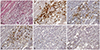
Immunohistochemical (IHC) staining for TIL subsets was performed as previously described.516 Briefly, paraffin-embedded PDAC tissue sections at a thickness of 4 µm were deparaffinized in xylene and rehydrated in decreasing concentrations of ethanol. Antigen retrieval was performed in citrate buffer in a microwave oven. Endogenous peroxidase activity was blocked by incubating the tissue with 3% hydrogen peroxide in methanol for 5 min. The sections were incubated for 60 min at room temperature with primary monoclonal antibodies against cluster of differentiation (CD)3 (Cat. No. RM-9107-S, 1:100, Lab Vision Corporation, Fremont, CA, USA), CD4 (Cat. No. NCL-L-CD4-1F6, 1:100, Novocastra™, Newcastle upon Tyne, UK), CD8 (Cat. No. IS62330, 1:100, Dako, Glostrup, Denmark), Foxp3 (Cat. No. ab20034, 1:100, Abcam, Cambridge, UK), and granzyme B (Cat. No. MS-1157-S, 1:100, Lab Vision Corporation), which were used to identify total numbers of T-cells, helper T-cells, cytotoxic T-cells, Treg, and activated cytotoxic T-cells, respectively. After washing the sections twice with 0.05 mol/L Tris-buffered saline with 0.2% Tween-20, the sections were incubated with horseradish peroxidase-conjugated secondary antibody (Dako EnVision® Detection system, Dako), followed by development with diaminobenzidine and counterstaining with hematoxylin (Fig. 1).
IHC staining was quantified by two experienced pathologists who were blinded to patient data. Three intense foci of staining in the tumor sections were selected and four high-power fields (magnification, ×400) from each slide were selected for calculation of IHC staining results. TILs tended to be distributed more in the interstitial area of the tumor microenvironment.16 Fields with necrosis or hemorrhage in the tumor portion were avoided.516 Patients were divided into low and high groups using the median value of absolute counts for positively stained cells and relative ratios between Treg and T-cells (CD3+, CD4+, CD8+, and granzyme B+ T-cell).
Statistical analysis
All statistical analyses were performed with SPSS 20.0 software (IBM, Corp., Armonk, NY, USA). Categorical variables were compared using χ2 or Fisher exact tests. Absolute counts of TIL subsets and the relative ratio between Treg and T-cells (CD3+, CD4+, CD8+, and granzyme B+ T-cells) were divided into two groups using cut-off values derived from the median value for χ2 or Fisher exact tests and survival analysis.5916 OS and DFS were calculated using the Kaplan-Meier method, and significance was evaluated by the log-rank test. Cox proportional hazard models were used for univariate and multivariate survival analysis. A p-value of 0.05 or less indicated statistical significance.
RESULTS
Patient demographics and general characteristics of resected left-sided PDAC
A total of 72 patients underwent curative distal pancreatectomy for left-sided PDAC. Among them, 13 patients who underwent neoadjuvant chemoradiation therapy were excluded. Nine patients were excluded because a paraffin-embedded tissue block was not available. Finally, 50 patients were enrolled in this study. The mean age was 62.8±9.4 years, and 20 patients (40%) were female. Only 3 patients (6%) underwent minimally invasive (laparoscopic or robotic) distal pancreatectomy. The mean operation time was 290±167 min. Combined organ resection was performed in 12 patients (24%). Thirteen patients (26%) received intraoperative transfusion. No postoperative mortality or severe complications (Clavien-Dindo classification≥IIIa) were recorded. The mean tumor size was 3.5±1.5 cm. Pathological T staging revealed T1 in 1 (2%), T2 in 5 (10%), T3 in 42 (84%), and T4 in 2 patients (4%). Pathologic N1 stage was observed in 23 patients (46%). Lymphovascular invasion and perineural invasion were observed in 10 patients (20%) and 23 patients (46%), respectively. Tumor differentiation was classified into one of four groups: 11 well differentiated, 34 moderately differentiated, four poorly differentiated, and one un-differentiated. Resection margin status was R0 in 45 (90%), R1 in 3 (6%), and R2 in 2 (4%) patients. All patients received postoperative adjuvant chemotherapy with gemcitabine.
Survival analysis according to clinicopathologic and operative findings
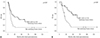
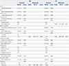
Survival outcomes based on univariate and multivariate analyses according to clinicopathologic parameters and operative findings are shown in Table 1. Univariate analysis showed patients satisfying Yonsei criteria (p=0.021), with fewer than two metastatic lymph nodes (LNs) (p<0.001), without combined organ resection (p=0.020), and without intraoperative transfusion (p=0.005) were associated with longer DFS. Univariate analysis also showed small tumor size (<3.5 cm, p=0.031), satisfying Yonsei criteria (p=0.001), fewer than two metastatic LNs (p=0.003), no combined organ resection (p=0.008) and no intraoperative transfusion (p=0.028) to be associated with longer OS. Multivariate analysis demonstrated that having more than two metastatic LNs [Exp(β)=3.354; 95% confidence internal (CI): 1.582–7.110; p=0.002] and intraoperative transfusion [Exp(β)=2.615; 95% CI: 1.248–5.482; p=0.011] to be independent risk factors for DFS. Not satisfying Yonsei criteria [Exp(β)=2.590; 95% CI: 1.151–5.829; p=0.021] and more than two metastatic LNs [Exp(β)=2.536; 95% CI: 1.083–5.936; p=0.032] were independent risk factors for OS (Table 1).
Survival analysis according to TIL subsets
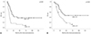
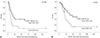
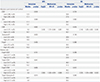
Univariate analysis showed that high CD8+ levels were significantly related to longer DFS (p=0.002) and OS (p=0.016). High granzyme B+ levels were significantly associated with longer DFS (p=0.033). Low levels of Foxp3+/CD4+, Foxp3+/CD8+, and Foxp3+/granzyme B+ were favorable prognostic factors in DFS (p=0.041, p=0.005, and p=0.014, respectively) and OS (p=0.034, p=0.012, and p=0.040, respectively) (Table 2). The Cox regression hazards model demonstrated that low levels of CD8+ [Exp (β)=3.436; 95% CI: 1.701–6.944; p=0.001] and high Foxp3+/CD8+ ratios [Exp(β)=2.505; 95% CI: 1.262–4.974; p=0.009] were independent prognostic factors for recurrence of resected left-sided PDAC. Low levels of CD8+ [Exp(β)=2.140; 95% CI: 1.035–4.426; p=0.040] and high levels of Foxp3+/CD8+ [Exp(β)=2.235; 95% CI: 1.074–4.650; p=0.032] were also independent prognostic factors for unfavorable OS (Table 2, Figs. 2 and 3).
Association between clinicopathologic factors and CD8+ counts and Foxp3+/CD8+ ratio
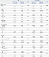
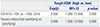
Among the preoperatively determined clinical factors used to predict patient survival, high levels of CA19-9 (p=0.022) and not satisfying Yonsei criteria (p=0.022) were significantly associated with a high Foxp3+/CD8+ ratio. Age, tumor size, gender, and NCCN resectability were not associated with CD8+ levels or Foxp3+/CD8+ ratio. Among the postoperative pathologic factors, more than two metastatic LNs were related with low tumor infiltrating CD8+ levels (p=0.020) and a high Foxp3+/CD8+ ratio (p=0.049). Mean LN ratios (LNR, the ratio of metastatic to retrieved LNs) were significantly higher in the high Foxp3+/CD8+ group (p=0.020). Pathologic tumor stage, nodal stage, tumor differentiation, lymphovascular invasion, and perineural invasion were not associated with CD8+ levels or Foxp3+/CD8+ ratio (Table 3). In multivariate analysis, high levels of CA19-9 and not satisfying Yonsei criteria were significantly associated with a high Foxp3+/CD8+ ratio [Exp(β)=3.558; 95% CI: 1.000–12.658; p=0.050] (Table 4). When the two factors, Yonsei criteria and CA19-9, were combined for analysis of factors affecting the CD8+ counts and Foxp3+/CD8+ ratio, high levels of CA19-9 and not satisfying Yonsei criteria were significantly associated with a high Foxp3+/CD8+ ratio, compared to other conditions (p=0.024) (Table 5).
DISCUSSION
In this study, we investigated the effects of TILs on the prognosis of left-sided PDAC and examined whether the Yonsei criteria were associated with differences in TIL distributions.
In addition to clinicopathologic factors, the host immune response against tumors plays a critical role in survival outcomes for numerous types of cancers.34581011 In terms of TIL subsets, a ratio of low Treg density to high T-cells (CD4+, CD8+, and granzyme B+ T-cell) has been reported to be a promising independent favorable factor in various tumors.459 In our previous study,16 we showed that patients with a low Foxp3+/granzyme B+ ratio had significantly improved DFS (25 months vs. 8 months; p=0.008) and OS (47 months vs. 17 months; p=0.003). Multivariate analysis of the data accumulated since our previous study supports that a high level of CD8+ cells and a low Foxp3+/CD8+ ratio are independent predictors for favorable DFS (p=0.001 and p=0.009, respectively) and OS (p=0.040 and p=0.032, respectively) (Table 2, Figs. 2 and 3).
In our previous study, we analyzed clinicopathologic factors associated with differences in the distribution of TIL subsets: only low levels of CA19-9 were significantly associated with a low Foxp3+/granzyme B+ ratio. Even though several studies have demonstrated that distributions of TILs are associated with survival outcomes in patients with PDAC,202122 only a few studies have described clinicopathologic factors associated with differences in TILs distribution. Fukunaga, et al.20 reported that positivity for both tumor infiltrating CD4+ T-cells and CD8+ T-cells was significantly associated with a lower grade of depth of invasion and tumor stage, compared with both negative group. Hiraoka, et al.23 demonstrated that a high prevalence of Treg in CD4+ T-cells was significantly correlated with distant metastasis, advanced tumor stage, and high tumor grade.
These studies have investigated pathologic findings that could only be analyzed postoperatively. To our knowledge, there are no other studies that have analyzed preoperative clinical factors associated with TILs to predict patient survival outcomes. In the present study, among the preoperatively detected clinical factors, high levels of CA19-9 (p=0.022) and not satisfying Yonsei criteria (p=0.022) were significantly associated with a high Foxp3+/CD8+ ratio. Among the postoperative pathologic factors, having more than two of metastatic LNs was also related to low CD8+ levels (p=0.020) and high Foxp3+/CD8+ ratio (p=0.049) (Table 3). In multivariate analysis, high levels of CA19-9 and not satisfying Yonsei criteria were associated with a high Foxp3+/CD8+ ratio [Exp(β)=3.558; 95% CI: 1.000–12.658; p=0.050] (Table 4). These two factors are preoperatively detectable clinical parameters. When we analyzed the survival outcomes between the patients with low and high CD8+ T cells and low and high FOXP3+/CD8+ T cells ratios in each subgroup of patients according to metastatic LN (<2 and ≥2) and CA19-9 (≤109 and >109), as well as Yonsei criteria (satisfying versus not satisfying), even the number of patients and the number of events in each group were too small to find meaningful results in all subgroups with Kaplan-Meier method, high CD8+ T cell and low FOXP3+/CD8+ T cell ratio were significantly associated with better survival in the subgroup with CA19-9 >109 (p=0.022 and p=0.022, respectively). Also, high CD8+ T cell levels and a low FOXP3+/CD8+ T cell ratio had marginal significance in the metastatic LN <2 subgroup (p=0.055 and p=0.069, respectively) (Supplementary Figs. 1, 2, and 3, only online).
Several clinicopathologic factors that can predict the prognosis of PDAC have been analyzed in various studies.124 Multivariate analysis in this study showed that more than two metastatic LNs [Exp(β)=3.354; 95% CI: 1.582–7.110; p=0.002] and intraoperative transfusion [Exp(β)=2.615; 95% CI: 1.248–5.482; p=0.011] were independent risk factors for DFS. Not satisfying Yonsei criteria [Exp(β)=2.590; 95% CI: 1.151–5.829; p=0.021] and more than two metastatic LNs [Exp(β)=2.536; 95% CI: 1.083–5.936; p=0.032] were also independent risk factors for OS (Table 1).
Slidell, et al.24 investigated the impact of nodal stage and LNRs affecting survival outcomes in a large population-based (with Surveillance, Epidemiology, and End Results, SEER data) study of patients with PDAC. In their study, N1 disease was associated with a worse 5-year survival rate, compared with N0 disease (4.3% vs. 11.3%, respectively, p<0.001). For N1 patients, LNR was one of the most powerful factors associated with survival (LNR >0–0.2, 15 months; LNR >0.2–0.4, 12 months; LNR >0.4, 10 months) (p<0.001). We did not find differences in survival outcomes according to pathologic nodal stage or LNR in N1 stage in this study; however, having more than two metastatic LNs was an independent risk factor for DFS and OS (Table 1).
Intraoperative transfusion has been shown to have an adverse oncologic impact on various kinds of tumors, including PDAC.25262728 Although the actual mechanism by which intraoperative transfusion negatively affects survival outcomes is unknown, it is thought to be an important factor in patient immune system suppression.29
Recently, although the number of patients diagnosed with early staged PDAC has increased due to routine medical checkup, it is still difficult to diagnose PDAC early, which is associated with a good prognosis. Furthermore, it is not easy to clearly define what is truly an early staged PDAC. The preoperative resectability definition of the National Comprehensive Cancer Network (NCCN) guidelines,30 which are based on imaging studies, are the most widely used method for determining surgical or non-surgical treatment strategies for PDAC. However, even in the NCCN guideline for ‘resectable’ status, it is difficult to distinguish early stage PDAC. In the present study, we were unable to discern any differences in survival outcomes according to NCCN guidelines. In our previous studies,171819 we found that the Yonsei criteria for left-sided PDAC, 1) tumor confined to the pancreas, 2) intact fascia layer between the distal pancreas and the left adrenal gland and kidney, and 3) tumor located more than 1–2 cm from the celiac axis, were valuable selection criteria for surgeons seeking to apply minimally invasive distal pancreatectomy. In the present study, patients that satisfied the Yonsei criteria showed favorable survival outcomes in regards to DFS (p=0.021) and OS (p=0.001) (Table 1, Fig. 4) and were significantly associated with favorable TIL subset distributions. Although we merely noted an association between preoperatively detectable parameters and host immune status against tumor, there are some reports203132 that suggest that patients with a high density of TILs (CD3+, CD4+, and CD8+ T cells) in pancreatic cancer have a better prognosis than those who have a low density of TILs. Accordingly, further well-designed translational research is warrant to better determine the biologic mechanisms for this phenomenon.
In conclusion, the host immune response against tumors plays a critical role in survival outcomes. High levels of tumor infiltrating CD8+ T cells and low Foxp3+/CD8+ ratios were significantly associated with long DFS and OS in this study. The Yonsei criteria can play a role as clinically detectable biologic marker with which to predict tumor-immunologic status and survival outcomes of patients with resected left-sided PDAC.
 XML Download