Journal List > Korean Circ J > v.50(5) > 1144307

Kim, Koo, and Lee: Coronary Arteriovenous Fistulas Mimicking Coronary Perforation After Chronic Total Occlusion Recanalization
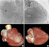
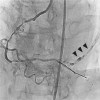
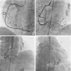
A 65-year-old female with stable angina presented with chronic total occlusion (CTO) of the distal right coronary artery (RCA) (Figure 1 and Supplementary Video 1). Coronary intervention of the distal RCA-CTO was performed via the antegrade approach. After successful crossing the CTO lesion gently with a soft guide wire (NEO's Sion black; Asahi, Aichi, Japan), the leakage of contrast media through multiple tiny channels at the posterior descending coronary artery was noted (Figure 2 and Supplementary Video 2). It was initially misinterpreted as coronary perforation, leading to prolonged ballooning of the leakage site without changes in leakage appearance. She was stable without evidence of pericardial effusion by echocardiography, indicating coronary arteriovenous fistulas. The RCA lesions were treated with two drug-eluting stents (Orsiro 2.5×40 mm; Biotronic, Bulach, Switzerland). Final angiogram showed arteriovenous fistulas between RCA and coronary sinus, which mimicked coronary perforation (Figure 3 and Supplementary Video 3).
Coronary artery fistulas can occur from any of the three major coronary arteries, and coronary artery dilatation is common because the fistulas drain into the low-pressure structures. However, when the fistulous communication arises in the distal part of a coronary artery, the artery diameter may remain small.1) In our case, the fistulas before CTO intervention were not recognized probably due to the insufficient driving pressure of collateral flow across tiny fistulas, but seen after restoration of brisk antegrade flow. We report an interesting image of arteriovenous fistulas with multiple tiny channels that can be mistaken for coronary artery perforation after CTO recanalization.

Figures and Tables

Figure 1

Diagnostic coronary angiography demonstrated total occlusion of the RCA (black arrow) (A) with good collaterals from the left anterior descending coronary artery (grey arrow) (B), and severe stenosis of the left anterior descending artery (B). Pre-procedure coronary computed tomography angiogram showed total occlusion of the distal RCA (white arrow) and there was no evidence of abnormal coronary artery drainage. (C, D).

RCA = right coronary artery.
kcj-50-464-g001
Figure 2

Coronary angiogram (left anterior oblique cranial view) showed contrast drainage through multiple channels after recanalization of chronic total occlusion, which mimicked coronary artery perforation (black arrow).

kcj-50-464-g002
Figure 3

(A, B) Two drug-eluting stents (Orsiro, 2.5×40 and 2.5×40 mm) were implanted in the proximal to middle segment and distal segment of the right coronary artery (A: LAO view, B: AP cranial view). Communication between posterior descending coronary artery and coronary sinus was prominently noted after antegrade flow restoration (C: AP cranial view, D: AP caudal view).

AP = anteroposterior; LAO = left anterior oblique.
kcj-50-464-g003

Notes

Conflict of Interest The authors have no financial conflicts of interest.

Author Contributions

  • Conceptualization: Lee CW.

  • Resources: Koo HJ.

  • Writing - original draft: Kim TO.

  • Writing - review & editing: Lee CW.

References

1. Gowda RM, Vasavada BC, Khan IA. Coronary artery fistulas: clinical and therapeutic considerations. Int J Cardiol. 2006; 107:7–10.
crossref

SUPPLEMENTARY MATERIALS

Supplementary Video 1

Baseline coronary angiography. Diagnostic coronary angiography demonstrated total occlusion of the RCA (A) with good collaterals from the left anterior descending coronary artery and severe stenosis of the left anterior descending artery (B).

Supplementary Video 2

Coronary angiogram after recanalization of chronic total occlusion. Coronary angiogram (left anterior oblique cranial view) showed contrast drainage through multiple channels after recanalization of chronic total occlusion, which mimicked coronary artery perforation.

Supplementary Video 3

Stent implantation at RCA and final result. Two drug-eluting stents (Orsiro, 2.5×40 and 2.5×40 mm) were implanted in the proximal to middle segment and distal segment of the RCA (A: LAO view, B: AP cranial view). Communication between posterior descending coronary artery and coronary sinus was prominently noted after antegrade flow restoration (C: AP cranial view, D: AP caudal view).
TOOLS
ORCID iDs

Tae Oh Kim
https://orcid.org/0000-0002-7275-9302

Hyun Jung Koo
https://orcid.org/0000-0001-5640-3835

Cheol Whan Lee
https://orcid.org/0000-0003-3987-2159

Similar articles