Abstract
The integrins αvβ1, αvβ3, αvβ6, and αvβ8 are known to be the natural receptors of foot-and-mouth disease virus (FMDV). Among them, integrin αvβ6 is considered a major receptor for FMDV. We performed protein expression of full-length bovine integrins αv, β3, and β6 and confirmed the high efficiency of bovine α vβ6 as the FMDV receptor in FMDV non-permissive SW 480 cells. Next, we established the black goat kidney (BGK) cell line, stably expressing bovine integrin β6 (BGK-β6-4). We observed that BGK-β6-4 cells had significantly enhanced sensitivity to FMDV compared with that of BGK cells (P<0.05). In addition, BGK-β6-4 cells had equal or higher sensitivity to several serotypes of FMDV compared with that of other FMDV permissive cell lines, such as BHK-21 and IBRS-2. In conclusion, we established a promising novel goat cell line, BGK-β6-4, which can be used to isolate or culture FMDV. Furthermore, the BGK-β6-4 cell line may serve as a promising tool for studying integrin α vβ6 receptor functions.
Go to : 
REFERENCES
2). Knowles NJ, Samuel AR. Molecular epidemiology of foot-and-mouth disease virus. Virus Res. 2003; 91:65–80.

3). Baxt B, Becker Y. The effect of peptides containing the arginine-glycine-aspartic acid sequence on the adsorption of foot-and-mouth disease virus to tissue culture cells. Virus Genes. 1990; 4:73–83.

4). Fox G, Parry NR, Barnett PV, McGinn B, Rowlands DJ, Brown F. The cell attachment site on foot-and-mouth disease virus includes the amino acid sequence RGD (arginine-glycine-aspartic acid). J Gen Virol. 1989; 70:625–37.

5). Berinstein A, Roivainen M, Hovi T, Mason PW, Baxt B. Antibodies to the vitronectin receptor (integrin α vβ3) inhibit binding and infection of foot-and-mouth disease virus to cultured cells. J Virol. 1995; 69:2664–6.
6). Amadori M, Volpe G, Defilippi P, Berneri C. Phenotypic features of BHK-21 cells used for production of foot-and-mouth disease vaccine. Biologicals. 1997; 25:65–73.

7). Johns HL, Berryman S, Monaghan P, Belsham GJ, Jackson T. A dominant-negative mutant of rab5 inhibits infection of cells by foot-and-mouth disease virus: implications for virus entry. J Virol. 2009; 83:6247–56.

8). Koyama T, Hughes RC. Functional integrins from normal and glycosylation-deficient baby hamster kidney cells. Terminal processing of asparagine-linked oligosaccharides is not correlated with fibronectin-binding activity. J Biol Chem. 1992; 267:25939–44.

9). Duque H, Baxt B. Foot-and-mouth disease virus receptors: comparison of bovine α v integrin utilization by type A and O viruses. J Virol. 2003; 77:2500–11.
10). Duque H, LaRocco M, Golde WT, Baxt B. Interactions of foot-and-mouth disease virus with soluble bovine α vβ3 and α vβ6 integrins. J Virol. 2004; 78:9773–81.
11). LaRocco M, Krug PW, Kramer E, Ahmed Z, Pacheco JM, Duque H, et al. A continuous bovine kidney cell line constitutively expressing bovine α vβ6 integrin has increased susceptibility to foot-and-mouth disease virus. J Clin Microbiol. 2013; 51:1714–20.
12). LaRocco M, Krug PW, Kramer E, Ahmed Z, Pacheco JM, Duque H, et al. Correction for LaRocco et al., A continuous bovine kidney cell line constitutively expressing bovine α vβ6 integrin has increased susceptibility to foot-and-mouth disease virus. J Clin Microbiol. 2015; 53:755.
13). Lee SY, Lee YJ, Kim RH, Park JN, Park ME, Ko MK, et al. Rapid engineering of foot-and-mouth disease vaccine and challenge viruses. J Virol. 2017; 91:e00155–17.

14). Reed LI, Muench H. A simple method of estimating fifty percent end points. Am J lEpidemiol. 1938; 27:493–7.
15). Kim SM, Lee KN, Lee SJ, Ko YJ, Lee HS, Kweon CH, et al. Multiple shRNAs driven by U6 and CMV promoter enhances efficiency of antiviral effects against foot-and-mouth disease virus. Antiviral Res. 2010; 87:307–17.

16). Hynes RO. Integrins: versatility, modulation, and signaling in cell adhesion. Cell. 1992; 69:11–25.

17). O'Donnell V, Pacheco JM, Gregg D, Baxt B. Analysis of foot-and-mouth disease virus integrin receptor expression in tissues from naï ve and infected cattle. J Comp Pathol. 2009; 141:98–112.
18). Jackson T, Sheppard D, Denyer M, Blakemore W, King AM. The epithelial integrin αvβ6 is a receptor for foot-and-mouth disease virus. J Virol. 2000; 74:4949–56.

19). Wang Y, Mao Q, Chang H, Wu Y, Pan S, Li Y, et al. Inability of FMDV replication in equine kidney epithelial cells is independent of integrin αvβ3 and αvβ6. Virology. 2016; 492:251–8.

20). Burman A, Clark S, Abrescia NG, Fry EE, Stuart DI, Jackson T. Specificity of the VP1 GH loop of foot-and-mouth disease virus for αv integrins. J Virol. 2006; 80:9798–810.

21). Neff S, Mason PW, Baxt B. High-efficiency utilization of the bovine integrin α vβ3 as a receptor for foot-and-mouth disease virus is dependent on the bovine β3 subunit. J Virol. 2000; 74:7298–306.
Go to : 
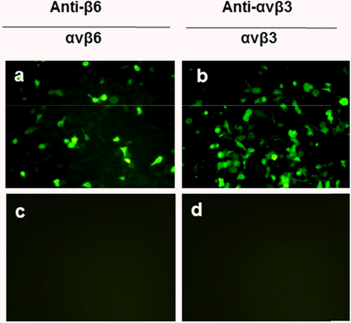
 | Figure 1.Detection of bovine integrins αvβ3 and αvβ6 heterodimer expression in SW480 cells using indirect immunofluorescence assay (IFA). SW-480 cells were co-transfected with vectors expressing bovine αv and β6 (a) or β3 (b) subunits. Protein expression was detected by IFA after 48 h of transfection using an anti-human β6 Mab (2076Z) (a) or an anti-human integrin αvβ3 Mab (LM609) (b). Untransfected cells treated with the same primary and secondary antibodies were used as negative controls (c and d). |
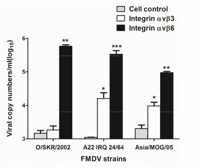
 | Figure 2.Comparison of viral infection efficiency in the cells expressing bovine αvβ3 and αvβ6 integrins. SW480 cells were co-transfected with vectors expressing bovine αv and β6 or β3 subunits. Twenty-four hours after transfection, the cells were infected with 104.5 TCID50 of FMDV O/SKR/2002, Asia 1/M0G/05, or A22/IRQ 24/64 viral suspensions. The supernatant was collected at 8 days after the infections, and viral RNA was extracted. FMDV quantification was performed using real-time RT PCR. An unpaired t-test was performed for statistical analysis by GraphPad InStat software (∗P< 0.05, ∗∗P< 0.01, ∗∗∗P < 0.001). Error bars indicate standard deviations from the mean. |
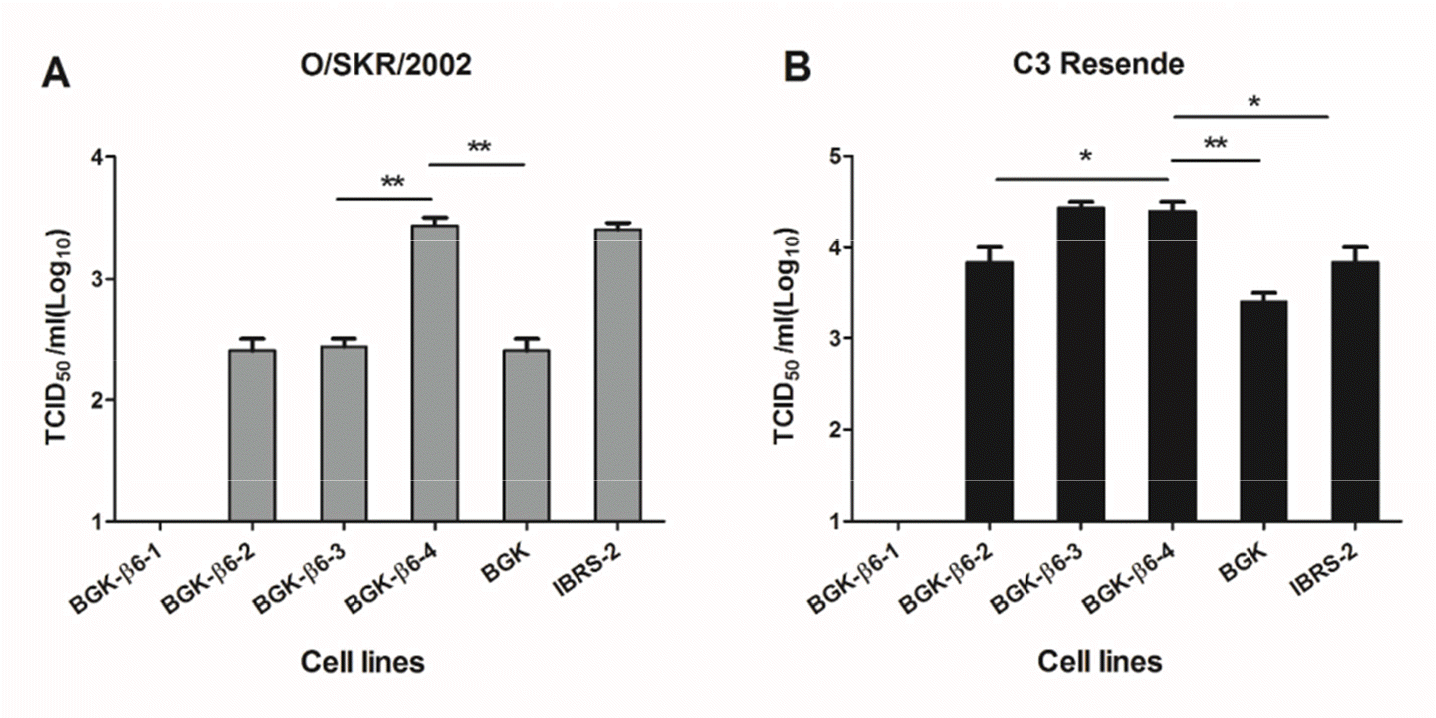
 | Figure 3.Comparative virus titration in BGK-β6 stable cell lines. Four stable BGK cell lines, expressing bovine integrin β6 (BGK-β6-1, BGK-β6-2, BGK-β6-3, and BGK-β6-4), in addition to BGK and IBRS-2 cell lines were used for titrating FMDV O/SKR/2002 or C3 Resende. Virus titration was measured 72 h post FMDV infection. TCID50 was calculated by Reed and Muench method (14). An unpaired t-test was performed for statistical analysis by GraphPad InStat software (∗P< 0.05, ∗∗P< 0.01). Error bars indicate standard deviations from the mean. |
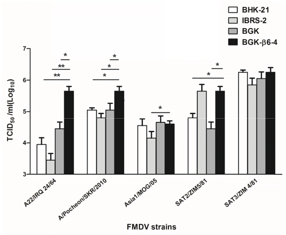
 | Figure 4.Comparative titration of various strains of FMDV in BGK-β6-4, BGK, IBRS-2, and BHK-21 cell lines. BGK-ß6-4, BGK, IBRS-2, and BHK-21 cell lines were used for titrating FMDV A22/IRQ 24/64, A/Pocheon/SKR/2010, Asia1/MOG/05, SAT2/ZIM 5/81, and SAT3/ZIM 4/81. Virus titration was measured 72 h post FMDV infection. TCIID50 was calculated by Reed and Muench method (14). An unpaired t-test was performed for statistical analysis by GraphPad InStat software (∗P< 0.05, ∗∗P< 0.01). Error bars indicate standard deviations from the mean. |
Table 1.
Passage history of FMDV strains used in this study


 PDF
PDF ePub
ePub Citation
 Print
Print


 XML Download