

 PDF
PDF ePub
ePub Citation
 Print
Print


Abstract
Purpose
To report a case of neovascular glaucoma after intraocular lens iris fixation, in which the neovascularization of the iris and the anterior chamber improved with only intraocular pressure (IOP) lowering agents, without treatment of the underlying cause.
Case summary
A 74-year-old woman who had undergone bilateral cataract surgery presented with left ocular pain and headache that started 3 days previously. At the initial examination, the best-corrected visual acuity was 0.9, and the IOP was 38 mmHg in the left eye. Slit-lamp examination of the left eye revealed diffuse iris neovascularization and several polypropylene suture knots fixated in the superior and inferior iris. Gonioscopic examination revealed angle neovascularization in all quadrants, with focal peripheral anterior synechia in the inferior quadrant. Fundus examination presented inferior neuroretinal rim thinning and an inferior retinal nerve fiber layer defect in the left eye. Fluorescent angiography showed no ischemic retinal lesions, with the exception of several retinal microaneurysms. Six months after topical IOP-lowering treatment in the left eye, the IOP was 10 mmHg, and neovascularization of the iris and angle had regressed completely.
Figures and Tables
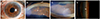 | Figure 1Initial anterior segment photographs of the left eye. (A) Slit-lamp examination showing diffuse iris neovascularization. (B) Note two polypropylene suture knots in the inferior part and (C) one in the superior part accompanying iris neovascularization (arrows). (D) Gonioscopic examination showing neovascularization (arrow) in all quadrants and the representative picture is shown in the temporal quadrant.
|
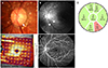 | Figure 2Initial examination of the patient. (A) Disc photograph showing an inferior neuroretinal rim thinning (arrow) and (B) red-free retinal nerve fiber layer (RNFL) photograph showing an inferior RNFL defect (arrowheads) in the left eye. Optical coherence tomography image presenting (C) inferior RNFL thinning and (D) corresponding retinal ganglion cell layer defect. (E) Fluorescein angiography image showing no definite ischemic retinal lesion, except several microaneurysms. NS = nasal-superior; TS = temporal-superior; T = temporal; TI = temporal-inferior; NI = nasal-inferior; G = global; N = nasal.
|
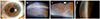 | Figure 3Anterior segment photographs of the left eye at six months after topical intraocular pressure lowering treatment. (A) Slit-lamp examination showing complete regression of the iris neovascularization. (B, C) The remaining polypropylene suture knots fixated in the iris with the compete regression of iris neovascularization. (D) Gonioscopic examination also showing complete regression of angle neovascularization.
|
References
1. Sivak-Callcott JA, O'Day DM, Gass JD, Tsai JC. Evidence-based recommendations for the diagnosis and treatment of neovascular glaucoma. Ophthalmology. 2001; 108:1767–1776.
2. Shazly TA, Latina MA. Neovascular glaucoma: etiology, diagnosis and prognosis. Semin Ophthalmol. 2009; 24:113–121.

3. Hayreh SS. Neovascular glaucoma. Prog Retin Eye Res. 2007; 26:470–485.
4. Jeong YC, Hwang YH. Etiology and features of eyes with rubeosis iridis among Korean patients: A Population-Based Single Center Study. PLoS One. 2016; 11:e0160662.
5. Cohen S, Kremer I, Yassur Y, Ben-Sira I. Peripheral retinal neovascularization and rubeosis iridis after a bilateral circular buckling operation. Ann Ophthalmol. 1988; 20:153–156.
6. Barile GR, Chang S, Horowitz JD, et al. Neovascular complications associated with rubeosis iridis and peripheral retinal detachment after retinal detachment surgery. Am J Ophthalmol. 1998; 126:379–389.
7. Aref AA. Current management of glaucoma and vascular occlusive disease. Curr Opin Ophthalmol. 2016; 27:140–145.
8. Wakabayashi T, Oshima Y, Sakaguchi H, et al. Intravitreal bevacizumab to treat iris neovascularization and neovascular glaucoma secondary to ischemic retinal diseases in 41 consecutive cases. Ophthalmology. 2008; 115:1571–1580. 80.e1–80.e3.
9. Wand M, Dueker DK, Aiello LM, Grant WM. Effects of panretinal photocoagulation on rubeosis iridis, angle neovascularization, and neovascular glaucoma. Am J Ophthalmol. 1978; 86:332–339.
10. Iliev ME, Domig D, Wolf-Schnurrbursch U, et al. Intravitreal bevacizumab (Avastin) in the treatment of neovascular glaucoma. Am J Ophthalmol. 2006; 142:1054–1056.
11. Batman C, Ozdamar Y. The effect of bevacizumab for anterior segment neovascularization after silicone oil removal in eyes with previous vitreoretinal surgery. Eye (Lond). 2010; 24:1243–1246.
12. Mason JO 3rd, Albert MA Jr, Mays A, Vail R. Regression of neovascular iris vessels by intravitreal injection of bevacizumab. Retina. 2006; 26:839–841.
13. Costa VP, Harris A, Anderson D, et al. Ocular perfusion pressure in glaucoma. Acta Ophthalmol. 2014; 92:e252–e266.
14. Rolle T, Tofani F, Brogliatti B, Grignolo FM. The effects of dorzolamide 2% and dorzolamide/timolol fixed combination on retinal and optic nerve head blood flow in primary open-angle glaucoma patients. Eye (Lond). 2008; 22:1172–1179.
15. Siesky B, Harris A, Cantor LB, et al. A comparative study of the effects of brinzolamide and dorzolamide on retinal oxygen saturation and ocular microcirculation in patients with primary open-angle glaucoma. Br J Ophthalmol. 2008; 92:500–504.
 XML Download