

 PDF
PDF ePub
ePub Citation
 Print
Print


Abstract
Purpose
We report a case of hemorrhagic lymphangiectasia of the conjunctiva with a 360° connected circumference, which recovered spontaneously.
Case summary
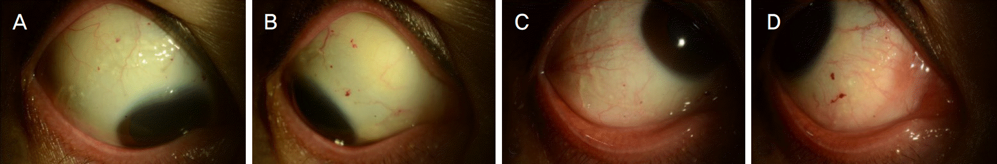
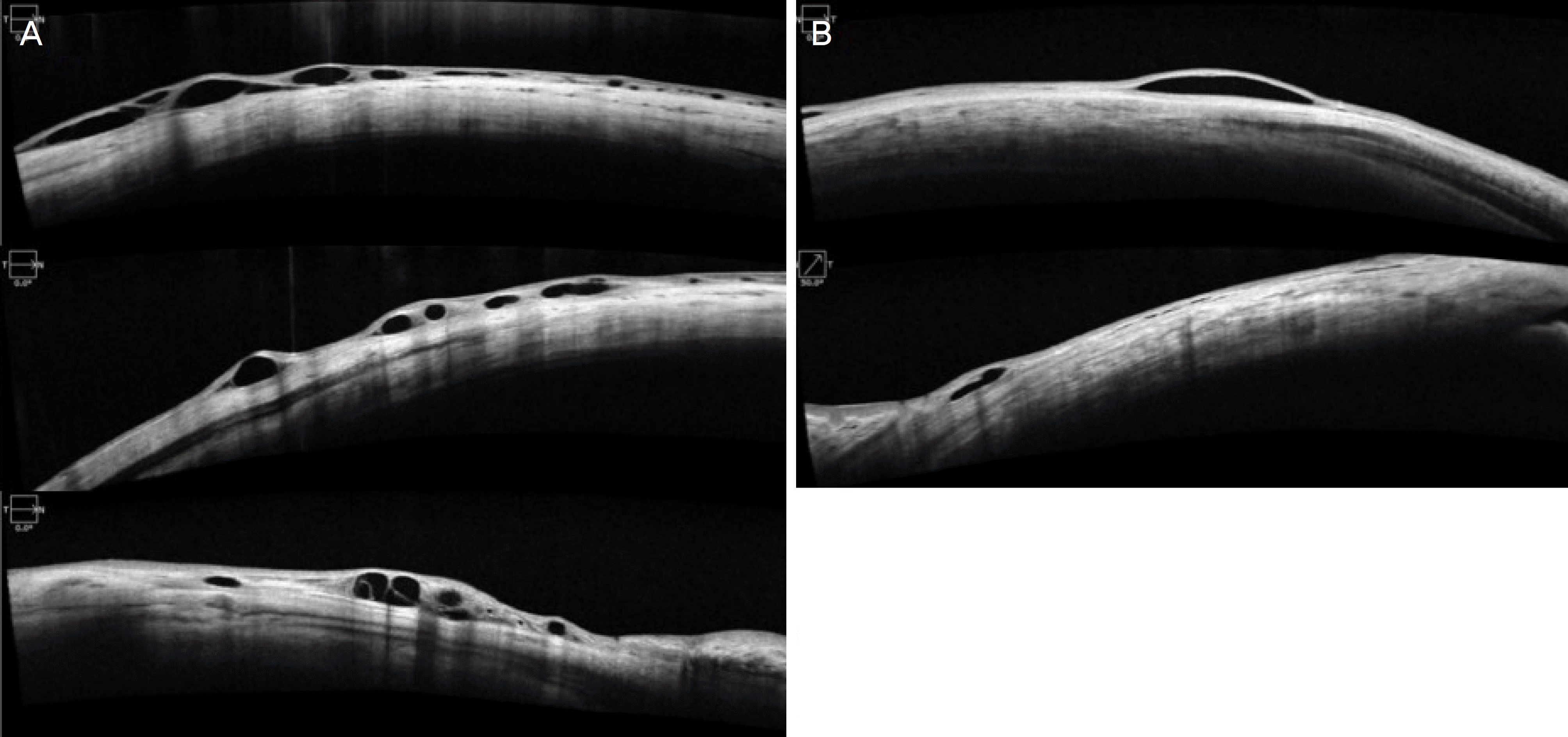
A 44-year-old female patient presented with congestion of the right eye 1 day prior to her visit. There was no history of any systemic disease or trauma, but she had experienced relapses of the same episode three times before the visit. There were no accompanying symptoms such as decreased vision or pain. Blood analysis, orbital computed tomography, and angiographic findings showed no remarkable finding. Slit lamp examination showed circumferential lymphatic dilatation extending 360° under the conjunctiva of the eye at a distance of about 6 mm behind the limbus of the right eye, which was accompanied by intralymphatic bleeding. Irregular local lymphatic dilatations were observed on the bulbar conjunctiva at 4 and 8 o'clock of the left eye. The bleeding spontaneously resolved in about 2 weeks, but the translucent enlarged lymphatic vessels were still observed on slit lamp examination and anterior segment optical coherence tomography.
Go to : 
References
1. Leber T. Lymphangiectasia haemorrhagica conjunctivae. Graefes Arch Ophthalmol. 1880; 26:197–201.
2. Stewart DE. System of Ophthalmology. 1st ed.8. St. Louis: Mosby;1965. p. 40.
3. Chelsky MP, Magnus DE. Conjunctival hemorrhagic lymphangiectasis. J Am Optom Assoc. 1988; 59:676–8.
4. Scott KR, Tse DT, Kronish JW. Hemorrhagic lymphangiectasia of the conjunctiva. Arch Ophthalmol. 1991; 109:286–7.

5. Welch J, Srinivasan S, Lyall D, Roberts F. Conjunctival lymphangiectasia: a report of 11 cases and review of literature. Surv Ophthalmol. 2012; 57:136–48.
6. Teichmann L. The lymphatic system. Leipzig: Engelmann;1861. p. 1–121.
7. Busacca A. The lymphatic vessels of the human bulbar conjunctiva studied by the method of in vivo injections of trypan blue. Arch Ophthalmol. 1948; 8:10–3.
8. Freitas-Neto CA, Costa RA, Kombo N, et al. Subconjunctival indocyanine green identifies lymphatic vessels. JAMA Ophthalmol. 2015; 133:102–4.
9. Lochhead J, Benjamin L. Lymphangiectasia haemorrhagica conjunctivae. Eye (Lond). 1998; 12(Pt 4):627–9.
10. Wang DH, Chung JK, Choi KS. A case of conjunctival hemorrhagic lymphangiectasia. J Korean Ophthalmol Soc. 2012; 53:1330–3.
11. Huerva V, Traveset AE, Ascaso FJ, Sánchez MC. Spontaneous resolution of a rare case of circumferential lymphangiectasia haemorrhagica conjunctivae. Eye (Lond). 2014; 28:912–4.
12. Chaudhry IA, Elkhamry SM, Al-Rashed WA, Bosley TM. Carotid cavernous fistula: ophthalmological implications. Middle East Afr J Ophthalmol. 2009; 16:57–63.
13. Krachmer JH, Mannis MJ, Holland EJ. Cornea. 3rd ed.1. St. Louis: Mosby;2011. p. 488–9.
14. Meisler DM, Eiferman RA, Ratliff NB, Burns CD. Surgical abdominal of conjunctival lymphangiectasis by conjunctival resection. Am J Ophthalmol. 2003; 136:735–6.
15. Jeong HK, Park HS, Seo KY, Lee J. The effects of surgery for abdominal lymphangiectasia using a high-frequency radio wave electrosurgical unit. J Korean Ophthalmol Soc. 2018; 59:314–8.
Go to :
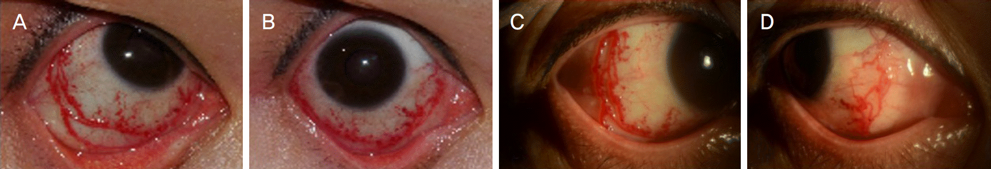 | Figure 1.Anterior segment photography (at first visit). Slit lamp microscopy of right eye reveals a circumferential (360°) engorgement of the bulbar conjunctival lymphatic vessels filled with blood. (A) Inferotemporal, (B) inferonasal, (C) temporal, (D) nasal. |
 XML Download