Abstract
Purpose
To investigate how the effect of bilateral rectus muscle recession changed by analyzing the effect/dose ratio of surgery according to the preoperative angle deviation.
Methods
We retrospectively studied the medical records of patients from January 2007 to March 2014 who underwent bilateral lateral rectus muscle recession and who visited our hospital for at least 2 years after surgery. We classified the patients into two groups: the preoperative large angle deviation group (35 prism diopters [PD] or more) and the small angle deviation group (20 PD or less). We observed exodrift patterns by measuring distant and near angle deviation according to the preoperative and postoperative times. The effect/dose ratio of recession was calculated at each visit. Surgical success was defined as an alignment between 10 PD of exodeviation and 5 PD of esodeviation, both at distance and at near.
Results
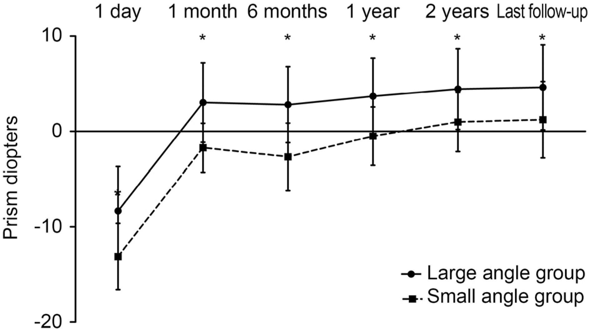
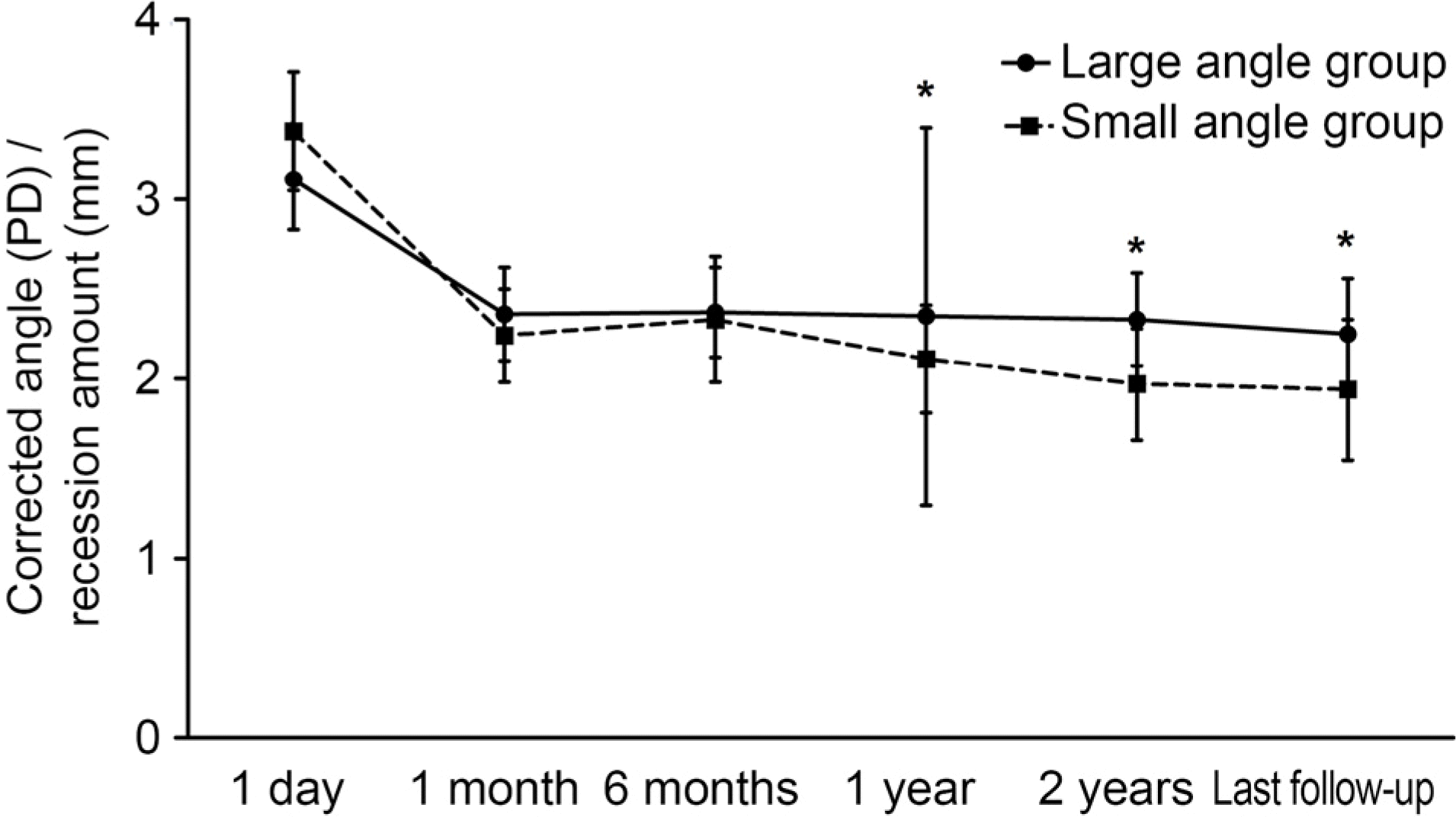
Among 165 patients, 84 patients were in the large angle deviation group and 81 patients were in the small angle deviation group. Preoperative angle deviation of the large angle deviation group was 39.34 ± 5.13 PD (range: 35–55 PD) and the small angle deviation group was 19.49 ± 1.62 PD (range: 18–20 PD) (p < 0.001). At postoperative 1 day, the alignments of eyes of the two groups were −8.32 ± 9.31 PD and −13.11 ± 6.94 PD; p < 0.001, respectively. At the date of the final follow-up, the alignments of eyes of the two groups were 4.63 ± 8.94 PD and 1.22 ± 8.01 PD; p = 0.011, respectively, and the effect/dose ratios were 2.25 ± 0.62 PD/mm and 1.94 ± 0.78 PD/mm, respectively, which meant that the effect of correction for the large angle deviation group was larger than that of the small angle deviation group (p = 0.005). Both groups showed postoperative exodrift patterns and similar success rates (75.0% and 80.2%, respectively), which showed no significant difference (p = 0.268).
Conclusions
The small angle deviation group showed a larger effect of correction and high exodrift pattern at the postoperative initial time and the large angle deviation group showed a smaller effect of correction and low exodrift pattern at the postoperative initial time. The preoperative angles of deviation and the operative success rates were not related.
Go to : 
References
1. Seo EJ, Chung SA. Variability of preoperative angle of deviation measured on the day of surgery in intermittent exotropia. J Korean Ophthalmol Soc. 2015; 56:1591–8.

2. Pratt-Johnson JA, Barlow JM, Tillson G. Early surgery in abdominal exotropia. Am J Ophthalmol. 1977; 84:689–94.
3. Kushner BJ. Selective surgery for intermittent exotropia based on distance/near differences. Arch Ophthalmol. 1998; 116:324–8.

4. Ing MR, Nishimura J, Okino L. Outcome study of bilateral lateral rectus recession for intermittent exotropia in children. Ophthalmic Surg Lasers. 1999; 30:110–7.

5. Kim WJ, Kim MM. Variability of preoperative measurements in intermittent exotropia and its effect on surgical outcome. J AAPOS. 2017; 21:210–4.

6. Jin KW, Choi DG. Outcome of two-muscle surgery for large-angle intermittent exotropia in children. Br J Ophthalmol. 2017; 101:462–6.

7. Wang L, Nelson LB. Outcome study of unilateral lateral rectus abdominal for small to moderate angle intermittent exotropia in children. J Pediatr Ophthalmol Strabismus. 2010; 47:242–7.
8. Maruo T, Kubota N, Sakaue T, Usui C. Intermittent exotropia abdominal in children: long term outcome regarding changes in binocular alignment. A study of 666 cases. Binocul Vis Strabismus Q. 2001; 16:265–70.
9. Han DH, Paik HJ. The minimal postoperative follow-up period to determine secondary surgery in patients with intermittent exotropia. J Korean Ophthalmol Soc. 2014; 55:711–8.

10. Kim M, Choi MY. Result comparison after reoperation in recurrent exotropia according to the type of first operation. J Korean Ophthalmol Soc. 2014; 55:726–33.

11. Kim MJ, Kim SH. Factors associated with improved surgical abdominals in recurrent exotropia. J Korean Ophthalmol Soc. 2017; 58:692–7.
12. Park KH, Kim SY. Clinical characteristics of patients that abdominal different rates of exodrift after strabismus surgery for abdominal exotropia and the effect of the rate of exodrift on final abdominal alignment. J AAPOS. 2013; 17:54–8.
13. Yam JC, Chong GS, Wu PK, et al. Prognostic factors predicting the surgical outcome of bilateral lateral rectus recession surgery for abdominals with infantile exotropia. Jpn J Ophthalmol. 2013; 57:481–5.
14. Parks MM, Mirchell P. Concomittant exodeviations. Duane TD, editor. Clinical Ophthalmology. 1. Philadelphia: JB Lippincott;1988. p. 1.
15. Choi JW, Lee SG. Surgical outcomes of large-angle exotropia. J Korean Ophthalmol Soc. 2011; 52:959–63.

16. Choi J, Kim SJ, Yu YS. Initial postoperative deviation as a predictor of long-term outcome after surgery for intermittent exotropia. J AAPOS. 2011; 15:224–9.

17. Lee JC, Lee YC, Lee SY. Comparison of postoperative outcomes according to deviation angle in moderate-angle intermittent abdominal of basic type. J Korean Ophthalmol Soc. 2013; 54:475–8.
18. Chung YR, Yang H, Lew HM, et al. The assessment of stereoacuity in patients with strabismus. J Korean Ophthalmol Soc. 2008; 49:1309–16.

19. Hatt SR, Gnanaraj L. Interventions for intermittent exotropia. Cochrane Database Syst Rev. 2013; (5):CD003737.

20. Cho YA, Kim SH. Postoperative minimal overcorrection in the abdominal management of intermittent exotropia. Br J Ophthalmol. 2013; 97:866–9.
21. Pineles SL, Deitz LW, Velez FG. Postoperative outcomes of abdominals initially overcorrected for intermittent exotropia. J AAPOS. 2011; 15:527–31.
22. Demer JL, Poukens V, Miller JM, Micevych P. Innervation of abdominal pulley smooth muscle in monkeys and humans. Invest Ophthalmol Vis Sci. 1997; 38:1774–85.
23. Sloper J. Clinical strabismus management: principles and surgical techniques. Br J Ophthalmol. 2000; 84:1333.

24. Demer JL, Miller JM, Poukens V, et al. Evidence for fibromuscular pulleys of the recti extraocular muscles. Invest Ophthalmol Vis Sci. 1995; 36:1125–36.
25. Clark RA, Demer JL. Posterior inflection of weakened lateral abdominal path: connective tissue factors reduce response to lateral rectus recession. Am J Ophthalmol. 2009; 147:127–33.e2.
Go to : 
 | Figure 1.Changes in angle of deviation at distance during post-operative follow-up. Mean angle of deviation showed significant difference at all dates. * Mann-Whitney U-test (p < 0.05). |
 | Figure 2.Change in amount of corrected prism diopters (PD) per lateral recession (mm) in large angle and small angle groups (dose-effect relationship). Mean PD/mm showed significant difference at postoperative 1 day, 1 year, 2 years and last follow-up. * Mann-Whitney U-test (p < 0.05). |
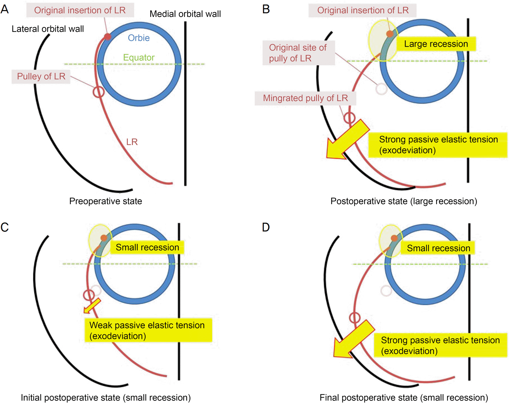
 | Figure 3.Diagram of lateral rectus (LR) muscle and other structures. (A) Pulley of LR is located 2–4 mm posterior to the equator originally. (B) After large amount of recession on LR, pulley is translocated largely to the lateral side and causes strong passive elastic tension, which has the exodeviation force. (C) After small amount of recession on LR, pulley is translocated smally to the lateral side and causes weak passive elastic tension initially. (D) At the final state after small recession of LR, as the pulley is deviated later-ally, passive elastic tension becomes strong. LR = lateral rectus muscle. |
Table 1.
Surgical dosages of bilateral rectus recession for exotropia
| Prism diopter | LR recession (mm) in BLR |
|---|---|
| 18–20 | 4.5–5.0 |
| 35–40 | 7.5–8.0 |
| 41–45 | 8.0–8.5 |
| 46–50 | 8.5–9.0 |
| 51–55 | 9.0–9.5 |
Table 2.
Preoperative characteristics of large angle and small angle group
| Charecteristic |
Large angle group (n = 84) |
Small angle group (n = 81) |
p-value | ||
|---|---|---|---|---|---|
| Average | Range | Average | Range | ||
| Sex (male/female) | 37/47 | – | 45/36 | – | 0.162* |
| Age at surgery (years) | 8.46 ± 8.77 | 2 to 44 | 7.58 ± 2.36 | 4 to 18 | 0.092† |
| Follow-up (months) | 42.03 ± 26.44 | 26 to 120 | 42.90 ± 23.25 | 24 to 112 | 0.824† |
| BCVA (logMAR) | 0.06 ± 0.24 | 0.00 to 0.18 | 0.06 ± 0.42 | 0.00 to 0.18 | 0.866† |
| Spherical equivalent (diopters) | –0.73 ± 1.92 | –5.50 to 1.25 | –0.78 ± 1.65 | –3.75 to 3.00 | 0.869† |
| Anisometropia (diopters) | 0.15 ± 0.32 | 0.00 to 1.75 | 0.18 ± 0.16 | 0.00 to 1.00 | 0.711† |
| Stereopsis (good/total) | 75/84 | – | 78/81 | – | 0.081* |
| Amount of recession (mm) | 7.68 ± 0.64 | 7.50 to 9.25 | 4.93 ± 0.32 | 4.50 to 5.00 | <0.001 |
| Preoperative angle deviation (PD) | 39.34 ± 5.13 | 35 to 55 | 19.49 ± 1.62 | 18 to 20 | <0.001† |
Table 3.
Angle of deviation at distance during postoperative follow-up
| Follow-up period | Large angle group (n = 84) | Small angle group (n = 81) | p-value* |
|---|---|---|---|
| 1 day (PD) | –8.32 ± 9.31 | –13.11 ± 6.94 | <0.001 |
| 1 month (PD) | 3.02 ± 8.33 | –1.72 ± 5.11 | <0.001 |
| 6 months (PD) | 2.80 ± 7.86 | –2.67 ± 7.05 | <0.001 |
| 1 year (PD) | 3.68 ± 8.05 | –0.50 ± 6.10 | 0.001 |
| 2 years (PD) | 4.44 ± 8.51 | 0.98 ± 6.21 | 0.008 |
| Last follow-up (PD) | 4.63 ± 8.94 | 1.22 ± 8.01 | 0.011 |
Table 4.
The amount of corrected prism diopters per lateral recession (mm) in large angle and small angle groups (dose-effect relationship)
| Follow-up period | Large angle group (n = 84) | Small angle group (n = 81) | p-value* |
|---|---|---|---|
| 1 day (PD/mm) | 3.11 ± 0.57 | 3.38 ± 0.67 | 0.006 |
| 1 month (PD/mm) | 2.36 ± 0.52 | 2.24 ± 0.53 | 0.124 |
| 6 months (PD/mm) | 2.37 ± 0.50 | 2.33 ± 0.70 | 0.664 |
| 1 year (PD/mm) | 2.35 ± 2.11 | 2.11 ± 0.60 | 0.005 |
| 2 years (PD/mm) | 2.33 ± 0.53 | 1.97 ± 0.63 | <0.001 |
| Last follow-up (PD/mm) | 2.25 ± 0.63 | 1.94 ± 0.79 | 0.005 |


 PDF
PDF ePub
ePub Citation
 Print
Print


 XML Download