

 PDF
PDF ePub
ePub Citation
 Print
Print


Abstract
Purpose
To quantify the size of commotio retinae and investigate its spontaneous resolution over time using ultra-wide field (UWF) color fundus photography.
Methods
We analyzed serial UWF color fundus photographs of 33 eyes of 33 ocular trauma patients with commotio retinae. Total visible retinal areas and the areas of commotio retinae were measured at baseline, 3 days, 1 week, and 4 weeks from the initial traumatic event.
Results
The median time of observation was 10.8 ± 12.1 (4–44) weeks. Spontaneous resolution of commotio retinae was observed in all patients, and no patients experienced any complications during the follow-up period. The mean percentage of commotio retinae at 3 days significantly decreased compared to the baseline (8.51 ± 9.66% versus 12.23 ± 10.39%; p < 0.001), and more decreased at 1 week (1.04 ± 2.75%; p < 0.001), but no significant differences were observed between 1 week and 4 weeks (0.00 ± 0.00%; p = 0.219). The spontaneous resolution percentages during the first 3 days, between 3 days and 1 week, and during the next 4 weeks were 12.97 ± 13.44%/day, 19.62 ± 9.22%/day, and 0.87 ± 1.87%/day, respectively (p = 0.192 and p < 0.001, respectively). The resolution rate was higher during the first 1 week.
Go to : 
References
1. Berlin R. On the so-called Commotio retinae. Klin Mbl Augenheilk. 1873; 11:42–78.
2. Sipperley JO, Quigley HA, Gass DM. Traumatic retinopathy in primates. The explanation of commotio retinae. Arch Ophthalmol. 1978; 96:2267–73.
3. Mansour AM, Green WR, Hogge C. Histopathology of commotio retinae. Retina. 1992; 12:24–8.

4. Sony P, Venkatesh P, Gadaginamath S, Garg SP. Optical coherence tomography findings in commotio retina. Clin Exp Ophthalmol. 2006; 34:621–3.
5. Hart J, Frank H. Retinal opacification after blunt non-perforating concussional injuries to the globe. A clinical and retinal fluorescein angiographic study. Trans Ophthalmol Soc U K. 1975; 95:94–100.
6. Oh J, Jung JH, Moon SW, et al. Commotio retinae with spectral-abdominal optical coherence tomography. Retina. 2011; 31:2044–9.
7. Ghasemi Falavarjani K, Wang K, Khadamy J, Sadda SR. Ultra-wide-field imaging in diabetic retinopathy; an overview. J Curr Ophthalmol. 2016; 28:57–60.
8. Witmer MT, Parlitsis G, Patel S, Kiss S. Comparison of abdominal-field fluorescein angiography with the Heidelberg Spectralis((R)) noncontact abdominalfield module versus the Optos((R)) Optomap((R)). Clin Ophthalmol. 2013; 7:389–94.
9. Hirano T, Imai A, Kasamatsu H, et al. Assessment of diabetic retinopathy using two abdominal-field fundus imaging systems, the Clarus(R) and Optos™ systems. BMC Ophthalmol. 2018; 18:332.
10. Tang M, Hui YN, Li YY, et al. Evaluation of traumatic retinopathy with abdominal field imaging under corneal scar or fixed small pupil. Int J Ophthalmol. 2018; 11:1371–6.
11. Kim DR, Hong EH, Shin YU, et al. Utility of abdominal fundus photography in patients with acute blunt ocular trauma. J Korean Ophthalmol Soc. 2019; 60:428–33.
12. Bunt-Milam AH, Black RA, Bensinger RE. Breakdown of the outer blood-retinal barrier in experimental commotio retinae. Exp Eye Res. 1986; 43:397–412.
13. Liem AT, Keunen JE, van Norren D. Reversible cone photo-receptor injury in commotio retinae of the macula. Retina. 1995; 15:58–61.
14. Itakura H, Kishi S. Restored photoreceptor outer segment in commotio retinae. Ophthalmic Surg Lasers Imaging. 2011; 42:Online:. e29–31.
15. Blanch RJ, Good PA, Shah P, et al. Visual outcomes after blunt abdominal trauma. Ophthalmology. 2013; 120:1588–91.
Go to :
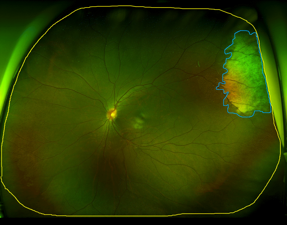 | Figure 1.Commotio retinae image analysis examples. The freehand tool in Image J was used to select the area of total visible retina, commotio retinae the yellow and blue line indicates the area of visible retinae, the commotio retinae, respectively. The percentage of commotio retinae was calculated by dividing the area of commotio retinae by the total visible retinal area and multiplying by 100. |
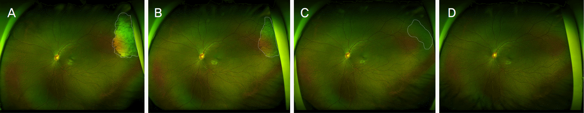 | Figure 2.Serial follow up ultra-wide field (UWF) color fundus photography from the left eye of 15-year old female patient. 15-year old female patient presented with decreased vision in her left eye after eyeball injury by wood. She referred to us the day of the trauma. Cells were observed markedly in the anterior chamber by slit lamp examination. (A) Baseline UWF color fundus photography demonstrated a localized whitish opaque lesion at superior-temporal side. (B) At 3 days, the area of commotio retinae was decreased and (C) at 1 week the area of commotio retinae was showed more decrement but remained (D) After 4 weeks, UWF color fundus photography revealed absence of whitish lesion. The white dot lines on (A-C) indicate the area of commotion retinae. |
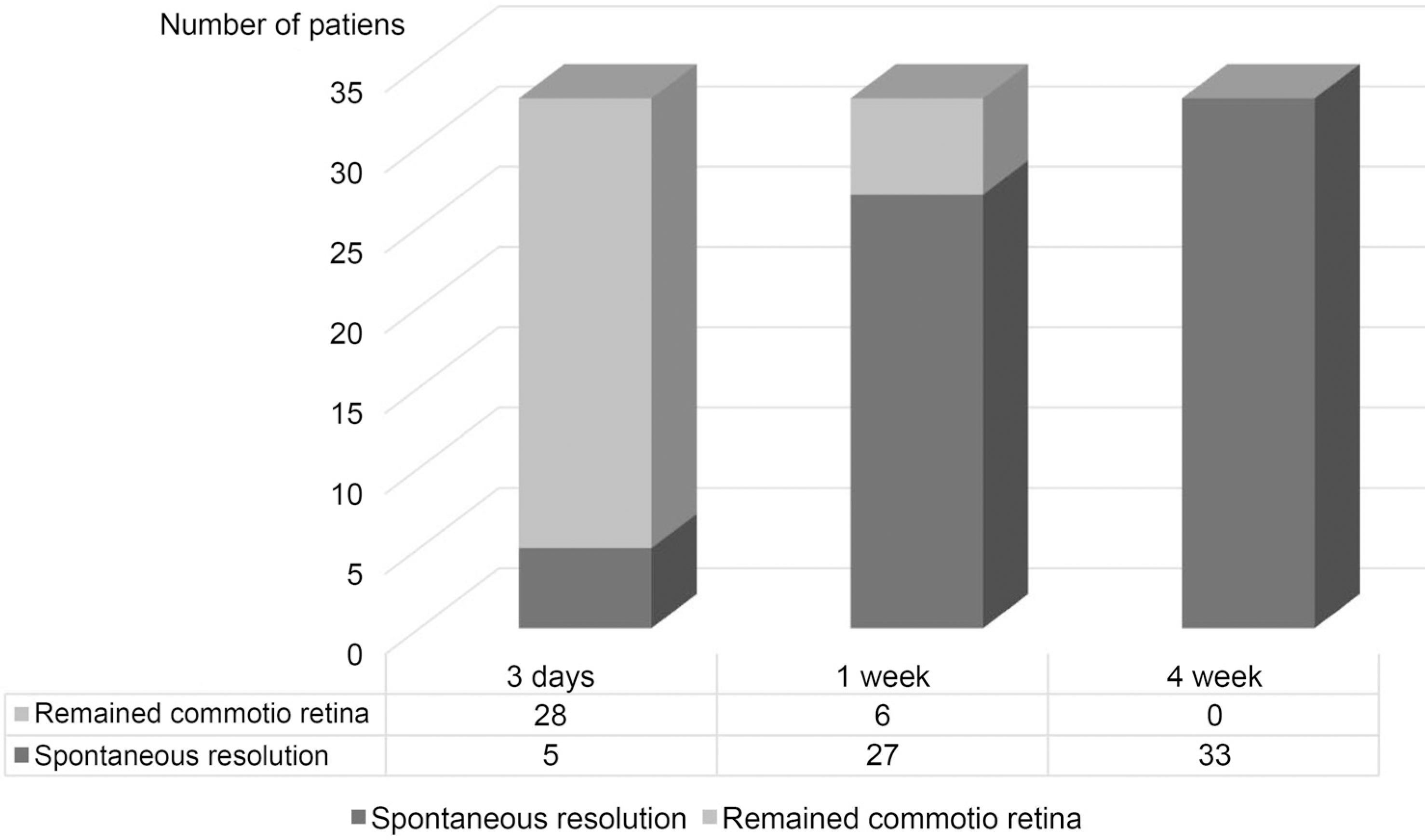 | Figure 3.Number of patients with spontaneous resolution in relation to observation time. After 3 days from the traumatic event, 5 patients presented spontaneous resolution, while 27 patients showed spontaneos resolution at 1 week. One month later, all of them presented spontaneous resoltuion. |
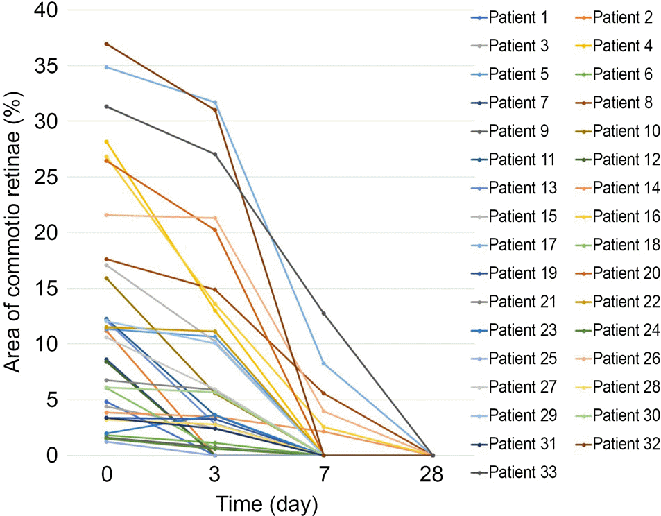 | Figure 4.Changes in area of commotio retina for individuals (%). Scatterplot showing the mean percentage of commotio retinae for 33 patients. Spontaneous resolution of commotio retinae was observed in all patients after 4 weeks and none of them had experienced any complication during follow-up. |
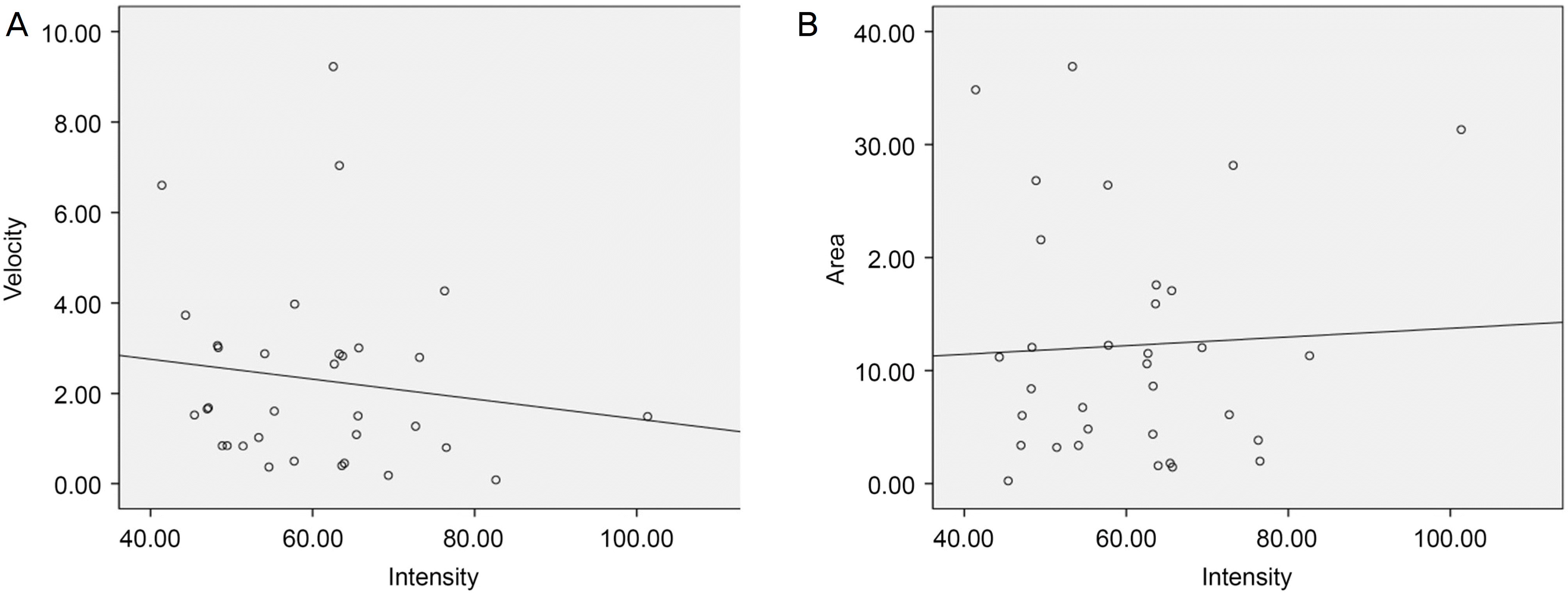 | Figure 5.Relationsip between intensity, resolution rate and area of commotio retinae. Scatterplot (A) showing the velocity versus the intensity (r = −1.34, p < 0.457) and (B) showing the area versus intensity (r = 0.47, p = 0.795). |
Table 1.
General characteristics in patients with commotio retinae
Table 2.
Causes of ocular trauma and location of commotion retinae
Table 3.
Repeated measures design for mean area of commotio retinae (%)
| Follow-up period | Mean area (%) | p-value* | ||
|---|---|---|---|---|
| Baseline | 12.23 ± 10.39 | <0.001† | <0.001‡ | <0.001§ |
| 3 days | 8.51 ± 9.66 | <0.001† | <0.001∏ | <0.001# |
| 1 week | 1.04 ± 2.75 | <0.001‡ | <0.001∏ | 0.219¶ |
| 4 weeks | 0.00 ± 0.00 | <0.001§ | <0.001# | 0.219¶ |
Table 4.
Repeated measures design for mean intensity of commotio retinae (%)
| Follow-up period | Mean intensity (%) | p-value | ||
|---|---|---|---|---|
| Baseline | 60.49 ± 2.22 | <0.001*† | <0.001*‡ | <0.001*§ |
| 3 days | 42.58 ± 4.42 | <0.001*† | <0.001*∏ | <0.001*# |
| 1 week | 8.58 ± 3.30 | <0.001*‡ | <0.001*∏ | 0.084¶ |
| 4 week | 0.00 ± 0.00 | <0.001*§ | <0.001*# | 0.084¶ |
Table 5.
Initial mean area of commotio retinae % with respect to the resolution period
| Group | Patient | Mean area (%) |
p-value |
|
|---|---|---|---|---|
| 1 | 5 (15.2) | 6.66 ± 4.24 | 0.665* | 0.021†‡ |
| 2 | 22 (66.7) | 10.65 ± 9.51 | 0.665* | 0.023†§ |
| 3 | 6 (18.2) | 22.66 ± 11.16 | 0.021†‡ | 0.023†§ |
 XML Download