

 PDF
PDF ePub
ePub Citation
 Print
Print


Abstract
Purpose
We evaluated the surgical prognoses of patients with advanced cataract who were unable to be evaluated by fundus imaging and their satisfaction with daily life.
Methods
We retrospectively reviewed 748 eyes of 480 patients who underwent cataract surgery from January 2015 to December 2017. Preoperative factors, surgical technique, degree of cataract, and the best-corrected visual acuity for 1 and 6 months after surgery were analyzed. Among 91 eyes of 78 patients with advanced cataract who were unable to be evaluated by fundus imaging, the degree of discomfort before surgery and postoperative satisfaction were evaluated.
Results
Hypertension was positively correlated with visual acuity after cataract surgery (p = 0.004). Low corneal endothelial cell count, primary open-angle glaucoma, a history of trabeculectomy due to glaucoma, corneal dystrophy or corneal opacity, advanced cataract unable to be evaluated by fundus imaging, hypermature cataract, extracapsular cataract extraction, and intracapsular cataract extraction and visual acuity <0.5 after 1 month showed negative correlations with the visual outcomes after 6 months (p = 0.019, p = 0.002, p = 0.037, p = 0.001, p = 0.004, p = 0.012, p = 0.00, and p = 0.00, respectively). The risk of a final visual acuity <0.5 after cataract surgery was 3.18-fold higher in cases of advanced cataract, unable to be evaluated by fundus imaging (p = 0.003). Ten patients with 10 eyes postponed surgery due to poor prognoses, which was expected, and six patients (60%) had a best-corrected visual acuity <0.5 after 6 months. Six patients (60%), expected to have a poor prognosis were satisfied after surgery and the postoperative satisfaction was high when compared with a poor visual outcome.
Conclusions
Poor surgical prognoses were expected in advanced cataract patients unable to be evaluated by fundus imaging. However, advanced cataract patients, who postponed surgery due to an unfavorable visual prognosis, showed a higher subjective satisfaction when compared with the postoperative visual acuity.
Go to : 
References
1. Resnikoff S, Pascolini D, Etya'ale D, et al. Global data on visual impairment in the year 2002. Bull World Health Organ. 2004; 82:844–51.
2. Hart WM. Adler's physiology of the eye. 9th ed. St. Louis, Mosby;1992. p. 348–90.
3. Minassian DC, Reidy A, Desai P, et al. The deficit in cataract abdominal in England and Wales and the escalating problem of visual abdominal: epidemiological modelling of the population dynamics of cataract. Br J Ophthalmol. 2000; 84:4–8.
4. Westcott MC, Tuft SJ, Minassian DC. Effect of age on visual abdominal following cataract extraction. Br J Ophthalmol. 2000; 84:1380–2.
5. Mönestam E, Wachmeister L. Impact of cataract surgery on the abdominal ability of the very old. Am J Ophthalmol. 2004; 137:145–55.
6. Muñoz B, West SK, Rubin GS, et al. Causes of blindness and visual impairment in a population of older American: The Salisbury Eye Evaluation Study. Arch Ophthalmol. 2000; 118:819–25.
7. Elliott DB, Trukolo-Ilic M, Strong JG, et al. Demographic characteristics of vision-disabled elderly. Invest Ophthalmol Vis Sci. 1997; 38:2566–75.
8. Mönestam E, Wachtmeister L. Dissatisfaction with cataract abdominal in relation to visual results in a population-based study in Sweden. J Cataract Refract Surg. 1999; 25:1127–34.
9. Stifter E, Sacu S, Weghaupt H, et al. Reading performance depending on the type of cataract and its predictability on the visual outcome. J Cataract Refract Surg. 2004; 30:1259–67.

10. Quintana JM, Arostegui I, Alberdi T, et al. Decision trees for indication of cataract surgery based on changes in visual acuity. Ophthalmology. 2010; 117:1471–8.
11. Minassian DC, Rosen P, Dart JK, et al. Extracapsular cataract abdominal compared with small incision surgery by abdominal: a randomised trial. Br J Ophthalmol. 2001; 85:822–9.
12. Blomquist PH, Rugwani RM. Visual outcomes after vitreous loss during cataract surgery performed by residents. J Cataract Refract Surg. 2002; 28:847–52.
13. Uusitalo RJ, Brans T, Pessi T, Tarkkanen A. Evaluating cataract surgery gains by assessing patients' quality of life using the VF-7. J Cataract Refract Surg. 1999; 25:989–94.
14. Mönestam E, Wachtmeister L. The impact of cataract surgery on low vision patients. A population based study. Acta Ophthalmol Scand. 1997; 75:569–76.
15. Hegde SP, Sekharreddy MR, Kumar MR, Dayanidhi VK. Prospective study of hypermature cataract in Kanchipuram district: causes of delayed presentation, risk of lens-induced glaucoma and visual prognosis. Kerala J Ophthalmol. 2018; 30:187–92.
16. Ionides A, Minassian D, Tuft S. Visual outcome following abdominal capsule rupture during cataract surgery. Br J Ophthalmol. 2001; 85:222–4.
Go to :
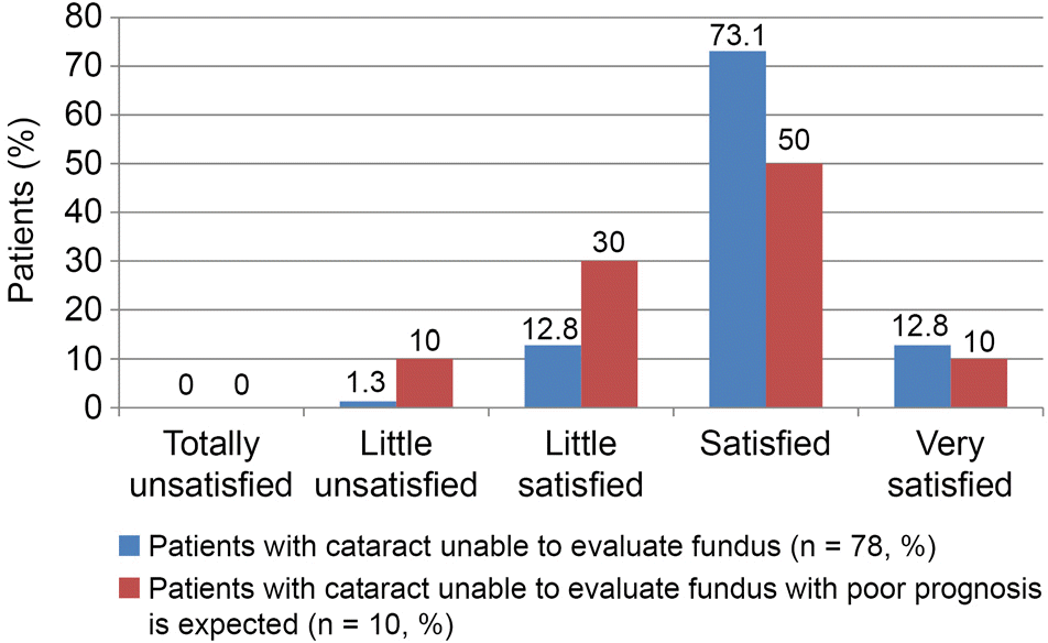 | Figure 1.Comparison of postoperative satisfaction of patients with advanced cataract unable to evaluate fundus. The whole patients were compared with patients with poor prognosis was expected. |
Table 1.
Baseline characteristics of patients
Table 2.
Univariate analysis of factors influencing visual outcome in cataract surgery
| Factor | Final VA less than 0.5 | Final VA over 0.5 | p-value |
|---|---|---|---|
| Age (years) | 70.28 ± 9.17 | 72.14 ± 8.75 | 0.246* |
| Location (right:left) | 18:16 | 366:348 | 0.863† |
| Sex (male:female) | 11:23 | 225:489 | 0.918† |
| Diabetes mellitus | 2 (5.9) | 85 (11.9) | 0.413† |
| Hypertension | 2 (5.9) | 196 (27.5) | 0.004† |
| Topography (with the rule:against the rule) | 15:19 | 413:301 | 0.115† |
| Preoperative ECC (cells/mm2) | 2,615 ± 272 | 2,730 ± 274 | 0.019* |
| History of glaucoma | |||
| Primary open angle glaucoma | 5 (14.7) | 26 (3.6) | 0.010† |
| History of angle closure glaucoma | 2 (5.9) | 7 (1.0) | 0.059† |
| History of trabeculectomy | 2 (5.9) | 5 (0.7) | 0.037† |
| Corneal dystrophy or corneal opacity | 4 (11.8) | 12 (1.7) | 0.001† |
| Pseudoexfoliation | 0 | 10 (1.4) | 0.378† |
| Advanced cataract unable to evaluate fundus | 13 (38.2) | 121 (17.0) | 0.004† |
| Hypermature cataract | 4 (11.8) | 17 (2.4) | 0.012† |
| Surgical technique including ICCE or ECCE | 6 (17.7) | 7 (1.0) | 0.000† |
| Posterior capsular rupture | 4 (11.8) | 30 (4.2) | 0.063† |
| Visual acuity under 0.5 after 1 months | 28 (82.4) | 43 (6.0) | 0.000† |
Table 3.
Multivariate analysis of advanced cataract influencing visual outcome in cataract surgery
| Advanced cataract | Exp (B, 95% CI) | p-value* |
|---|---|---|
| Advanced cataract unable to evaluate fundus | 3.18 (1.48–6.86) | 0.003 |
| Hypermature cataract | 7.42 (2.25–24.48) | 0.001 |
Table 4.
Causes of poor visual outcome (under 0.5) after 6 months in advanced cataract unable to evaluate fundus (multiple factors on each cases are counted separately)
Table 5.
Response to the questions of daily discomfort before cataract surgery of patients who had advanced cataract unable o evaluate fundus
| Discomfort in daily life before cataract surgery | Value (n = 78) |
|---|---|
| Very discomfort | 23 (29.5) |
| Discomfort | 46 (59.0) |
| Little discomfort | 7 (9.0) |
| Not discomfort at all | 2 (2.5) |
Table 6.
Response to the questions of reason for delayed cataract surgery of patients who had advanced cataract unable to evaluate fundus (multiple answers were permitted)
 XML Download