

 PDF
PDF ePub
ePub Citation
 Print
Print


Abstract
Purpose
Generally, the treatment of ankle trimalleolar open fractures is divided into two stages: external fixation and debridement; and secondary internal fixation. On the other hand, this two-stage operation takes considerable treatment time and is challenging in procedures requiring reduction. The purpose of this study was to evaluate the radiologic and clinical results of an immediate one-stage internal fixation operation considering the wound conditions to overcome two stage operation disadvantages.
Materials and Methods
From September 2009 to January 2018, 24 cases of ankle trimalleolar open fractures, who underwent immediate internal fixation and were followed up for at least one year, were studied retrospectively. The open wound was divided into the Gusti-lo-Anderson classification. Open reduction and internal fixation were performed on every medial and lateral malleolar fracture. On the other hand, with posterior malleolar fractures, surgical or conservative treatment was performed depending on the fragment size. The radiologic outcome was evaluated using the Burwell and Charnley criteria and American Orthopaedic Foot and Ankle Society (AOFAS) scores, and complications, such as infection and posttraumatic arthritis, were used for the clinical evaluation.
Results
The wound was classified into eight cases (33.3%) of type I, 11 cases (45.8%) of type II, and five cases (20.8%) of type IIIa. The degree of reduction was anatomical, fair, and poor in 16 cases (66.7%), six cases (25.0%), and two cases (8.3%), respectively. The mean AOFAS score was 79 points, and there were complications, such as infection in three cases (12.5%) and post-traumatic arthritis in two cases (8.3%).
Go to : 
References
1. Hulsker CC, Kleinveld S, Zonnenberg CB, Hogervorst M, van den Bekerom MP. Evidence-based treatment of open ankle fractures. Arch Orthop Trauma Surg. 2011; 131:1545–53. doi:. DOI: 10.1007/s00402-011-1349-7.

2. Bray TJ, Endicott M, Capra SE. Treatment of open ankle fractures. Immediate internal fixation versus closed immobilization and delayed fixation. Clin Orthop Relat Res. 1989; 240:47–52.
3. Gustilo RB, Anderson JT. Prevention of infection in the treatment of one thousand and twenty-five open fractures of long bones: retrospective and prospective analyses. J Bone Joint Surg Am. 1976; 58:453–8. doi:. DOI: 10.2106/00004623-197658040-00004.
4. De Vries JS, Wijgman AJ, Sierevelt IN, Schaap GR. Long-term results of ankle fractures with a posterior malleolar fragment. J Foot Ankle Surg. 2005; 44:211–7. doi:. DOI: 10.1053/j.jfas.2005.02.002.
5. McDaniel WJ, Wilson FC. Trimalleolar fractures of the ankle. An end result study. Clin Orthop Relat Res. 1977; 122:37–45.
6. Sachs W, Kanat IO, McLaughlin E, Burns DE. A surgical approach to a displaced ankle fracture. J Foot Surg. 1984; 23:302–7.
7. Talbot M, Steenblock TR, Cole PA. Posterolateral approach for open reduction and internal fixation of trimalleolar ankle fractures. Can J Surg. 2005; 48:487–90.
8. Lee GC, Lee JY, Ha SH, You JW, Lee SH, Sohn HM, et al. Treatment of the trimalleolar fracture using posterolateral approach: minimum 2-year follow up results. J Korean Fract Soc. 2011; 24:328–34. doi:. DOI: 10.12671/jkfs.2011.24.4.328.
9. Burwell HN, Charnley AD. The treatment of displaced fractures at the ankle by rigid internal fixation and early joint movement. J Bone Joint Surg Br. 1965; 47:634–60.
10. Toolan BC, Wright Quinones VJ, Cunningham BJ, Brage ME. An evaluation of the use of retrospectively acquired preoperative AOFAS clinical rating scores to assess surgical outcome after elective foot and ankle surgery. Foot Ankle Int. 2001; 22:775–8. doi:. DOI: 10.1177/107110070102201002.
11. Kitaoka HB, Alexander IJ, Adelaar RS, Nunley JA, Myerson MS, Sanders M. Clinical rating systems for the ankle-hindfoot, midfoot, hallux, and lesser toes. Foot Ankle Int. 1994; 15:349–53. doi:. DOI: 10.1177/107110079401500701.
12. Roberts CS, Pape HC, Jones AL, Malkani AL, Rodriguez JL, Giannoudis PV. Damage control orthopaedics: evolving concepts in the treatment of patients who have sustained orthopaedic trauma. Instr Course Lect. 2005; 54:447–62.
13. Melvin JS, Dombroski DG, Torbert JT, Kovach SJ, Esterhai JL, Mehta S. Open tibial shaft fractures: I. Evaluation and initial wound management. J Am Acad Orthop Surg. 2010; 18:10–9. doi:. DOI: 10.5435/00124635-201001000-00003.
14. Scharfenberger AV, Alabassi K, Smith S, Weber D, Dulai SK, Bergman JW, et al. Primary wound closure after open fracture: a prospective cohort study examining nonunion and deep infection. J Orthop Trauma. 2017; 31:121–6. doi:. DOI: 10.1097/BOT.0000000000000751.
15. Franklin JL, Johnson KD, Hansen ST Jr. Immediate internal fixation of open ankle fractures. Report of thirty-eight cases treated with a standard protocol. J Bone Joint Surg Am. 1984; 66:1349–56.
16. Ovaska MT, Madanat R, Honkamaa M, Mäkinen TJ. Contemporary demographics and complications of patients treated for open ankle fractures. Injury. 2015; 46:1650–5. doi:. DOI: 10.1016/j.injury.2015.04.015.
17. Tho KS, Chiu PL, Krishnamoorthy S. Grade III open ankle fractures: a review of the outcome of treatment. Singapore Med J. 1994; 35:57–8.
18. Joshi D, Singh D, Ansari J, Lal Y. Immediate open reduction and internal fixation in open ankle fractures. J Am Podiatr Med Assoc. 2006; 96:120–4. doi:. DOI: 10.7547/0960120.
19. Johnson EE, Davlin LB. Open ankle fractures. The indications for immediate open reduction and internal fixation. Clin Orthop Relat Res. 1993; 292:118–27.
Go to :
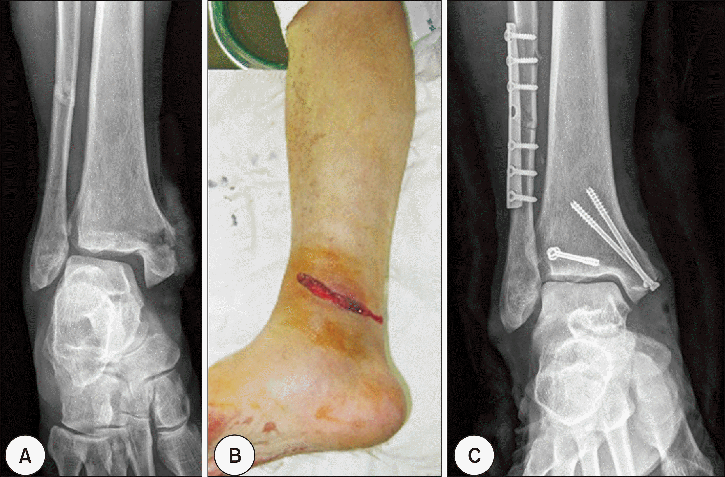 | Figure 1.(A) The initial film shows a trimalleolar fracture of the tibia with medial open wound. (B) Gustilo-Anderson II exposition. (C) Postoperative radiograph shows satisfactory position of plate and 4.0 mm cannulated screws. |
 | Figure 2.(A) A 55-year-old male sustained a right trimalleolar fracture with medial open wound after traffic accident. (B) First, lateral malleolar fracture was fixed using 1/3 semitubular plate, and 4.0 mm cannulated screws were applied. After 2 weeks, Wound infection was observed. (C) After 5 weeks, incision and debridement was done on medial malleolar infective wound. |
Table 1.
Patient Data
Table 2.
Comparison with Other Open Ankle Fracture Article
 XML Download