

 PDF
PDF ePub
ePub Citation
 Print
Print


INTRODUCTION
Alpha-1 antitrypsin is a single chain 52 kD glycoprotein belonging to the serpin (serine protease inhibitor) family. It consists of 394 amino acids and is primarily synthesized in the hepatocytes, in the lung epithelium and in macrophages, and released into the blood [1]. The main function of alpha-1 antitrypsin is to protect the lung epithelium from the non-specific release of proteases (such as neutrophil elastase) during inflammation. Additionally, alpha-1 antitrypsin inhibits the release of these proteases in the lung epithelium [2].
Alpha-1 antitrypsin is encoded by a gene (SERPINA1) located on the distal long arm of chromosome 14 (14q31-32.2) [3]. To date, more than 200 allelic variants have been defined, some of them leading to severe liver disease in childhood and pulmonary emphysema in adulthood. The most common allele is PiM, which is found in ~95% of the population and encodes normal functions and levels of alpha-1 antitrypsin. PiS (expressing 50–60% of alpha-1 antitrypsin) and PiZ (expressing 10–20% of alpha-1 antitrypsin) alleles are associated with low and aberrant alpha-1 antitrypsin expression [4]. Rare alleles such as MMalton and MPalermo are also associated with low alpha-1 antitrypsin levels [3]. Alpha-1 antitrypsin deficiency (A1ATD) is a genetic proteinopathy that affects the lungs and the liver through different mechanisms and shows an autosomal codominant inherence pattern. Pulmonary emphysema develops when alpha-1 antitrypsin is low or absent due to the presence of mutant alleles (PiS and PiZ) for the inhibition of the serine proteinases, that destroy the lung tissues. In contrast, aberrant protein accumulation inside the endoplasmic reticulum of hepatocytes causes hepatic inflammation, fibrosis, and cirrhosis by triggering a cascade of hepatocellular apoptosis, regeneration, and injury [56].
A1ATD is the most common genetic reason for liver transplantation in children in Northern Europe [78]. The prevalence of A1ATD in newborns was 1/1,639 in USA and 1/1,575 (PiZZ genotype) in Sweden [910]. Conversely, the incidence of A1ATD has been reported to be very low in Iranian patients with neonatal cholestasis [11]. The incidence of this disease seems to be low in our country, Turkey, based on data from the pediatric transplantation center [12]; it was reported in 3.6% of infants with cholestasis [13]. In this study, we aimed to analyze the (i) demographic and clinical characteristics and (ii) outcome of the patients with A1ATD from five pediatric hepatology units in Turkey.
MATERIALS AND METHODS
This study included patients diagnosed with A1ATD since 2005 and selected from five pediatric hepatology units (n=25). Demographic characteristics, laboratory parameters (serum albumin, alanine aminotransferase, aspartate aminotransferase, gamma-glutamyl transferase, total/direct bilirubin, and alpha-1 antitrypsin levels), clinical findings (presenting symptoms and physical examination findings), genetic analyses, and outcome of the patients were collected from hospital record files.The diagnosis of A1ATD was made based on low alpha-1 antitrypsin levels (normal range; 90–200 mg/dL, using the nephelometric method) and positive genotype analysis (either homozygous or heterozygous). Patients with: (i) secondary A1ATD such as the nephrotic syndrome or protein-losing enteropathy; (ii) low levels of A1ATD and normal genotype; or (iii) inadequate file records, were not included in this study. All other causes of liver diseases, such as infectious, anatomic and metabolic conditions, were excluded in all cases. Outcome of the patients in the long-term were defined as chronic liver disease (classified according to the Child-Pugh score), chronic hepatitis (only elevated liver enzymes without any signs of chronic liver disease such as growth retardation, organomegaly, and osteopenia), and asymptomatic (normal liver enzymes without any signs of chronic liver disease). Patients were symptomatically treated according to the presence of complications such as portal hypertension (treated using beta blockers or band ligation) or ascites (treated using diuretics).
All calculations in our study were performed using IBM SPSS Statistics for Windows, Version 23.0 (IBM Co., Armonk, NY, USA), and the continuous variables were expressed as mean±standard deviation (SD) and categorical variables as percentage (%). Comparison of the quantitative data between the groups was performed using Student t-test in the normally distributed variables, and the Mann-Whitney U-test in the non-normally distributed variables. Qualitative data were compared using the Chi-squared test. A p-values ≤0.05 were considered statistically significant.
The study was approved by the Karadeniz Technical University, Faculty of Medicine, Scientific Research Ethics Committee, reference number 2018/236.
RESULTS
Demographic and clinical findings, as well as outcome of the patients have been shown in Table 1. Twelve (48.0%) of the 25 patients were female and the mean±SD age of the patients at the time of diagnosis was 20.1±2.5 months. A 84% of the patients were diagnosed in the infancy period (≤24 months old age) [14].
Table 1
Demographic and clinical characteristics of the patients during initial admission
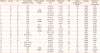
ALT: alanine aminotransferase, AST: aspartate aminotransferase, GGT: γ-glutamyl transferase, T/D: total/direct, INR: international normalized ratio, M: male, F: female, ITE: isolated transaminase elevation, CH: chronic hepatitis, C: coagulopathy, IH: intracranial hemorrhage, CLD: chronic liver diseases, NC: neonatal cholestasis, UB: umbilical bleeding, FS: family screening, HMG: hepatomegaly, HSMG: hepatosplenomegaly, FTT: failure to thrive, I: icterus, A: asymptomatic, D: death, CAS: Child A cirrhosis.
Normal values for ALT, AST, and GGT were 5–44 U/L, 6–46 U/L, and 8-64 U/L, respectively. Normal values for INR was 0.9–1.2 and for T/D bilirubin was 0.2–1.2 mg/dL.
The main indications for the screening for alpha-1 antitrypsin level were isolated transaminase elevation (ITE) in ten patients (40.0%), etiology for chronic liver disease in four patients (16.0%), etiology for neonatal cholestasis in three patients (12.0%), bleeding due to coagulopathy in two patients (8.0%), and chronic hepatitis in two patients (8.0%). One patient had intracranial hemorrhage and another had umbilical bleeding due to coagulopathy at the time of admission. Alpha-1 antitrypsin level and genotype analyses were performed for family screening in four patients (16.0%) (two patients had a sister with a PiZZ-associated chronic liver disease and the others had a PiMZ-associated bleeding disorder in siblings). Overall, eight patients (32.0%) had a homozygous PiZZ genotype while 17 (68.0%) had a heterozygous genotype (eight PiMZ and one MMalton) (Table 1). Patient 25 had chronic hepatitis and a low alpha-1 antitrypsin level, and targeted mutation analysis revealed a PiMM genotype. After exclusion of all other causes of chronic hepatitis, full gene sequencing of the SERINA1 gene was performed for A1ATD and reported as a heterozygous MMalton mutation. At the time of admission, organomegaly was present in eight patients (32.0%), icterus in four patients (16.0%), and failure to thrive was observed in two patients (8.0%).
Comparison of the patients with the PiZZ and the PiMZ genotypes revealed that alpha-1 antitrypsin level was significantly lower in patients with the PiZZ genotype [mean±SD, median (range): 37.6±7.7 mg/dL (37.5 mg/dL, 29–50 mg/dL) vs. 66.5±22.7 mg/dL (76 mg/dL, 30–87 mg/dL), p=0.0001]. Patients with the PiZZ genotype were diagnosed earlier than patients with the PiMZ genotype but this was not significantly different (13±6.8 months vs. 23.7±30.1 months, p=0.192) (Table 2).
Table 2
Association of genotype with the age at time of diagnosis and the alpha-1 antitrypsin level

Liver biopsy was performed in six patients (three PiMZ, two PiZZ, and one MMalton) (24.0%) during the diagnostic process. Histopathological examination revealed globules resistant to the periodic acid-Schiff positive diastase in all patients except one patient with MMalton. All patients had portal inflammation and hepatic fibrosis, and four had steatosis.
Median (range) duration of follow-up of patients with homozygous and heterozygous mutations were 49 months (6–144 months) and 38 months (4–97 months), respectively. One patient (12.5%) with a homozygous mutation died during the follow-up (patient no 4) due to decompensated liver disease and pulmonary emphysema at age of 11 years. Liver transplantation could not be performed, because this patient had severe neurological sequelae due to intracranial hemorrhage. One patient had Child A cirrhosis and five patients (62.5%) had chronic hepatitis. Another patient (12.5%) with a homozygous mutation was asymptomatic (normal liver function tests). Nine of the 17 patients with a heterozygous mutation had chronic hepatitis (52.9%), two patients had Child A cirrhosis (11.7%), and six (35.2%) patients were asymptomatic (four of these patients were diagnosed during the family screening and had transient liver enzyme elevation during the follow-up, but all had normal enzyme levels in the last visit). Overall, 7 (28.0%) of the 25 children were asymptomatic with normal liver enzymes in the final visit.
DISCUSSION
In this study, we analyzed the clinical features and outcome of children with A1ATD in Turkey. We found that: (i) patients may be admitted with a variety of liver pathologies ranging from asymptomatic or ITE to chronic liver diseases from early infancy to the adolescent age; (ii) alpha-1 antitrypsin level was lower in patients with the PiZZ genotype compared to the heterozygous genotype; (iii) full-gene sequencing of the SERPINA1 gene must be performed in children with low alpha-1 antitrypsin level and normal genotypes with targeted mutation analysis; and (iv) A1ATD caused a liver pathology (ranging from chronic hepatitis to cirrhosis) in approximately 75% of the children during long-term follow-up.
Liver pathology in children with an A1ATD has bimodal characteristics. It is generally presented as neonatal hepatitis or prolonged cholestasis in the neonatal period and transient elevation of liver enzymes or chronic hepatitis in the adolescent period. Comba et al. [15] studied 20 children with A1ATD, and found that 65% of the children presented with elevated liver enzymes and 15% had neonatal hepatitis. Prolonged cholestasis in the neonatal period was reported in 11% of the Swedish children who were homozygous with the mutation (n=122), while 6% of the patients presented with the clinical signs of liver disease without jaundice in the adolescents [1016]. Although we could not make an analysis due to the small number of patients, it seems that patients with homozygous mutations presented with an advanced disease at an early age. However, patients with heterozygous mutations are generally asymptomatic and this may cause delays to diagnosis. Additionally, we found that the alpha-1 antitrypsin level was lower in patients with a homozygous mutation. The pathogenesis of liver involvement in A1ATD is related to the accumulation of defective protein inside the endoplasmic reticulum of hepatocytes, which triggers apoptosis and injury [56]. The level of alpha-1 antitrypsin is only related with the pulmonary involvement, but the role of low alpha-1 antitrypsin levels must be investigated in the severity of pathophysiology of liver diseases in homozygous patients. It may be related to the difference in severity of apoptosis and hepatocellular injury at various levels of alpha-1 antitrypsin.
Bleeding problems may be the initial presentation of A1ATD, as found in our study. van Hasselt et al. [17] analyzed the vitamin K deficiency and risk of bleeding in infants with A1ATD and found that the risk of bleeding due to a vitamin K deficiency in infants with A1ATD is as high as infants with biliary atresia, and is higher in breastfed-compared to formula-fed infants. However, the risk of bleeding is mainly related to the presence of cholestasis, and not related to low levels of alpha-1 antitrypsin. Early diagnosis of A1ATD and vitamin K treatment in these patients may decrease the risk of bleeding complications. Life-threatening late hemorrhagic diseases due to a vitamin K deficiency, such as umbilical bleeding, was also reported in patients with A1ATD, showing that 2.8% of the total patients in a Swedish study and 5% of the total patients in a Polish study were affected [718].
Screening of other siblings for A1ATD is important for an early diagnosis. The heterozygous mutation with low alpha-1 antitrypsin level was found in the other siblings in our study, but liver enzymes were within the normal limits at initial screening. However, transient elevation of liver enzymes was seen during follow-up in all patients. A detailed and timely follow-up of these patients will prevent the complications related to liver diseases.
The diagnosis of A1ATD is based on the presence of low alpha-1 antitrypsin levels and identification of causal mutations. The analysis of the causal mutations is important in order to distinguish the genetic causes from the non-genetic causes of A1ATD. Current routine genetic analysis consists of only the most common mutations including M, S, and Z alleles. Rare mutations associated with A1ATD have been reported in recent years [3]. Graham et al. [19] reported that 42 (1.2%) of the 3,523 patients with low alpha-1 antitrypsin levels had normal genotype with routine genetic analysis, and full-gene sequencing revealed rare mutations, such as MMalton, MHeerlen, and PLowell, in 16 of the 42 patients. They suggested performing full-gene sequencing in patients with low alpha-1 antitrypsin levels and with a normal genotype in routine analyses. One of our patients was diagnosed after full-gene sequencing.
Liver biopsy is not needed for the diagnosis but may be performed for assessing the progression of liver disease. The Periodic acid-Schiff positive diastase-resistant globules are diagnostic markers for A1ATD but may not be seen in liver biopsies if performed during an early infancy, in biopsies taken with Menghini or tru-cut, or in patients with advanced cirrhosis that may be distributed irregularly [20].
Long-term prognosis of A1ATD is mainly dependent on the genotype; patients with the PiZZ genotype exhibited more severe prognosis and developed chronic liver disease in the long-term, whereas it is rare in patients with the heterozygous genotype. Liver disease developed only in a minority of the patients with heterozygous genotype. In population-based studies, it was shown that heterozygous patients have an increased risk (2–3-fold) of developing a chronic liver disease and cirrhosis in the long-term. Genetic (obesity), environmental factors (viral diseases, alcoholism, and drugs), and comorbid conditions predispose patients to the development of a chronic liver injury [21]. Teckman et al. [22] analyzed the long-term prognosis of 269 patients with A1ATD in the US and Canada, and reported that most of the liver abnormalities may improve spontaneously during the follow-up. In their cohort study, severe liver disease was reported in 121 patients (44.9%) in the long-term. None of the factors, such as age at the onset of symptoms, sex, ethnicity, and race, were associated with the development of the severe liver disease. In contrast, genotype was not associated with the outcome in previous studies. Presence of jaundice at initial presentation and high liver enzymes were predictors of the development of a portal hypertension. A small percentage of patients with A1ATD require liver transplantation by the time they proceed with the follow-up. A1ATD accounted for 3.5% of all pediatric liver transplants in the UNOS database, with a decreasing trend in recent years [8]. Contrary to previous studies, we found that the majority of our patients had liver pathology in the long-term follow-up. One patient required liver transplantation but this could not be performed due to severe neurological sequelae.
Our study is a descriptive study about the demographic and clinical findings and highlights the outcome of liver diseases in patients with A1AT. The limitations of our study are: (i) the small number of patients, which may cause a relatively high percentage in some studies, and (ii) we may have missed some patients with A1ATD, because it was not routinely tested in all patients with liver pathology (depends on the experience of a physician).
In conclusion, we report our findings about A1ATD. Alpha-1 antitrypsin levels should be checked in patients with liver disorders. Patients with bleeding disorders may also be admitted, and full-gene sequence analyzing may be needed in some cases. Moreover, long-term follow-up is essential because most patients had a liver pathology, the incidence of which was high compared to that reported in the previous studies.
 XML Download