

 PDF
PDF ePub
ePub Citation
 Print
Print


Dear Editor:
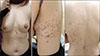
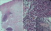
Cutaneous plasmacytosis (CP) is a rare skin disorder characterized by multiple reddish-brown patches and nodules that mainly occur on the trunk in adults1. Histologically, there is a dermal infiltrate of mature plasma cells. We report a case of 35-year-old female who presented with multiple red to brownish macules and patches on the left side of the trunk with a neuronal distribution (Fig. 1). The lesion appeared 4 years ago without any symptoms of pain or itching. A histopathological examination revealed a dense dermal patch-like cellular infiltration surrounding the follicles and the nerves (Fig. 2). The infiltrate was composed of lymphocytes, histiocytes, and plasma cells. Immunohistochemical staining was positive for CD138, CD3, and CD20. Serum protein electrophoresis and immunoelectrophoresis revealed polyclonal hypergammaglobulinemia. Based on these findings, a diagnosis of CP was made. The patient underwent treatment with systemic corticosteroids and narrowband ultraviolet B for 3 months till date. However, the response to the treatment has been minimal.
In our review of the literature, skin manifestations of CP were considerably uniform among patients, with multiple reddish to brownish colored scaly patches, nodules, and infiltrated plaques. Moreover, they were usually persistent, asymptomatic, or mildly pruritic and were mainly distributed on the chest or back in a symmetrical or “Christmas tree” like pattern, which may require precise differential diagnosis including pityriasis rosea and small patch parapsoriasis12. Several other cases with facial or scalp involvement demonstrated a typical trunk involvement pattern as well2. Interestingly, in our patient, the distribution of the lesion showed an atypical pattern, affecting only the left side of the body and showing neuronal pattern dermatoses.
The histopathologic findings characteristically show dermal nodular and perivascular/periadnexal cell infiltrations with predominance of plasma cells admixed with variable numbers of lymphocytes and histiocytes3. Recently, Honda et al.3 reported 6 cases of CP all showing the perineural and intraneural distribution of plasma cells. Similarly, in our patient's punch biopsy specimen, we observed perineural plasma cell infiltration. In this case, we hypothesize that the numerous plasma cells aggregated around the nerve fiber may have a correlation with the unilaterally neuronal pattern of the skin lesions. Furthermore, the typical symmetric pattern of CP on the chest or back may be a phenotypic expression reflecting its close relationship to the spinal nerve tract distribution.
Protein electrophoresis and immunoelectrophoresis generally show polyclonal increase in the gamma globulin fraction in major portion (84%) of the CP patients2. However, the direct relationship between polyclonality and etiology of CP is unclear. If monoclonality is observed, it is necessary to exclude the possibility of marginal zone B cell lymphoma. Polymerase chain reaction for immunoglobulin heavy chain gene rearrangement and B-cell lymphoma 2 staining would be helpful to make the diagnosis clear4.
In summary, a diagnosis of CP should be considered when dermatologists encounter cases of asymptomatic neuronal pigment dermatosis. We speculate that further investigations on the association of the neural pathway with respect to the pathophysiology would be helpful to improve our understanding about CP.
 XML Download