

 PDF
PDF ePub
ePub Citation
 Print
Print


Dear Editor:
MK-7684, an antagonistic agent targeting T-cell immunoreceptor with immunoglobulin and immunoreceptor tyrosine-based inhibitory motif domains (TIGIT), is on phase 1 trial for the treatment of advanced solid tumors sinceDecember 2016. We present a case of psoriasiform dermatitis after treatment with MK-7684. Until date, there exists no reported case in English literature about psoriasiform dermatitis associated with TIGIT inhibitor.
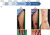
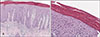
A 63-year-old male with non-small-cell lung cancer (NSCLC) failed to respond to classic chemotherapy and was subsequently treated with pembrolizumab (Fig. 1A). After nine months, he developed erythematous plaques on his arms and legs without changes in his nails (Fig. 1B, C). The skin lesion was mild and controlled by topical steroids. Five months later, due to the progression of NSCLC, pembrolizumab was discontinued and MK-7684 was started subsequently. After one month, he developed erythematous scaly plaques on his whole body (Fig. 1D, E). He also developed trachyonychia on his twenty nails (Fig. 1F). He was a non-smoker and had been taking metformin and atorvastatin for more than two years. He denied a past medical history of psoriasis. After a skin biopsy, the lesion was histologically diagnosed as psoriasiform dermatitis (Fig. 2). He was treated with topical steroids and phototherapy. MK-7684 was stopped after the second injection due to exacerbation of psoriasiform dermatitis. No new lesions had developed after the discontinuation.
Drug-related psoriasis is a side effect of several drugs, such as NSAIDs, antimalarial agents, lithium, beta-blockers and anti-programmed cell death 1 (PD-1) antibodies1. These drugs can induce psoriasis or exacerbate preexisting psoriasis1. The time interval from drug administration to the appearance of the lesion varies from less than four weeks to three months or more1.
TIGIT is a new checkpoint receptor target for cancer immunotherapy and thought to be upregulated on T-cells in multiple cancer models2. By blocking TIGIT, the interaction of CD155 and CD112 with the costimulatory receptor CD226 can be enhanced in association with activation of T-cell mediated immune response against cancer cells3.
In our case, although mild psoriasiform eruption was induced during treatment with pembrolizumab, skin lesions progressed to biopsy-proven psoriasiform dermatitis during treatment with MK-7684. It has been hypothesized that over-activation of T-helper 1 and 17 cells through anti-PD-1 could contribute towards the development of psoriasis4. Although the pathological mechanisms of developing psoriasiform dermatitis by TIGIT inhibitor are unknown, increased production of cytokines related to psoriasis could be one of the mechanisms5. Wang et al.5 reported about TIGIT+ CD4+ T-cells and cytokine levels in patients with psoriasis. The researchers reported that the activation of TIGIT inhibited psoriatic CD4+ T-cell proliferation, decreased the production of IFN-γ and IL-17A, and increased the IL-10 level5. They also reported an increase in the production of IFN-γ and IL-17A subsequent to treatment with TIGIT inhibitor5. We believe that downregulation of TIGIT on CD4+ T-cells might have contributed towards the pathogenesis of psoriasiform dermatitis in our case5. Therefore, we claim that clinicians should be aware of the potential development or aggravation of psoriasiform dermatitis when opting for TIGIT inhibitor treatment.
 XML Download