

 PDF
PDF ePub
ePub Citation
 Print
Print


Dear Editor:
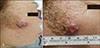
A 62-year-old female presented with a 4-month history of a rapidly growing dome-shaped nodule on the right side of the face. Physical examination revealed a 1.8×1.5-cm, well-demarcated, erythematous, dome-shaped, ulcerated nodule arising within a diffuse, yellow to brown-colored verrucous plaque on the right cheek (Fig. 1). The verrucous plaque had been present since infancy. The patient had no previous relevant medical history, including local trauma, burn, or radiation exposure.
An incisional biopsy of the nodule was performed. Histologic examination revealed aggregates of atypical spindle cells with a fascicular or irregular pattern in the dermis (Fig. 2A). Spindle cells had a large hyperchromatic vesicular nucleus and scanty eosinophilic cytoplasm (Fig. 2B). There was variable pleomorphism with many mitotic figures. An area of epidermal hyperplasia and increased number of sebaceous glands were observed adjacent to the tumor. Immunohistochemical analysis of the spindle cells was positive for cytokeratin (CK) (Fig. 2C), p40 (Fig. 2D), and vimentin (Fig. 2E), but negative for melan-A, HMB-45, S-100, cluster of differentiation (CD) 31, CD34, and desmin. The patient was diagnosed with cutaneous spindle cell squamous cell carcinoma (SpSCC) superimposed on a nevus sebaceous (NS). Computed tomographic scan of the brain and 99mTechnetium bone scintigraphy revealed no evidence of metastasis.
The patient underwent a wide excision with a 10-mm margin from the periphery of the nodule. There has been no evidence of recurrence or metastases during 8 months of follow-up.
Numerous secondary tumors can arise in NS, including syringocystadenoma papilliferum, trichoblastoma, basal cell carcinoma, and squamous cell carcinoma (SCC). The development of an SCC in NS is extremely rare. Our case is an even more unusual variant of SCC (spindle cell) arising in NS. There is only one case report to date1.
Cutaneous SpSCC is a rare, aggressive variant of SCC that presents as a raised or exophytic nodule accompanied by bleeding and central ulceration. Generally, SpSCC lesions are associated with sun-damaged skin, burn scar, local trauma, and prior radiation exposure2. Histopathologically, SpSCC comprises atypical spindle cells with elongated vesicular nuclei. The presence of squamous differentiation, dyskeratotic cells, and continuity with the epidermis may assist in the diagnosis. However, as these characteristics are not always detected, immunohistochemistry can help distinguish this form from other spindle cell tumors such as atypical fibroxanthoma and dermatofibrosarcoma protuberans. SpSCC cells stain positively for CK, CK5/6, AE1/3, CAM5.2, and vimentin, whereas other nonsquamous spindle cell tumors are negative for CKs3.
Because of its lack of differentiation, SpCC exhibits more aggressive characteristics and accounts for >33% of cutaneous metastatic SCC. Typically, malignancies that arise in association with NS are considered non-aggressive lesions. However, sudden growth of a nodular tumor within NS and a large developed nodule as in our patient are regarded as an indication of aggressive behavior4.
Herein, we described a case of SpSCC arising within NS. Clinicians should be aware that aggressive malignancies can develop in NS, and early excision should be considered when clinical signs of malignancy are seen, such as rapid nodular growth or ulceration within NS.

 XML Download