

 PDF
PDF ePub
ePub Citation
 Print
Print


Abstract
The authors performed clinical study particularly on the skeletal changes in 20 patients with congenital syphilis who visited the department of orthopedic surgery or were tansferred from the department of pediatrics for the past two and a half years from January 1972 to July 1974.
The results were as follows:
1. Age distribution disclosed from 3 days to 14 months with mean 55 days, and the male to female ratio was 8.12.
2. Chief complaints showed buccal and nasal hemorrhage in 7 cases (35%). the commonest one, upper respiratory infection in 6 cases(30%), abdominal distention 4(20%), rash and jaundice 2(10%) respectively, excoriation and high fever in 1(5%) each.
3. Physical examination showed hepatomegaly in 11 cases (55%), excoriation, edema, cyanosis in 2 cases each (10%)
4. Blood study revealed anemia in 13 cases (65%)» leukocytosis in 14(70%)
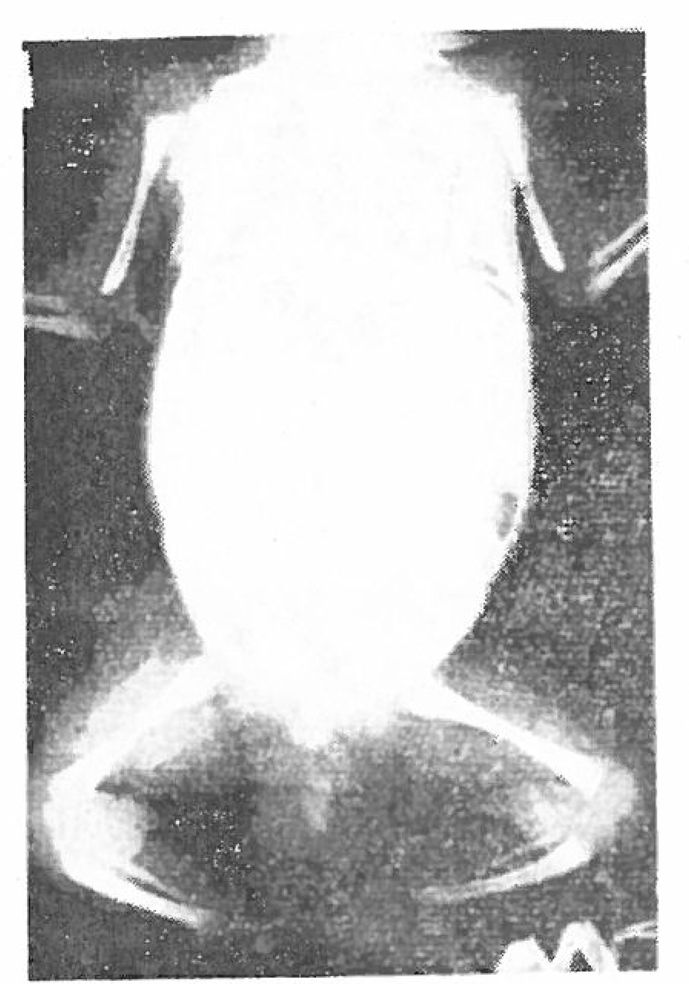
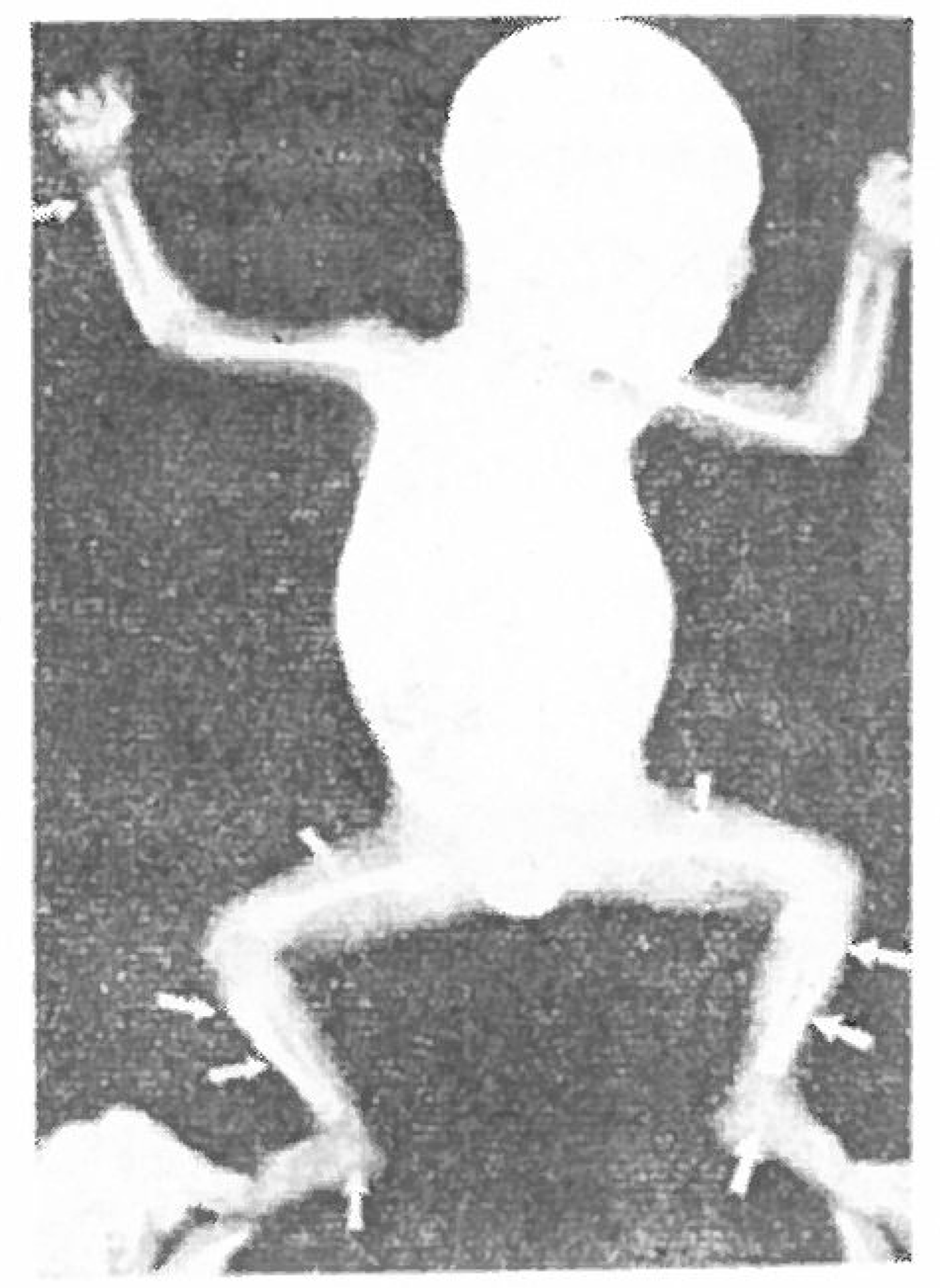
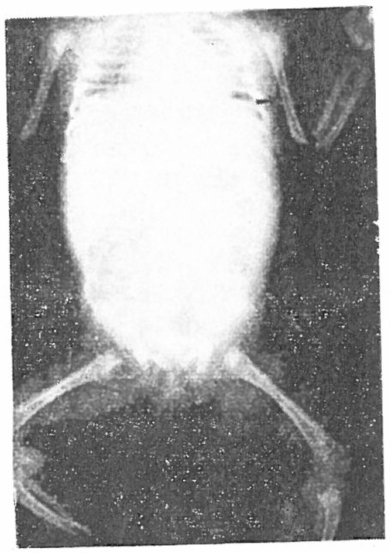
5. X-ray findings showed positive changes in 16 cases(80%), most of which were involved bilaterally
1) Osteochondritis 11 cases (55%) ‘
The involved bones were radius, ulna, and tibia in 10 cases(50%) each, humerus, in 8(40 %), femur in 7(35%), fibula in 2(10%), tarsal phalanges in 1(5%).
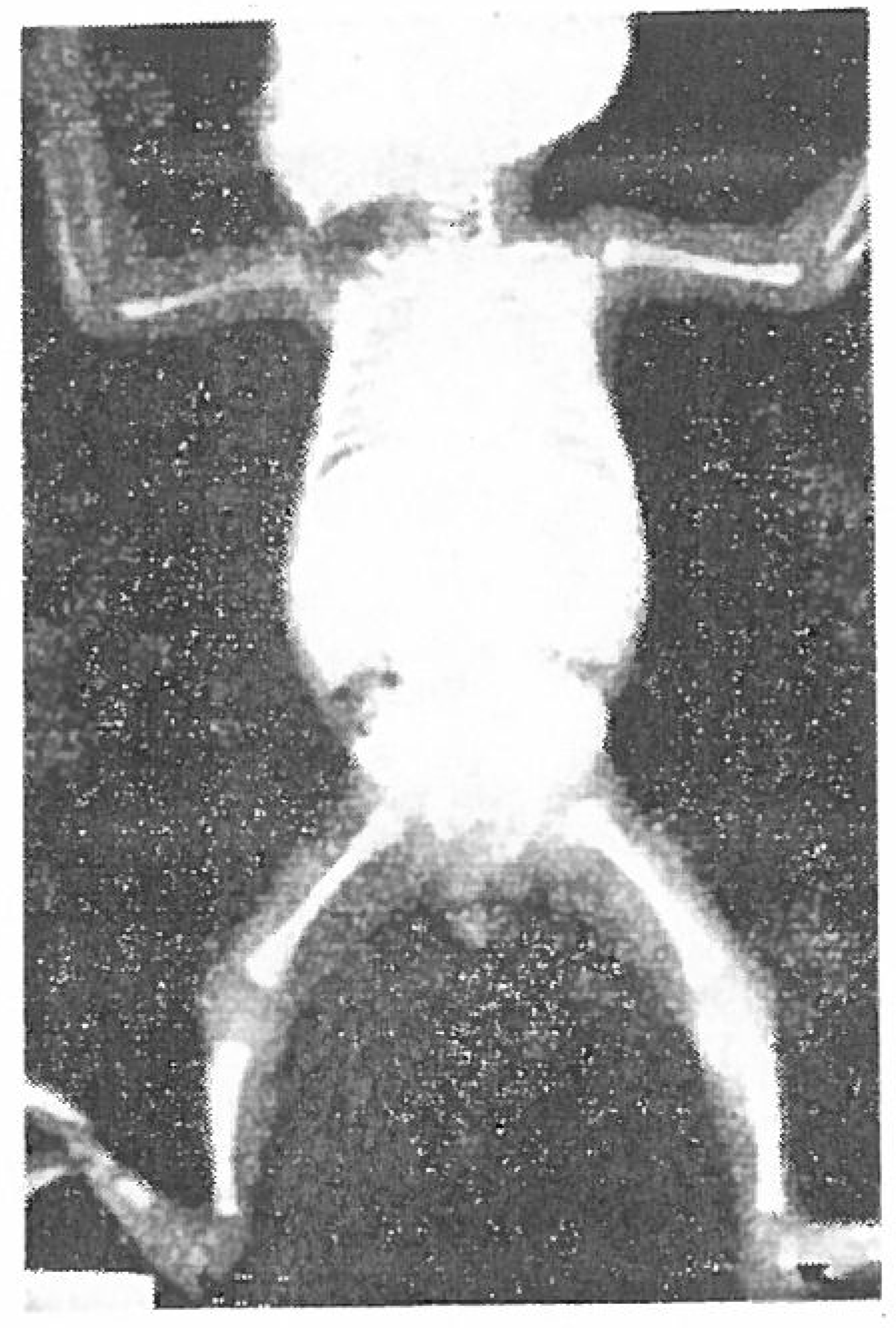
2) Periosteal reaction 9 cases (45%) ‘
The involved bones were ulna and femur in 9 cases(45%) respectively, humerus and tibia 8 cases(40%) each, fibula 6 cases(30%), radius 5 cases(25%), and clavicle in 1 case (5%).
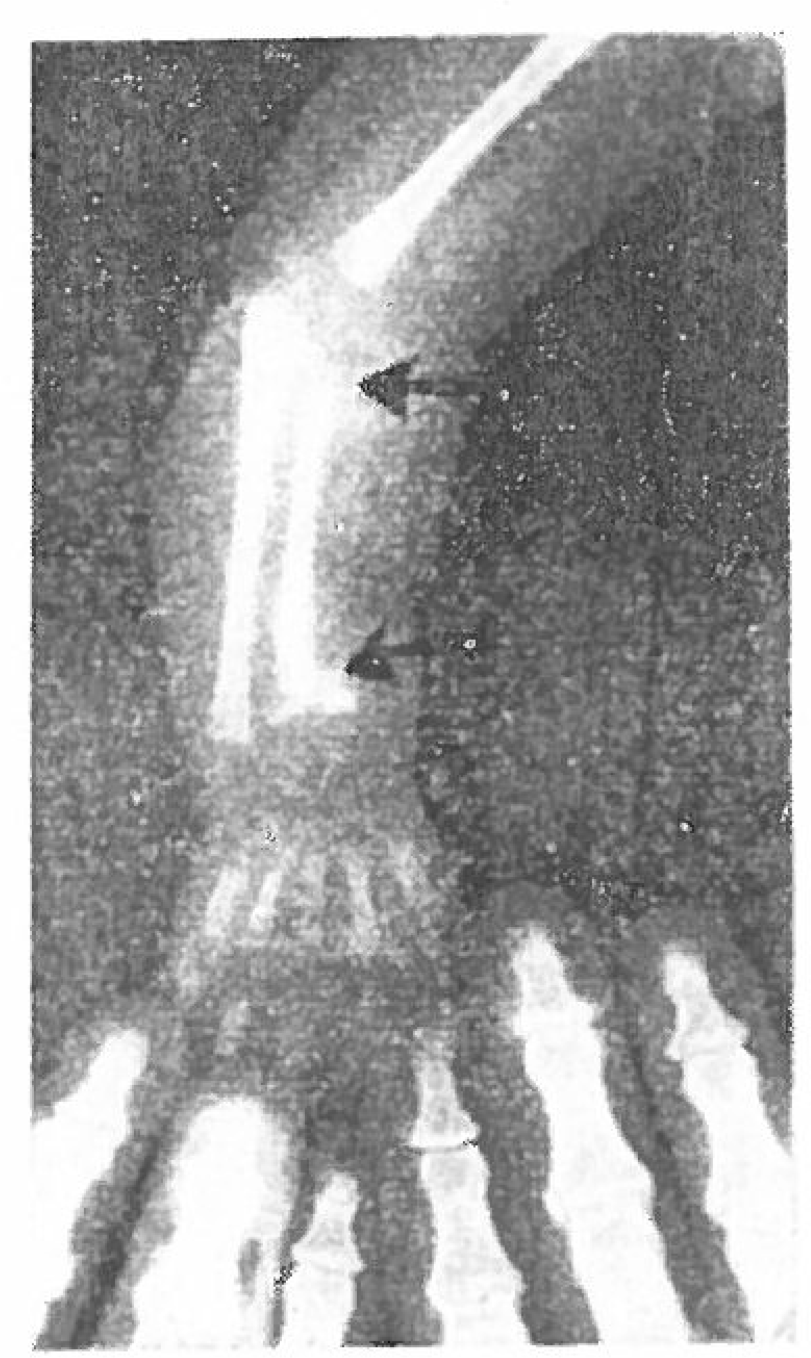
3) Wimberger's sign 6 cases(30%) :
4) Sigmoid notch sign 3 cases (30%) :
5) Double ring shadow in tarsal bone 3 cases(15%):
6) Metaphyseal fracture 1 case (5%) æ
REFERENCES
1.Aegerter E.., Kirkpatrick J.A.Orthopedic Disease,. p. 611–622. Philadelphia, W.B. Saunders;1968.
2.Barnett H.L.Pediatrics, pp. 652–654. .1972.
3.Beeson P.B.., McDermott W.Textbook of Medicine,. p. 662–667. Philadelphia, W.B. Saunders;1971.
4.Brown W.J.., Moor M.B.Congenital Syphilis in the U.S.A. Clinical Pediatrics. 2:220. 1963.
5.Davis J. A.., Dobbing J.Specific Foundation of Pediatrics.
6.Duthie R.B.., Ferguson A.B.Mercer's Orthopedic Surgery,. p. 509–512. London, Edward Arnold;1973.
7.Edeiken J.., Hodes P.J.Roentgen Diagnosis of Diseases of bone,. p. 611–622. Baltimore, the Williams and Willkins;1973.
8.Fiumara N. J.Legacy of Syphilis. Arch Dermatology. 92:676. 1965.
9.Fleming T.C., Bardenstein M.B.Congenital Syphilis. J. Bone and Joint Surg. 53-A:1648–1651. December 1971.

10.Harris W.D.., Lave V.G.Congenital Syphilis in the Newborn: Diagnosis and Treatment. J.A.M.A. 194:1312. 1965.
11.Isadore Meschan M.A.Analysis of Roentgen Signs in General Radiology,. p. 266–269. Philadelphia, W. B. Saunders;1973.
12.Jacobson H.G.., Murray R. O.The Radiology of Skeletal Disorders. Baltimore. 1972.
13.Jaffe H.L.Metabolic, Degenerative, and Inflammatory Disease of Bone and Joints. 908–924.
14.Levin E. J.Healing in Congenital Osseous Syphilis. Am. J. Roengenol. 110:591–597. 1970.
15.Nelson W.E.Textbook of Pediatrics, pp. 522–529. 1964.
16.Sante L. R.Principles of Roengenological Interpretation. Michigan, Edwards Brothers. 1965.
17.Tachdjian M.O.Pediatric Orthopedics, pp. 522–529. 1964.
18.Turek S.L.Orthopedics,. p. 100–111. Philadelphia, J. B. Lippincott;1967.
 XML Download