

 PDF
PDF ePub
ePub Citation
 Print
Print


INTRODUCTION
Ulnar impaction syndrome (UIS) can be treated by a variety of conservative and surgical methods. In patients with positive ulnar variance (UV), decreased load-sharing by ulnar recession is necessary to relieve ulnar side pain. Surgical treatments include ulnar shortening osteotomy (USO) and the arthroscopic wafer procedure (AWP); although both are effective at relieving pain, AWP is not complicated by hardware and nonunion problems12. AWP seems to unload the ulnocarpal joint in the wrists with neutral and positive UV, although this is slightly less effectively at severe positive UV3. At the completion of resection, −2 or −3 mm of UV is recommended45; too much wafer resection is likely to increase the pressure in the distal radioulnar joint (DRUJ) due to the decrease in DRUJ contact surface area6. However, it is not clear yet whether the final UV after AWP affects the outcomes.
We have experienced some patients who require secondary USO following AWP due to persistent pain. While USO has been reported to stabilize the DRUJ in addition to unloading the ulnocarpal joint, as yet, there have been no reports for DRUJ ligament tension associated with AWP7.
We aim to identify risk factors for secondary USO following AWP in terms of the degree of DRUJ subluxation. Our objective was to evaluate the results of AWP for UIS at our institution and identify preoperative factors and the degree of DRUJ subluxation that could predict outcomes.
MATERIALS AND METHODS
This was a retrospective case control study performed at a tertiary medical center, and approved by the research ethics committee at our institution. We searched our institutional database for patients who underwent AWP for UIS, by a single surgeon, from March 2005 to February 2018. This study was approved by the Institutional Review Board of the Soonchunhyang University Bucheon Hospital (IRB No. SCHBC 2018-09-015-002).
The inclusion criteria were as follows: 1) Age ≥18 years old and 2) patients receiving AWP for UIS. Diagnosis of UIS was based on the patients' history, physical examination, imaging studies, and the triangular fibrocartilage complex (TFCC) wear type. The diagnostic criteria for a UIS were as follows: 1) patient presenting with ulnar side wrist pain, 2) positive result for the ulnocarpal stress test8, and 3) negative results for provocative tests for other causes of ulnar side wrist pain such as the lunotriquetral shear test9 and the extensor carpi ulnaris synergy test10, and 4) TFCC wear or perforation observed by magnetic resonance imaging (MRI). Conservative treatments were provided by various doctors prior to the patients visit to our clinic. To standardize the conservative treatment, we treated them again conservatively using our own protocol, even though they had enough time for treatment. We initiated conservative treatment using a short-arm wrist brace with intermittent use of non-steroidal anti-inflammatory drugs for at least 3 months. The indication of surgery was UIS patients with less than 2.5 mm positive UV who failed to respond to conservative treatment for more than 3 consecutive months. Exclusion criteria were as follows: 1) fracture or fracture dislocation of the wrist, 2) lunotriquetral instability or Palmer Class 2 D, 3) DRUJ instability or arthritis, 4) degenerative or inflammatory arthritis of the wrist, and 5) follow-up less than 12 months.
1. Demographics and patient flow
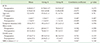
We identified a total of 19 consecutive cases that were eligible for this study. Of these, eight cases were excluded because of a short follow-up period in three cases, and the absence of a follow-up MRI or computed tomography (CT) in the remainder. Thus, 11 wrists of 9 patients were included in the study cohort. Among them, five cases were converted to secondary USO due to persistent pain and were categorized to group A. The average conversion duration from AWP to secondary USO was 1.3 years (6 months to 3.5 years). Group A included 5 cases with a median age of 53.8 years (range, 41–67 years) and included one female. The remaining 6 cases were categorized as group B (AWP only), with a median age of 51 years (range, 43–65 years) and all of the cases were female. There were no significant differences in the demographic and clinical characteristics between the groups, with the exception of sex (p>0.05, Table 1). The average follow-up was 22 months (12–48) and the average symptom duration was 18.4 months.
2. Outcome measures, data sources, and bias
An independent examiner performed all of the pre- and post- AWP (before USO) evaluations. The outcome assessments included measurement of the wrist range of motion and the grip strength, Mayo wrist score, and the Disability of Shoulder, Arm and Hand (DASH) questionnaire1112. The wrist range of motion was assessed with a hand-held goniometer, and the grip strength was measured using a Jamar dynamometer (Samo Preston, Bolingbrook, IL, USA).
The UV was measured using the perpendicular method13 on the wrist posterior-anterior view X-ray taken in the neutral rotation position of the wrist before and after the AWP. Neutral rotation was judged based on the extensor carpi ulnaris groove criterion14. The UV was measured twice and the average was taken using semi-automated image analysis software installed in the picture archiving and communication system (Deja View; DEIT, Bucheon, Korea).
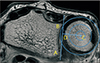
CT or MRI scans were obtained with the shoulder in 90° abduction, elbow in 90° flexion, forearm in neutral rotation, and wrist in neutral alignment for accurate measuring. To diagnose the DRUJ dorsal translation based on CT or MRI scans, we measured the translation of the ulnar head with respect to the radius using the radioulnar ratio (RUR) method by Lo et al.15 before and after AWP. To obtain the RUR, the center of the ulnar head was identified using a transparent template marked with concentric circles. From this point, a perpendicular line was drawn to the line connecting the volar and dorsal margins of the sigmoid notch. The distance from the intersection of these two lines to the volar margin of the sigmoid notch was measured, and the ratio of this distance to the length of the sigmoid notch was calculated (Fig. 1). To determine the interobserver reliability of measurements, the intraclass correlation coefficients (ICCs) were calculated using a two-way random-effects model and absolute agreement. The first measurement value of each observer was used for the interrater reliability assessment in order to control for any learning effect. The following criteria were used to determine the reliability coefficients: very low (<0.20), low (0.21–0.40), moderate (0.41–0.60), good (0.61–0.80), and excellent (0.81–1.00)16. The intraobserver reliability was calculated using both the first and second measurements from each observer
MRI scans were obtained in all cases and the MRI evidence of mechanical impaction, such as edema, cystic changes, or sclerosis in the proximal ulnar or lunate, was investigated.
3. Arthroscopic techniques
Under general anesthesia upper arm tourniquet exsanguination, the wrist is distracted with 10 pounds (4.5 kg) of distal traction via finger traps, with the upper arm secured to a padded arm board. The standard 3–4, 4–5, DRUJ arthroscopy portals are used for the procedure. The radiocarpal joints are distended with sterile saline inflow via gravity. A 2.4 mm arthroscope is inserted into the 3–4 portal for visualization. A small-joint mechanical shaver is introduced through the 4–5 (n=7) or distal DRUJ portal (n=4) after the TFCC defect is confirmed. Scar tissue and border flaps are debrided from the central TFCC wear, as well as any irregularities in the lunotriquetral ligament, if it is found to be torn. Following thorough debridement, the prominent ulnar head is visible through the defect in the central disk. The shaver is replaced with a 2.9 mm motorized burr, and the articular cartilage and 2–3 mm of subchondral bone are resected from the distal ulna. It is important to have an assistant rotate the forearm into full pronation, neutral rotation, and full supination during burring to ensure that the full circumference of the ulnar head is resected to the medullary bone. Intraoperative fluoroscopic examination of the wrist is performed during burring to ensure that adequate bone is resected from the ulna. The portals are closed with 4-0 nylon sutures. The wrist is bandaged and immobilized for 1 week in a palmar splint.
4. Aftercare/follow-up
Range of motion exercises are begun at 7 days following surgery, with the wrist protected by a short arm brace during activities. Strengthening exercises start at 6 weeks following the wafer procedure.
5. Statistical analysis
Correlation coefficient was calculated to compare the continuous variables between the groups and prevs. postoperative outcomes in the same group. The chi-square test or Fisher's exact test were used to compare dichotomous variables between the groups, as appropriate. The significance level was set at p<0.05.
RESULTS
Most of the preoperative outcome measures were comparable between group A and group B (p>0.05, Table 1). Six cases (4 in group A and 2 in group B) were found to have MRI evidence of mechanical impaction, such as edema, cystic changes, or sclerosis in the proximal ulnar or lunate. The MRI evidence did not differ significantly between groups A and B (p=0.175)
The mean preoperative DASH score improved from 47.6 (group A: 49.5, group B: 46.3) to 16.8 (group A post AWP: 24.0, group A post USO: 10.6, group B: 9.5). The mean grip strength of the contralateral side improved from 77.1% (group A: 76.7, group B: 77.8) preoperatively to 85.2% (group A post AWP: 74.2, group A post USO: 88.2, group B: 93.3) postoperatively. The Mayo wrist score was excellent in 6 cases, good in 3 cases, and fair in 2 cases, and improved from 69.1 (group A: 68.0, group B: 70.0) preoperatively to 85.5 (group A post AWP: 76.0, group A post USO: 92.4, group B: 93.3) postoperatively.
All the patients in group A showed substantial pain and disability that persisted for more than 6 months after the AWP; they underwent revision surgery with USO at 24 to 175 weeks following the index surgery. All patients showed improved outcomes following the USO.
The intraobserver ICC, as determined by CT measurements, for observer A was good (0.80) and slightly lower for observer B, but still good (0.70). The interobserver ICC values were good between observers A and B (0.72).
The average UV changed from 1.6 ± 0.6 mm before the AWP to −1.5±0.9 mm after the AWP. The average RUR, as measured from the axial image, was 0.68±0.17 before the AWP and 0.54±0.10 after the AWP. There was no significant difference in the preoperative RUR between group A (0.70±0.11) and group B (0.65±0.22) (p=0.275). However, the difference between the preoperative RUR and postoperative RUR in group B (0.49±0.09) was significant (p=0.027, Pearson correlation coefficient=0.862); this indicates that DRUJ dorsal translation persisted in group A after AWP (Fig. 1)
Second-look arthroscopy was performed in 4 out of 5 wrists at the time of USO (group A), and fibrocartilaginous healing of the ulnar head was confirmed in all of the wrists.
DISCUSSION
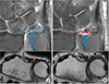
The degree of DRUJ dorsal translation was reduced in group B after the AWP. Resection of ulnar head dorsal prominence seems to move ulnar head center toward to volar side of the sigmoid notch even though real position of ulnar head is not changed. Conversely, it was not reduced in secondary USO (group A). The fact that secondary USO improved residual symptom in group A is not sufficient to explain that the reason for failure is residual ulnar impaction symptom because final UV was not different between group A and B. If the DRUJ ligament iatrogenic injury occurred at the time of AWP, the degree of DRUJ dorsal translation will be increased. However the DRUJ ligament volar and dorsal limbs, as well as the radial and ulnar attachments, were carefully protected during the AWP. In addition, the ulnar fovea was not burred so as to leave the attachment of the deep limb of the TFCC intact at the time of the AWP. The DRUJ ligament tension could be affected by the UV because sudden disappearance of the ulnar head pole reduces contact pressure between the TFCC proximal surface and the ulnar head pole (Fig. 2). A cadaveric study revealed that the DRUJ joint reaction force increases significantly after USO and did not increase after ulnar wafer resection; this suggests that the tightening effect of the DRUJ after USO does not exist after AWP17. The USO can stabilize the DRUJ by increasing the intrastructural suspension effect of the TFCC when the radioulnar ligaments attach to the ulnar fovea7. While a biomechanical study demonstrated increasing amounts of wafer resection led to a linear increase in pressure in the DRUJ, it seems to be related to a decrease in the DRUJ contact surface, not with radioulnar ligament tension6. However, to the best of our knowledge there have been no previous studies that have reported DRUJ ligament tension associated with AWP.
At the completion of the resection, −2 or −3 mm of UV is recommended4. The postoperative UV in our series was −1.5 mm on plain radiographs, since we tried to remove only the protruding 2–3 mm dome of the ulna in order to preserve the DRUJ contact surface area as much as possible. Our study shows that the average UV changed to −1.4 mm in group A and −1.5 mm in group B after the AWP (not significant, p>0.05). The UV did not reach −2 mm in either group after the AWP. Inadequate ulna resection alone was not sufficient to explain the reason for failure of unloading the ulnocarpal joint in group A. A further biomechanical study and prospective randomized study is required to examine the optimal target UV and the dynamic relationship between residual DRUJ instability and ulnocarpal loading.
In reporting predictors of good outcomes of UIS, Meftah et al.18 demonstrated that MRI evidence, such as cystic changes, edema, or sclerosis, was associated with better pain relief, using Gartland and Werley's demerit point system in their series19. However, we could not detect any difference in the presence of MRI signs between group A and group B in our series.
If we could detect persistent DRUJ dorsal translation immediately after the AWP, we could prevent secondary USO using additional protective measures, such as immobilization with supination.
The present study had some limitations. Firstly, it was a retrospective case series. Secondly, some of the patients in group A underwent revision secondary USO at 24 weeks following AWP, which is a relatively short follow-up. However, we believe that 24 weeks is a sufficient amount of time to judge the progression of symptom resolution after AWP for UIS. Despite the fact that none of the patients in our study had DRUJ instability symptoms before the AWP, asymptomatic DRUJ dorsal translation may be associated with UIS in our studies. Thus, we confirmed that the radial and ulnar attachments of the DRUJ ligament were intact arthroscopically during the AWP. There is large normal variation in DRUJ translation. Normal values derived with RUR methods for measuring translation of the DRUJ have been reported as 0.32–0.6920. Scanning of both wrists might be helpful to prevent the radiological overdiagnosis of DRUJ translation because the uninjured wrist will reflect the normal laxity of the DRUJ.
Lastly, numbers of the patients underwent revision USO is limited to achieve sufficient statistical power for RUR, even though the power for sex is over 90%.
 XML Download