

 PDF
PDF ePub
ePub Citation
 Print
Print


INTRODUCTION
Non-ischaemic cardiomyopathies (NICMs) encompass a heterogeneous group of diseases of the myocardium which may manifest in ventricular dilatation, systolic impairment or hypertrophy. By definition, these cardiomyopathies occur in the absence of contributory coronary artery disease or abnormal loading conditions (such as valvular heart disease). NICM therefore broadly captures multiple distinct morphological and functional phenotypes which can be broadly separated into genetic disease processes and acquired disorders which are often secondary to inflammation, infiltration or toxins.
Ventricular arrhythmias (VAs) in the form of premature ventricular complexes (PVCs), ventricular tachycardia (VT) and, less commonly, ventricular fibrillation (VF) have a distinctly increased prevalence in NICM and are a significant driver of morbidity and mortality. Their occurrence often triggers a cascade of management strategies, including initiation of anti-arrhythmic drugs, insertion of implantable cardioverter-defibrillators (ICDs) and referral for catheter ablation. VT in NICM differs fundamentally from those in ischemic cardiomyopathy (ICM) based on the location distribution of substrate, the need for advanced catheter ablation strategies and the influence of the underlying aetiology on long-term outcomes.
The use of catheter ablation to manage VAs is becoming increasingly prevalent in the past decade, accompanied by progresses in clinical outcomes driven by further technological advances and our growing understanding of the disease processes involved. Achieving non-inducibility, which is the most common marker of procedural success, ranges from 38% to 74%. VT recurrence can be as high as 58% in NICM, where outcomes have been found to be worse in sarcoidosis and better in arrhythmogenic right ventricular cardiomyopathy (ARVC). Beyond preventing recurrence, catheter ablation has been shown to reduce anti-arrhythmic drug use and device shock burden, as well as an effective tool for aborting electrical storm. Earlier referral for catheter ablation has been found to improve outcomes; either prior to the second episode of VT or failure of any antiarrhythmic drug.
The purpose of this review is to explore the fundamental pathophysiology underlying VT in NICM and the inherent challenges that this poses with respect to electroanatomic characterisation of substrate and eventual catheter ablation, including the use of adjunctive techniques that are gaining increasing traction.
NON-ISCHEMIC CARDIOMYOPATHY: A HETEROGENEOUS GROUP
Though NICM (deemed at the time “non-coronary cardiomyopathies”) was first characterised as a disease process in 1957 by Brigden1), it was in 1961 that Goodwin et al.2) developed the first classification system, separating them into congestive (dilated), hypertrophic and constrictive (restrictive) groups. Since this categorisation, there have been many attempts to develop a satisfactory classification system for NICMs.3)4)5) Advances in cardiac imaging (in particular, cardiac magnetic resonance imaging [cMRI]), genetic studies and histopathological and biomarker analyses have not only aided in classifying subtypes of NICM, but have also allowed early diagnoses in patients yet to manifest overt ventricular dilatation or systolic dysfunction.
Whilst a description of VAs within all subgroups of NICM is beyond the scope of this review, it is important to understand key differences in their manifestation. Not only does the frequency of VT vary according to subtype; the pattern and distribution of myocardial fibrosis causing arrhythmogenic substrate varies by aetiology. Furthermore, outcomes of ablation are affected by the underlying disease process.
Dilated cardiomyopathies
Dilated cardiomyopathies (DCMs) are characterised by left ventricular or biventricular dilatation and systolic impairment in the absence of aforementioned coronary artery disease or abnormal loading. There are both genetic (titin, lamin A/C [LMNA], phospholamban and desmin) and acquired causes of DCM (alcohol, myocarditis, peripartum, drug mediated and autoimmune).6) Despite the heterogeneity of DCM, most studies characterising ablation outcomes in NICM have included a mixed group of DCM which might explain the disparity in outcomes compared to VT ablation of ICM.
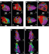
LMNA mutations are associated with a higher rate of atrial and VAs compared to idiopathic DCM with a more aggressive disease course characterised by faster progression to both end stage heart failure and malignant VT.7)8) In a prospective study of 19 patients with LMNA mutation who required permanent cardiac pacing and had prophylactic insertion of an ICD, 42% (8 patients) received appropriate therapy for VAs in a follow up period of 33 months9) with the majority having a relatively preserved left ventricular ejection fraction (>45%). Unfortunately, these same patients have poor catheter ablation outcomes and in a case series of 25 patients with LMNA cardiomyopathy, acute success was only attained in 25% of patients even after multiple procedures with a complication rate of 25% (including risk of stroke, atrioventricular block and cardiogenic shock).10) Likely contributing to this is the challenging sites from which VT in LMNA cardiomyopathy often originates, including from intramural scar in the septum and basal left ventricle, as well as the basal inferior wall and sub-aortic mitral continuity (Figure 1A and B).10)
Figure 1
Electroanatomic voltage maps in nonischemic cardiomyopathy by disease aetiology. (A) Bipolar (top) and unipolar (bottom) electroanatomic voltage maps of the LV in a patient with Lamin A/C cardiomyopathy showing deep intramural anteroseptal substrate (light green, yellow, and red). Red dots mark points of radiofrequency ablation. (B) Epicardial voltage maps demonstrating extensive inferolateral low-voltage substrate (red) in the RAO (top) and LAO (bottom) views. (C) Bipolar voltage map (top) of the LV of a patient with sarcoidosis demonstrating low-voltage substrate (red) in the basal peri-valvular region and focal substrate in the anteroseptum. Unipolar mapping (bottom) showing more extensive deep intramural anteroseptal substrate (light-green, yellow, and red). (D) Superior view of both endocardial & epicardial voltage maps of the RV & LV showing significant basal-perivalvular, septal and inferolateral epicardial substrate (red) (top). Bottom panel shows confluent and patchy low-voltage inferolateral epicardial substrate from an inferior view due to the patchy inflammatory involvement of the myocardium in sarcoidosis (light green, yellow, and red). (E) Endocardial bipolar voltage map (top panels) of the RV in the RL and PA (F) views showing low-voltage substrate (red) at the superior aspect of the TA and free wall of the RVOT in a patient with ARVC. Unipolar voltage mapping (bottom panels) showing more extensive low-voltage (light-green) extending from the TA to the RVOT, indicating a larger area of far-field scar, likely to be epicardial given the natural history of ARVC.
ARVC = arrhythmogenic right ventricular cardiomyopathy; INF = inferior; LAO = left anterior oblique; LMNA = lamin A/C cardiomyopathy; LV = left ventricle; LVOT = left ventricular outflow tract; MA = mitral annulus; PA = postero-anterior; RAO = right anterior oblique; RL = right lateral; RV = right ventricle; RVOT = right ventricular outflow tract; TA = tricuspid annulus.
Myocarditis (which can be diagnosed based on clinical presentation, cardiac MRI and endomyocardial biopsy) is another well recognised cause of DCM. Unlike LMNA mutation, VT in myocarditis is much easier treated by catheter ablation. In a study of 26 patients with post myocarditis VT and preserved LV function, catheter ablation proved to be effective with 77% VT free-survival after a 24-month follow-up.11) Arrythmogenic substrate in myocarditis is often mapped to the epicardium.12) This raises its own challenges including the risk of damage to coronary arteries and the phrenic nerve.
Inflammatory cardiomyopathies
Sarcoidosis is a multisystem disease with unknown aetiology characterised by non-caseating granulomas. An acquired cause of NICM, cardiac sarcoidoisis is associated with a high incidence of sudden cardiac death13) as well as conduction abnormalities, VAs and heart failure. It is an inflammatory condition and the most common mechanism of VT is postulated to be re-entrant arrhythmias around areas of granulomatous scar although non re-entrant triggered activity and abnormal automaticity have been described in patients with cardiac sarcoidosis with frequent ventricular ectopy.14) cMRI has demonstrated myocardial scar in sarcoidosis predominantly involves the basal and paravalvular left ventricle with subepicardial rather than subendocardial involvement (Figure 1C and D).15)
In patients with known cardiac involvement and ICD device in-situ, sustained VAs occur at a rate of up to 15% per year.16) Further, compared to mixed NICM cohorts, catheter ablation outcomes in cardiac sarcoidosis is associated with higher rates of VT recurrence, requirement of transplantation and mortality.14) In the first published case series of catheter ablation in patients with cardiac sarcoidosis in incessant, Koplan et al.17) demonstrated a VT recurrence rate of 75% within 6 months. In the largest case series of catheter ablation in cardiac sarcoidosis, following 21 patients for 58 months, electroanatomic voltage mapping demonstrated confluent right ventricular epicardial and endocardial scar with patchy left ventricular scar favouring the septum, anterior wall and perivalvular areas.18) This study also reinforced poor outcomes in catheter ablation of cardiac sarcoidosis with freedom from VT 37% at one year follow up even after multiple procedures.
Arrhythmogenic cardiomyopathies
ARVC is characterised by fibro-fatty inflammation of the right and left ventricles. Surviving myocardial fibers embedded within this network of fibrofatty tissue form complex connection with neighbouring healthy myocardium increasing potential for re-entrant circuits that classically progresses from the epicardium to endocardium (Figure 1E and F). Patients with ARVC typically have a high burden of ventricular ectopy with risk of sudden cardiac death. In an analysis of the North American ARVC Registry, Link et al.19) report that of 108 patients who had an AICD placed, 44% had sustained VAs requiring ICD treatment, the majority of which were treated with anti-tachycardia pacing. Although there have been mixed results in outcomes of VT ablation in ARVC, more recent studies have demonstrated that aggressive substrate modification with combined endo-epicardial ablation have significantly improved VT free survival. Garcia et al.20) reported in a cohort of 13 patients a 77% rate of freedom from VT in a follow up of 18 months whilst Bai et al.21) have demonstrated 84% VT free survival in a combined endo-epicardial approach (compared to 52% in endocardial only ablation). Further, in a long term follow up of 62 patients over a median of 55 months, Santangeli report 71% VT free survival with limited need for anti-arrhythmic drugs.22)
Hypertrophic cardiomyopathy (HCM) is a genetic sarcomeric cardiomyopathy that is well-recognised for its strong association with sudden cardiac death that is ameliorated with ICD insertion is often a catalyst for familial screening.23) Whilst VAs in HCM have classically been thought to arise from VF and polymorphic VT, monomorphic VT has been increasingly described in more recent literature.24) This has been found to correlate with late gadolinium enhancement (LGE), similar to scar substrate in other forms of NICM.25)26) 7 out of 10 patients in one case series had apical LV substrate, with 3 being found to have an apical aneurysm,24) which, in itself, is considered a harbinger for arrhythmic events.27) 80% of cases had epicardial substrate on mapping, with a longer average endocardial ablation time of 13 seconds attributable to difficulty penetrating thickened myocardium to ablate deep intramural substrate. Ablation was able to achieve a recurrence-free survival of 78% on 3 years of follow-up, demonstrating the utility of catheter ablation in this disease process as a means of reducing device therapy burden and improving quality of life. Igarashi et al.'s series28) of 12 patients with an apical aneurysm in HCM demonstrated the efficacy of catheter ablation in eliminating VT arising from this region, achieving freedom from VT in 87% on 12 months' follow-up.
Non-compaction cardiomyopathy is another clinical entity that has a distinct predilection for VAs. Classified as a form of structural and genetic cardiomyopathy,5) the most commonly proposed mechanism is that of inappropriate arrest of the compaction process of the foetal myocardium, resulting in hypertrabeculation and prominent intratrabecular recesses.29) Prevalence of VAs and sudden cardiac death are as high as 50%30) and 18%29) respectively, although no dedicated guidelines exist to assist with risk stratification for ICD implantation. It has been commonly presumed that VAs arise from structural abnormal noncompacted myocardium, although this has not been consistently reproduced in case reports and case series over recent decades.31) Nonetheless, VT in noncompaction cardiomyopathy has been shown to be amenable to catheter ablation,32) most often from an endocardial approach, but with successful epicardial approaches being documented in case reports.33)
MECHANISMS OF VENTRICULAR TACHYCARDIA
There are three major mechanisms of VT: triggered activity, automaticity and scar-related re-entry.34) The underlying cardiac disease can often suggest a potential mechanism and also predict the likelihood of success with discrete radiofrequency ablation. Locations of triggered activity or automaticity, or very small re-entry circuits, are more likely to respond to focal ablation, whereas macro-re-entrant circuits due to ventricular scar are often more complex requiring endocardial and epicardial substrate homogenization.34)
Whilst NICM encompasses multiple varying aetiologies, the majority of VA has shown to be from scar-related re-entry. This was demonstrated by Hsia et al.,35) who studied 19 patients with NICM. They were successfully able to induce VT in all patients with programmed stimulation (without provocation) and the majority (74%) were also entrained with over-drive pacing, suggesting underlying re-entry. Similar to infarct scar, inflammatory processes associated with NICM are thought to create fibrosis dispersed within surviving muscle bundles, causing poor electrical coupling, functional and anatomic block and a subsequent macro-reentrant circuit that can span all layers of the myocardium.36) VT in ICM arises from myocardium involved in specific vascular territories, where the degree of ischemia progresses from subendocardial to transmural in complete infarction. Hence, endocardial scar surface area is often greater than that found in NICM. In contrast, the underlying disease process in NICM may result in different distributions of scar, with a greater proportion of patients with intramural and epicardial substrate that may only be evident with unipolar endocardial electroanatomic mapping. Combined with the progressive nature of this scar37) and its predilection for the interventricular septum,38) it may be difficult to ablate targets in deep regions with limitations such as its proximity to the native conduction system. Substrate may also be progressive, possibly driven by the underlying disease process, as demonstrated in a case series of 13 patients, with total unipolar low-voltage substrate increased from 6 to 46% over a period of 32 months.37)
Recent histopathological studies have validated electroanatomic correlation with myocardial fibrosis. In 2018, Glashan et al.38) published work analysing whole heart histopathology in 8 deceased or transplanted patients with ICM and monomorphic VT, concluding similar distribution of subendocardial, mid-myocardial and transmural patterns. The dominant histopathological characteristics were patchy and interstitial fibrosis, and only 3% of total biopsies demonstrated the compact fibrosis that is usually seen in ischemic scar. Two distinct patterns of scar distribution have been delineated in NICM based on electroanatomic mapping and necropsy studies.35)38)39)40)41) The first is basal anteroseptal, which tends to cluster around the peri-aortic left ventricle, aorto-mitral continuity and basal septum. The deep, intramural nature of this scar, combined with its relative shielding by the right ventricular outflow tract (RVOT), epicardial fat and proximal coronary arteries makes it challenges to access for mapping and ablation. The second substrate pattern is that of basal inferolateral scar which is primarily epicardial, with limited access due to overlying epicardial fat and its proximity to the phrenic nerve.
Less commonly, VT in CM originates from triggered activity and automaticity. Triggered activity relates to non-physiological derangements to the membrane potential either during, called early after-depolarisations (EADs), or immediately following, called delayed after-depolarisations (DADs). EAD are thought to be as a result of acquired or hereditary causes of prolonged QT syndrome, however the mechanism of premature ventricular ectopic beats, which are the targets for ablation, remains unclear.34) DAD is related to transient intracellular calcium overload, which may be from tachycardia or beta-adrenergic stimulation. Idiopathic RVOT tachycardia is an example of triggered activity, and thus may be successfully terminated with vagal manoeuvres, intravenous adenosine or calcium channel blockade. Classically, VAs from triggered activity are difficult to induce with programmed stimulation or with isoprenaline provocation.34) Tachycardias which occur from automaticity within the ventricle arise due to increased adrenergic stimulation. Automaticity can often be suppressed solely through beta or calcium channel blockade. The provoking factors are often stress, or isolated isoprenaline provocation in the electrophysiology lab, however similar to triggered activity, programmed stimulation usually fails to evoke these tachycardias.
CLINICAL CHARACTERIZATION OF VT SUBSTRATE
In conjunction with the patient's clinical history, there are several readily available clinical tools may be used to further characterise the anatomical location of VT substrate, as well as the likelihood of substrate being non-endocardial (i.e. intramural or epicardial) that may dictate the approach taken on initial mapping and ablation.
12-Lead electrocardiogram
Traditional localization techniques may be employed to determine the anatomical location of scar based on bundle branch morphology, superior/inferior axis, positivity in I/aVL and precordial transition or concordance. However, beyond this, distinct VT electrocardiogram (ECG) morphologies have been shown to correlate with the two aforementioned scar patterns in NICM. Anteroseptal scar can be predicted with the use of Oloriz et al.'s algorithm42): 1) PR>230 ms; 2) QRS>170 ms; and 3) r≤0.3 mV in lead V3. Intramural anteroseptal scar is more likely in those with longer PR and QRS intervals. Oloriz40) further demonstrated VT with a left bundle branch block morphology and inferior axis to have a 100% positive predictive value for anteroseptal substrate. A right bundle branch block morphology with a superior axis was also found to have an 89% positive predictive value for inferolateral scar, which tended to be epicardial in location.
12-lead ECG VT morphology may also suggest epicardial exits and assist with planning for upfront epicardial mapping and ablation in certain patients with a high degree of suspicion for epicardial disease. A prolonged time to peak Q or R wave with overall slurring of the QRS complex is indicative of more epicardial than endocardial activation. Valles et al.43) have suggested particular features that support epicardial substrate, including 1) absence of q waves in the inferior leads; 2) a pseudo-delta wave (the interval from the earliest ventricular activation [or from the stimulation artefact] to the onset of the earliest fast deflection in any precordial lead) and; 3) a Q wave in lead I (specificity 95%).
Cardiac magnetic resonance imaging
cMRI is being used increasingly to help further delineate the etiology of non-ICM, as well as characterise VT substrate to assist with mapping and ablation planning. In particular, the presence of LGE has been shown to correlate with scar that gives rise to re-entrant VT. The pattern of LGE distribution of cMRI can also correlate with the two broad types of scar patterns within NICM, with almost 90% of cases in one particular series being classified as anteroseptal or inferolateral.39) Detection of intramural scar is also possible on cMRI, further conferring the likelihood of success with traditional endocardial ablation strategies and the potential need for adjunctive measures. In fact, mid-myocardial scar may even remain undetected on electroanatomic bipolar voltage mapping if there is >2 mm of healthy surrounding myocardium, with such substrate only being revealed with either unipolar mapping or with LGE on cMRI.44) Although traditionally seen as a contraindication to using MRI, patients with intracardiac devices, including those that are not MRI-conditional, may still undergo cMRI for substrate characterization.45)
Electroanatomic mapping
Beyond non-invasive imaging, invasive assessment of arrhythmogenic substrate requires electroanatomical voltage mapping. In sinus rhythm, unipolar and bipolar voltage mapping is employed to identify scar substrate. Unipolar voltage mapping has a larger field of view and can help determine intramural and sub-epicardial scar whilst bipolar voltage mapping detects subendocardial scar. Low voltages have been shown to correlate to areas of scar on histopathological analysis of a porcine model.46) Originally, normal unipolar voltage values were determined after mapping in 6 healthy controls (36 years±18) with 95% of all collected voltages greater than 8.27 mV.47) Subsequent refinement of expected values for unipolar and bipolar voltage has been possible after correlation with late gadolinium enhanced MRIs. Suggested values for endocardial scar are unipolar voltage less than 8.01 mV and bipolar voltage less than 2.04 mV.39) Epicardial scar cut-offs are unipolar voltage less than 7.95 mV and bipolar voltage less than 1.81 mV.48) However, Piers et al.48) have used computed tomography (CT) and MRI to demonstrate that unipolar and bipolar voltage criteria are ineffective in determining scar substrate from viable myocardium in areas covered by more than 2.8 mm of epicardial fat. Further, studies comparing electroanatomical mapping to human histological data have demonstrated that voltage is affected not only by wall thickness of the myocardium but also the amount of fibrosis.38)
Substrate mapping can also extend beyond voltages through the use of the electrogram to identify delayed activation channels of surviving myocardial bundles. That is, fractionated, poorly coupled and late potentials can be considered indicative of arrhythmogenic substrate. Unfortunately, differing patterns in fibrosis between ischemic and non -ischemic scar may also contribute to the difficulty in mapping and ablation of NICM.
Mapping during VT requires the use of activation mapping to localize the earliest activation of the arrhythmia.49) Subsequently, entrainment (acceleration of the tachycardia to the paced cycle length) helps to determine the mechanism of VT as re-entry and subsequently to define the key components of the circuit. Once mapped, the central isthmus represented by mid-diastolic potentials during VT and entrainment is ablated to terminate the tachycardia. Extracorporeal membrane oxygenation could be considered in the event of multiple hemodynamically unstable VAs.
Intra-cardiac echocardiography
In recent years, intra-cardiac echocardiography (ICE) has become an important imaging modality used in invasive cardiology procedures. In comparison to cardiac MRI, ICE does not require normal renal function and is compatible with intra-cardiac devices. It can be used by the operator with conscious sedation precluding the need for a transesophageal echocardiogram. However, it is associated with increased cost and requires the insertion of an 8 or 10 French steerable catheter.
In a case series of 18 patients, Bala et al.50) describe the use of ICE to identify abnormal echogenicity in the lateral wall of the left ventricle comparing echocardiographic characteristics to patients with structurally normal ventricles. The echogenicity could be identified to either the epicardium or epicardium and mid-myocardium and correlated to areas of low voltage identified during epicardial mapping, assisting in the decision to proceed to an endocardial and epicardial ablation in 12 of the 18 patients. More recently, Keiko et al.51) have also demonstrated a role for ICE to assess ventricular substrate with the signal intensity units on ICE correlating with electroanatomic mapping and LGE on cMRI.
ABLATION TECHNIQUES
There are many considerations that must be factored in when planning for ablation of VAs in NICM, with several causes for procedural failure. These can range from inadequate mapping and ablation, presence of intramural substrate that is more difficult to target, prohibitive surrounding structures such as coronary vessels and the phrenic nerve, as well as anatomical barriers such as epicardial fat which prevent adequate lesion formation.52)53)
Intramural substrate
Insufficient radiofrequency ablation lesion depth is a major cause for procedural failure in patients with NICM and intramural substrate, where maximal ablation energy of up to 50 Watts(W) may be inadequate. Extending ablation times to over 3 minutes may help target mid-myocardial scar, or even epicardial scar, from an endocardial approach, and has been found to be particularly helpful in VT arising from intramural substrate located in the LV summit.40)
Sequential or simultaneous unipolar or bipolar ablation can also increase ablation lesion depth and allow for successful targeting of intramural substrate. Sequential or simultaneous unipolar ablation involves combined endocardial and epicardial access and ablation from both aspects with the circuit set up in a unipolar configuration (current flow from catheter tip to skin patch).54)55) A bipolar configuration involves current flow between two sequentially placed catheters, with bipolar ablation achieving more transmural lesions in tissue up to 25 mm in thickness. Hence, bipolar ablation may be considered in the setting of failure with sequential unipolar ablation, as has been demonstrated in a small case series of 9 patients.56) Nonetheless, deeper lesion formation can increase the risk of ventricular septal defect formation, as well as thromboembolism.53)
Using half-normal saline, rather than normal saline, as the catheter tip irrigant may also increase lesion size, where the reduced osmolality and charge density results in increased radiofrequency energy delivery to the targeted tissue, thereby producing larger lesions.57)58) Unipolar ablation with half-normal saline has been found to produce lesions with depth comparable bipolar ablation.59) Relatively high acute non-inducibility rates (83%) have been demonstrated with half normal saline-irrigated catheter ablation in one recent multicenter study.58) The use of a retractable needle has been demonstrated to be another feasible solution for mapping and ablating intramural substrate in a small study of eight patients.60) More recently, Stevenson et al.61) conducted a larger series of 31 patients, of which 22 had NICM, demonstrating its utility to abolish at least 1 inducible VT arising from intramural substrate in 73% of cases, with 48% remaining arrhythmia-free at 6 months.
The coronary vasculature can also be harnessed to target otherwise unreachable substrate. Transcoronary ethanol ablation is perhaps the most commonly used technique to date, involving the injection of ethanol into the arterial wall to promote occlusion and subsequent transmural infarction that is homogenous, akin to substrate/scar homogenization.62) The largest series to date, involving 42 patients of which over half had NICM, demonstrated a significant reduction in ICD shocks and anti-arrhythmic burden, albeit with a higher rate of atrioventricular block when targeting septal substrate.53) Coronary arterial disease may be a significant barrier for this approach, which may be overcome by performing coronary venous ethanol ablation, thereby providing more unobstructed access to the myocardial capillary bed. Experience with the venous approach is, however, significantly limited, with only one small case series of seven patients demonstrating its use in ablation of substrate in the LV summit.63)
EPICARDIAL ABLATION
Percutaneous epicardial approach
Current evidence favours the use of a combined endocardial and epicardial mapping and ablation approach for VAs. A recent meta-analysis of 22 randomised control trials has shown clear benefit in induction and prevention of VT using this combined approach in ICM.64) The same study showed a trend to benefit in NICM which did not reach significance, however a randomised controlled trial in 2016 by Gokoglan et al.65) clearly demonstrates higher arrhythmia freedom with combined endo-epicardial homogenization when compared to limited substrate ablation. Piers et al.39) investigated a small group of patients with cMRI and ventricular ablation to correlate substrate location, and demonstrated that a significant portion (47%) had predominantly an inferolateral scar, best targeted via an epicardial approach. Therefore, irrespective of left ventricular wall thickness, epicardial ablation is a vital adjunctive technique in abolishing VA in NICM. Whilst ablation via the coronary sinus is an option, there are restrictions with venous anatomy. Hence, the non-surgical subxiphoid percutaneous epicardial approach in the electrophysiology laboratory is the preferred option. Infrequently, due to prior cardiac surgery and related adhesions, this method is contraindicated, and a surgical pericardial window or thoracotomy is preferred more suitable.66)
The traditional method of the percutaneous subxiphoid nonsurgical epicardial access was described by Sosa et al.67) more than 20 years ago, and it involves the use of a single blunt Touhy needle (17 or 18 gauge) to enter the skin and pericardium, followed by a guidewire and a deflectable tip catheter for mapping and ablation. An alternative “needle-in-needle” technique aims to minimise the risk of right ventricular puncture and hemopericardium by using a two-needle approach: a short 18 gauge (18g) Cook needle for percutaneous puncture followed by a smaller bore but longer 21 gauge (21g) Cook needle through the previous needle to enter the pericardium. There are two major studies comparing the techniques; Kumar et al.68) was a smaller study which found no statistically significant difference between the two groups with respect to significant pericardial bleeding (>80 mL). In contrast, Gunda et al.69) found a statistically significant difference in their cohort of 404 patients, with lower rates of large pericardial effusion with smaller bore needles (8.1% vs. 0.9%). Other methods of improving safety of epicardial access include the use of a needle with real-time pressure recordings, as well as intentional coronary venous perforation and subsequent carbon dioxide insufflation to help guide pericardial access.70)
Epicardial ablation is associated with larger radiofrequency lesions compared to an endocardial approach, believed to be primarily due to the absence of circulating blood in the epicardium, and lack of the “heat sink effect”71) seen in the endocardium. Ablation within the epicardial space is most commonly via irrigated (cooled) radiofrequency, helping to create deeper lesions, however this may cause significant fluid volumes being delivered into the epicardial space which can paradoxically hinder lesion formation. Ideal ablation is performed using reduced irrigation flow, close to 5 mL/min, within a “dry” pericardial space, minimising the “heat sink effect,” and this can be achieved with regular pericardial drainage.72) A unique challenge of epicardial ablation, however, is the presence of epicardial fat, which negatively impacts pacing, recording, impedance as well as the delivery of radiofrequency energy. The poor transfer of heat through the fat can cause weaker and more superficial lesions, and ablation through epicardial fat greater than 10 mm was shown to completely ineffective in one small study.73) Whilst low voltage areas may suggest areas of epicardial fat, the use of pre-procedure cardiac CT can definitively identify such areas where epicardial ablation is unlikely to be beneficial.73)
Complications
The overall complication rate of epicardial VT ablation is 5–10%,68) relating to the complexity of the procedure and proximity to nearby anatomical sites. Inadvertent right ventricular puncture is a recognised major complication, with an incidence of 4.5–17%.74) Most commonly, this causes mild hemopericardium (< 80 mL) which is self-resolving, however early recognition can prevent sheath advancement and subsequent exacerbation of bleeding. Continual aspiration of blood, runs of PVCs, lack of thin-walled contrast lining the pericardium, inappropriate positioning of guidewire on fluoroscopy projections, and direct visualisation via ICE can all help identify inadvertent right ventricular puncture or laceration.74) Other causes of hemopericardium include perforation of coronary vessels as well as concurrent use of anticoagulation. It is important to escalate to surgical intervention in cases of uncontrolled bleeding, and it is recommended to have anticoagulation reversal on standby in the electrophysiology lab.
Left phrenic nerve injury is another complication specific to epicardial ablation, as the nerve courses over the lateral epicardial left ventricular wall. The left phrenic nerve may be identified prior to ablation through high voltage pacing with capture of diaphragmatic stimulation either clinically, electroanatomically or via fluoroscopy.75)76) If warranted, the phrenic nerve can be displaced through the use of air and saline, or a separate pericardial sheath or balloon.74)
As mentioned, coronary vessels lie in the epicardial space, and may also be damaged through epicardial ablation which can result in coronary artery stenosis (0.6%) or myocardial infarction (0.6%).76) For this reason, it is routine to perform a coronary angiogram prior to ablation or concurrently use ICE, to clearly isolate and avoid the major epicardial vessels.34)76)
Mild pericarditis is experienced routinely by patients undergoing epicardial ablation, and it is most frequently self-limiting and treated with anti-inflammatory agents and regular analgesia.76) However, in a small minority (0.6%), patients may experience severe refractory pericarditis. There is animal data suggesting the use of intra-pericardial steroids may be useful to minimise post-procedure inflammation and reduce adhesion formation, and certain centres use pericardial methylprednisolone or triamcinolone.77)
SURGICAL EPICARDIAL APPROACH
In select patients where percutaneous epicardial access is contraindicated due to prior cardiac surgery and resulting adhesions, access to the pericardial space to facilitate mapping and ablation can be successfully compared through a surgical epicardial window.53)78) Through pericardiotomy, the surgeon is able to manually remove adhesions and allow entry of a sheath to help facilitate mapping and ablation in the epicardium. If the patient requires concomitant cardiac surgery, such as bypass grafting or valvular intervention, surgical cryoablation may be carried out in an operating theatre. However, it is important to note that inducibility of VA is significantly compromised by general anaesthesia and any cardioplegic techniques, and thus this approach requires meticulous preoperative planning, including imaging and electroanatomic mapping. There are similar risks associated with surgical epicardial access when compared to percutaneous access, including ventricular or vessel puncture, phrenic nerve injury and pericarditis, however the rate of complications may be significantly higher, with one study suggesting 36%.53)
STEREOTACTIC BODY RADIATION THERAPY
Borrowing from the use of precise radiation therapy to control tumour with minimal toxicity to healthy tissue, cardiac electrophysiologists can collaborate with radiation oncologists to ablate difficult to access arrhythmogenic substrate that is resistant to traditional catheter ablation techniques. Stereotactic body radiation therapy (SBRT) can be used to provide non-invasive, electro-anatomically guided high-dose radiation therapy to myocardial scar for complete abolition of substrate. A pilot study in 2017 demonstrated the use of a novel anatomical and electrophysiological mapping system using non-invasive electrocardiographic imaging combined with pre-procedural traditional imaging in the form of cardiac CT and MRI.79) Ablation was performed with 25 Gray radiation delivered to the arrhythmogenic target. In a case series of five patients with refractory VT there was a 99.9% reduction in VA burden with no effect on left ventricular systolic function and mild inflammatory pulmonary changes which resolved at one year.
More recently phase 1/2 data from the ENCORE-VT study has further supported interest in SBRT.80) In a prospective cohort of 19 patients (17 with VT and 2 with PVC mediated cardiomyopathy) VT episodes and PVC burden was reduced in 17 of 18 evaluable patients. The median number of VT episodes was reduced from 119 (4–292) in the 6 months' prior ablation to 3 (0–31) in the 6 months afterwards. Incidence of ICD shocks and anti-tachycardia pacing was similarly reduced. In an unwell high-risk cohort, overall survival was 89% at 6 months and 72% at 12 months. In terms of safety outcomes, pericardial effusions were found 6 times in 5 patients (28%). There was also one patient who developed pericarditis that was deemed probably secondary to the SBRT. Overall, these data suggest promise for SBRT although currently it is best likely targeted at patients with refractory VT who have failed traditional catheter ablation.
NEURAXIAL MODULATION
The autonomic nervous system is a key player in the initiation and maintenance of VAs. In fact, it is well recognised increased sympathetic tone via the neuraxis can generate electrical storm in patients with arrhythmogenic substrate.81) Methods to reduce sympathetic outflow include thoracic epidural analgesia with bupivacaine to reduce sympathetic outflow from T1–T5, used predominantly as a bail-out strategy for electrical storm due to its rapid onset of action and relative preservation of haemodynamic stability. Similarly, cervical stellate ganglion blockade can be used. Longer term and more definitive strategies to modulate the neuraxis include surgical cardiac sympathetic denervation via resection of bilateral stellate and thoracic ganglia82) or via renal denervation,83) although outcomes have only been modest in current trials of patients with structural heart disease.
CONCLUSION
VT in NICM poses unique challenges for catheter ablation that arise from the inherent nature of the underlying disease process. Whilst long-term outcomes in the group of patients remains limited, this has fuelled the ingenuity of utilising both traditional diagnostic clinical tools, such as ECG and cMRI, as well as advanced adjunctive ablation techniques to characterise and target deep intramural anteroseptal and diffuse epicardial inferolateral substrate. As our understanding of VA and NICM develops with time and further research, it is hoped that these novel techniques will continue to refine and promote procedural safety whilst improving long-term outcomes.
 XML Download