

 PDF
PDF ePub
ePub Citation
 Print
Print


Avulsion injuries in unossified elbow fractures are complicated in both diagnosis and management. Subtle radiological findings may be misinterpreted or overlooked and there are differences in opinion as to the indications for conservative or surgical treatment12.
Avulsion fractures around the unossified elbow can usually be treated with a splint or cast. However, avulsion fractures with elbow subluxation and radial head dislocation must undergo surgical treatment3. If proper treatment is not administered, there may be complications such as loss of joint motion, chronic pain, and arthritis of the elbow.
Likewise, unossified elbow avulsion fractures are complicated in both diagnosis and treatment, and special attention and magnetic resonance imaging (MRI) examination needed for accurate diagnosis and appropriate treatment. To this end, we would like to report two experiences with unossified elbow avulsion fracture.
CASE REPORT
1. Case 1
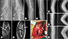
An 11-year-old boy was admitted to the hospital due to right elbow pain caused by a bicycle fall. Radiographs showed a right ulnar coronoid process fracture and radial subluxation (Fig. 1A–D). We applied a long arm splint and performed a MRI. The MRI finding showed fracture of the capitulum with common extensor tendon and lateral collateral ligament injuries (Fig. 1E, 1F). Surgical treatment was performed to correct the radial head subluxation. During the operation, we found a bony fragment with lateral collateral ligament injury and unstable radial head (Fig. 1G). Vicryl (Ethicon Inc., Somerville, NJ, USA) was used for sealing and a long-arm cast was applied for 4 weeks. Four months postoperatively, he had a partial limitation of elbow range of motion but no discomfort in active daily life (Fig. 1H–J).
2. Case 2
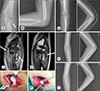
An 8-year-old boy was admitted to the emergency room due to left elbow pain caused by a 1 m fall. Radiographs showed a bony fragment around a radial head (Fig. 2A, 2B). We applied a long arm splint and followed up him at the outpatient clinic. According to the MRI, it showed a fracture with epiphyseal separation of the left radial head and neck along with a partial tear of ulnar collateral ligament and common flexor tendon located at the inferior aspect of medial epicondyle (Fig. 2C, 2D). Surgical treatment was performed for radial head reduction (Fig. 2E, 2F). As a result, the radial head had been completely reducted without causing damage to the soft tissue surrounding it to avoid postoperative necrosis (Fig. 2G). A long arm cast was applied for 4 weeks. One, five months postoperatively, he recovered fully the range of motion of the elbow (Fig. 2H, 2I).
DISCUSSION
Secondary ossification of the elbow begins at the capitulum and radial head at the age of 1–2 years and ends with fusion of the medial epicondyle and trochlea at the age of 17–18 years. The different parts of the elbow in pediatric patients are ossified and fused at different times, which explains the varying points of injury and fracture depending on the age of a patient. Elbow ossification occurs at the six elbow ossification centers in a reproducible order. The order of appearances of the elbow ossification centers is highly reliable and in most individuals, is consistent: capitellum, radial head, medial epicondyle, trochlea, olecranon, and lateral epicondyle. The age interval of appearance and fusion were, respectively: capitulum (0 to 1 year; 10 to 15 years), radial head (2 to 6 year; 12 to 16 years), medial epicondyle (2 to 8 years; 13 to 17 years), trochlea (5 to 11 years; 10 to 18 years), olecranon (6 to 11 years; 13 to 16 years), lateral epicondyle (8 to 13 years; 12 to 16 years)4. The elbow is primarily composed of cartilage prior to ossification and is harder to visualize injuries in this area on radiographs. For this reason, there is a high risk of mistaking an elbow injury for a simple fracture in children. Especially, medial epicondyle fractures associated with ligament injury or radial head fracture with dislocation, prior to ossification, may not be obvious on simple radiographs as in this case, even with associated dislocation or subluxation. Supportive treatment in such cases may lead to serious complications such as chronic pain, restricted range of motion, and traumatic arthritis.
The epicondyle in pediatric patients is too small to be visualized on a simple radiography, even if it is displaced, and may overlap with distal humeral metaphysis or be confused with the ossification center of the trochlea5.
Tanabe and Miyamoto6 stated that medial epicondylar fracture is difficult to detect by simple radiography. They presented a case of a 9-year-old girl patient with complete separation of her medial epicondyle and metaphysis which was successfully treated with surgery. Simple radiography and computed tomography failed to show any fracture or dislocation in this case, but MRI showed the lesion6. Also, Pudas et al.7 reported that MRI is a sensitive and accurate diagnostic tool for elbow fractures. They found that, in seven out of nine pediatric cases, MRI successfully revealed occult fractures, which were depicted as effusions on simple radiographs7. According to Beltran et al.8, because ossification centers present in pediatric patients, it is challenging to diagnose fractures with a simple radiograph. Hence, until ossification occurs by the age of 11–12 years, MRI is a better diagnostic tool than simple radiography to accurately delineate the fracture line8.
MRI is recommended if positive fat pad sign is the only radiological finding, if there is a lateral condyle fracture, or if there is elbow dislocation. In these cases, MRI helps in making the correct diagnosis, in making it easier to assess the extent of the damage, and in helping the clinician choose the optimal therapy7.
Based on my experience, simple fractures show gradual improvements in symptoms and swelling with time. On the other hand, fractures involving cartilage and those associated with joint dislocations or subluxations result in persistent discomfort, swelling, and anxiety in patients. In such patients with persistent symptoms, it is necessary to thoroughly examine the adjacent ligaments and consider the possibility of epiphyseal plate injury or dislocation of unossified regions by MRI.
 XML Download