

 PDF
PDF Citation
 Print
Print


INTRODUCTION
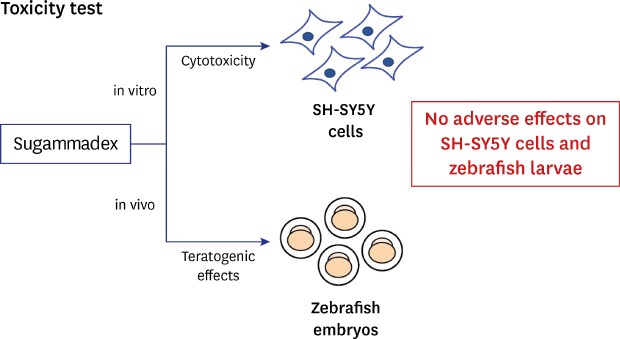
Sugammadex is a modified γ-cyclodextrin designed to encapsulate rocuronium bromide, which leads to rapid reversal of neuromuscular blockade.12 Traditionally, acetylcholinesterase inhibitors, such as neostigmine and pyridostigmine, have been widely used to reverse non-depolarizing neuromuscular blockers. However, these drugs have limitations, such as limited and unpredictable efficacy, undesirable autonomic response, and reverse mechanism of reversal. On the contrary, sugammadex is associated with fast recovery, predictable reversal of any degree of block in general anesthesia, increased patient safety, reduced incidence of residual block on recovery, and shortened hospital stay.13456 A γ-cyclodextrin ring in sugammadex reverses this blockage in a mechanism different from that of cholinesterases. It reverses muscle relaxants without causing side effects as caused by neostigmine. Therefore, sugammadex is widely used in patients under general anesthesia.7
Recently, Büyükfırat et al.8 analyzed the cytotoxicity, genotoxicity, and apoptotic effects of sugammadex and neostigmine on human embryonic renal cells (HEK-293) using 3-(4,5-dimethylthiazol-2-yl)-2,5-diphenyltetrazolium bromide (MTT) assay, comet assay, and flow cytometric annexin-V methods. The results showed that 50, 100, 250, and 500 µg/mL of neostigmine is more cytotoxic than the equivalent doses of sugammadex. Moreover, 500 and 1,000 µg/mL of neostigmine was found to be more genotoxic, and 500 µg/mL of neostigmine showed a significantly higher risk of causing necrosis and apoptosis than sugammadex. Neostigmine administered in vitro showed higher cytotoxic, genotoxic, and apoptotic effects on HEK-293 cells than those by sugammadex at the same dose.
Sugammadex, which has been used since 2008, has demonstrated a higher level of safety and effectiveness than that by pyridostigmine or neostigmine, when used to antagonize the muscle relaxation effects caused by steroid non-depolarizing neuromuscular blockers.9 Even in special cases, such as elderly people, children over 2 years of age, and patients with kidney, liver, or lung disease, it is proven to be effective.1011121314 In contrast, using sugammadex is challenging in fetuses and pediatric patients less than 3 years of age. Therefore, we tried to demonstrate the feasibility of using sugammadex during the fetal period by using zebrafish larvae.
METHODS
Cell culture and cell number determination
The human neuroblastoma cell line SH-SY5Y was cultured in a Gibco DMEM (Thermo Fisher Scientific, Inc., Waltham, MA, USA) medium, supplemented with 10% fetal bovine serum (FBS; Thermo Fisher Scientific, Inc.) and 1% penicillin-streptomycin (Hyclone, Logan, UT, USA), in an incubator at 37°C and 5% CO2 concentration. For cell number determination, the cell suspension (100 μL/well), was inoculated in a 96-well culture plate and maintained at 37°C in an incubator at 5% CO2 concentration. Further, 10 μL of the CCK-8 solution (Dojindo Laboratories, Kumamoto, Japan) was added to each well and incubated for 2 hours. Subsequently, the absorbance was measured at 450 nm using a microplate reader (SpectraMax M2; Molecular Devices, San Jose, CA, USA).
Cell cytotoxicity assay
The CCK-8 assay was used to determine the SH-SY5Y cell viability. The SH-SY5Y cells were seeded into a 96-well culture plate at a density of 5 × 10³ cells/well and cultured for 24 hours. The cells were treated with different concentrations of sugammadex (50, 100, and 200 μg/mL) for 6 hours at 37°C, in an incubator. Subsequently, 10 μL of CCK-8 solution was added to each well, and the cells were cultured at 37°C for an additional 2 hours. The absorbance of the CCK-8 solution was measured at 450 nm using a microplate reader.
Cell viability
The cell viabilities were measured by cell counting. The cells were removed from the culture media and placed in test tubes. The samples were diluted four times, stained with trypan blue, loaded in a hemocytometer, and observed under an optical microscope. Finally, cells with an intact membrane were counted.
Zebrafish maintenance and embryos
Zebrafish of the WT strain were provided by the Biomedical Research Center, Korea University Ansan Hospital. All zebrafish were bred in separate chambers, in a zebrafish auto-system (ZebTec Active Blue zebrafish housing, Tecniplast, Italy). The zebrafish were maintained in water at 27.0°C ± 1°C in a 14:10 light:dark cycle, and fed twice a day with brine shrimp.
Adult male and female zebrafish were housed separately in each cage. Two male and female (at a ratio of 1:1) adult zebrafish were paired in a breeding group, in spawning tanks overnight. Spawning and fertilization took place within 20–30 minutes after the onset of light. The embryos were collected after spawning and transferred into blue water (methylene blue in E3 medium), in a Petri dish. The unfertilized embryos were identified under a light microscope (Zeiss Stemi 508, Oberkochen, Germany) and discarded. The fertilized embryos (normal development of a blastula) were placed in a Petri dish filled with E3 medium (5 mM NaCl, 0.17 mM KCl, 0.33 mM CaCl2, and 0.33 mM MgSO4; pH, 6.9–7.2).
Embryonic toxicity experiments
The embryos were cultured in an incubator (Daihan Scientific, Wonju, Korea) at 28.0°C ± 1°C. To improve the permeability of the drug, a small hole was drilled and processed using forceps in the amnion of all embryos, except those in the negative control group. Sugammadex (MSD, Seoul, Korea) solutions of varying concentrations (0 [control], 25, 50, 75, 100, and 200 μg/mL) were prepared using E3 medium. About 1–2 hours post-fertilization (hpf), the embryos were randomly transferred into the sugammadex solution in 12-well plates until 120 hpf. The exposure solution was refreshed every 24 hours to maintain the appropriate concentration of sugammadex and quality of the E3 medium. The number of dead embryos and the state of development were observed every day. The dead embryos were removed when changing the exposure solution. Morphological development of the embryos was observed under a microscope. The larvae hatched 72 hpf, and body length was measured 96 hpf. The lethality was measured 120 hpf. The hatching and malformation of embryos were checked daily before changing the exposure solution. The body length and heart shapes were measured using a microscope (Nikon SMZ18, Tokyo, Japan). All experiments were performed in triplicates.
Statistical analysis
The absorbance and body length data were analyzed using GraphPad Prism v.5.0 (San Diego, CA, USA) with one-way analysis of variance, followed by Tukey post-test to compare all pairs of columns. The cell viability data were analyzed using GraphPad Prism v.5.0 with Kruskal–Wallis test followed by Dunn's post-test. All data were presented as means and standard error of the mean, and P value < 0.05 indicated statistical significance.
RESULTS
Cell cytotoxicity and viability
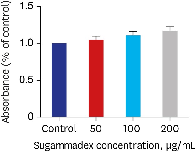
The role of sugammadex on neuronal cell cytotoxicity was studied using CCK-8 assays. No difference in absorbance was observed upon incubation with sugammadex at different concentrations (0, 50, 100, and 200 μg/mL) for 24 hours, as compared to the controls (Fig. 1).
Fig. 1
Change in the absorbance of CCK-8 at different sugammadex concentrations.
Neuronal cells were treated with different concentrations of sugammadex, and accumulated absorbance of CCK-8 was measured. Data are presented as mean ± standard error of the mean. All groups n = 12.
*P < 0.05.
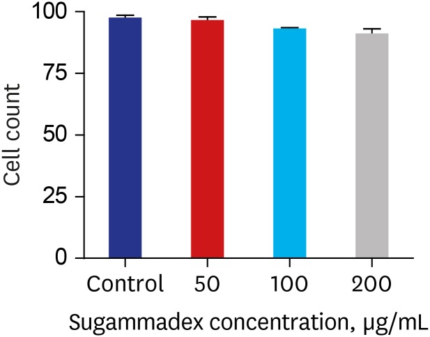
The cell viability of neuronal cells was also measured to confirm the toxicity of sugammadex at different concentrations (0, 50, 100, and 200 μg/mL). No significant difference in live-cell counts was observed at different concentrations of sugammadex (Fig. 2).
Zebrafish larvae: body length and cardiac morphology
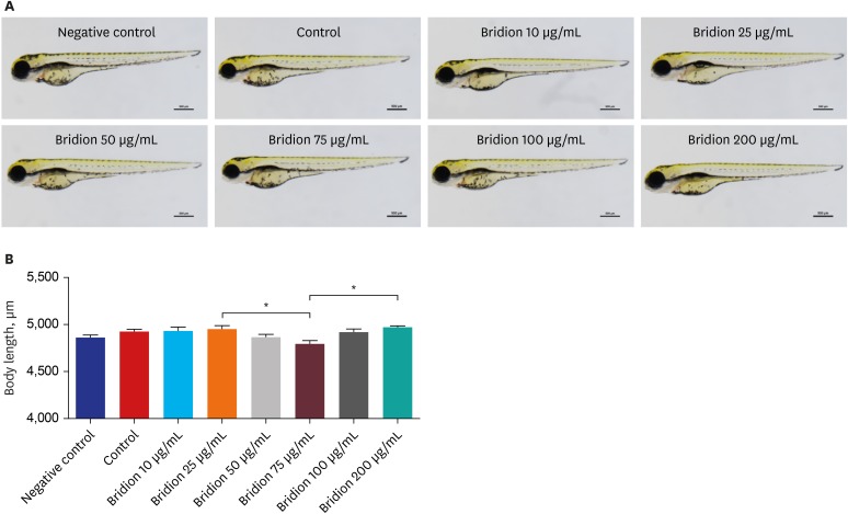
To determine the toxicity, zebrafish embryos were exposed to 0, 25, 50, 75, 100, and 200 μg/mL sugammadex for 96 hours (0–96 hours postfertilization, hpf), and the body length and cardiac morphology were measured at the endpoint (96 hpf). No remarkable change was observed in the body length of embryos exposed to different concentrations of sugammadex (Fig. 3A). Additionally, no morphological abnormalities, such as bent spine, tail, yolk, head, or eyes, were observed. However, larvae exposed to 75 μg/mL of sugammadex showed a smaller body length than those exposed to 25 μg/mL and 200 μg/mL of sugammadex (P < 0.05) (Fig. 3B).
Fig. 3
Morphology and body length of zebrafish embryos exposed to sugammadex.
Zebrafish embryos were treated with different concentrations of sugammadex, and accumulated body length were measured 96 hpf. Data are presented as mean ± standard error of the mean. Negative control (n = 30), control (n = 30), sugammadex 10 μg/mL (n = 30), sugammadex 25 μg/mL (n = 30), sugammadex 50 μg/mL (n = 30), sugammadex 75 μg/mL (n = 29), sugammadex 100 μg/mL (n = 30), and sugammadex 200 μg/mL (n = 30).
hpg, hours post-fertilization.
*P < 0.05.
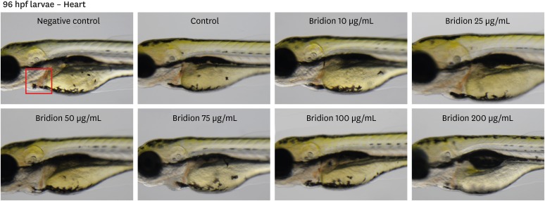
No abnormalities such as pericardial edema, heart hemorrhage, or morphological malformation were observed in sugammadex-treated larvae. The ventricle and atrium overlapped each other in the lateral view, showing a normal looping shape in the control and sugammadex-treated larvae (Fig. 4).
DISCUSSION
Sugammadex is a new reversal agent that acts more rapidly, effectively, and safely, with a faster recovery time than that by acetylcholine esterase inhibitors. Many studies are available on the safety of using sugammadex in children, adolescents, and adult patients.14 Recently, Plaud et al.10 reported that sugammadex rapidly, effectively, and safely reverses rocuronium-induced neuromuscular blockade in children, adolescents, adults, and a small number of infants. However, several adverse effects such as bradycardia and hypoglycemia were observed in infants treated with sugammadex. Bradycardia is considered to be the worst adverse effect on a fetus as fetal circulation is maintained by the heart rate. Bhavani15 reported bradycardic events caused by sugammadex; however, the teratogenic effect of sugammadex has been rarely studied.
Sugammadex is administered to patients in a wide range of doses from 0.5 to 16.0 mg/kg IV because it reverses rocuronium-induced neuromuscular blockade in a dose-dependent manner.1617 Hsu et al.18 reported the half maximal inhibitory concentration (IC50) of sugammadex as 30 µM in electrophysiologic cell experiments. However, the effective dose of sugammadex in zebrafish has not been estimated. In this study using zebrafish larvae, we selected sugammadex doses similar to those used in previous cell experiments. The range of sugammadex concentrations was 0–200 µg/mL. Considering the molecular weight of sugammadex, 70 µg/mL of sugammadex is similar to 30 µM sugammadex.
We noted some adverse effects such as mortality, abnormal body length, and heart morphology, as well as developmental malformations, after zebrafish embryos were exposed to sugammadex. None of the previous studies conducted on zebrafish have tested sugammadex toxicity at varying concentrations. Tulgar et al.19 reported that rats treated with 16 mg/kg sugammadex bolus (total dose of 12.4 mL/kg) showed increased survival of cardiotoxicity from toxic dose of verapamil. The present study indicated that sugammadex did not induce cardiac toxicity in zebrafish embryos 96 hpf. Additionally, mortality and developmental malformations were not seen in zebrafish embryos at any concentration of sugammadex. Another study showed potential neuronal toxicity in cortical neurons at clinically relevant concentrations (75 μg/mL) of sugammadex.20 In this study, lower body length was observed in 75 μg/mL sugammadex-treated zebrafish larvae as compared to the 25 and 200 μg/mL sugammadex-treated zebrafish larvae, 96 hpf. At higher concentrations (100 and 200 μg/mL), however, the body length was not lower than that in the control. We randomly treated embryos with sugammadex but, nonetheless, it was thought that individual differences occurred. It seems that the statistically significant all drug-treated group was not equivalent to the control. Considering that this data showed no meaning, there seems to have been a technical problem.
Zebrafish are a modern experimental animal model used worldwide. This animal model is widely used in toxicology and biomedical research, from embryonic stages to adulthood.21 The zebrafish model is widely used owing to its small size, rapid development, high reproducibility, transparency of embryos, and genetic chemical screens, as well as suitability for viewing reactions to chemical screens.222324 Zebrafish eggs hatch faster, and the larvae can start feeding 120 hpf, the point at which researchers can begin experimenting, which translates to rapid experimentation.25 It is also useful because the organs and tissues can be easily visualized and tested in vivo.2627 The zebrafish animal model is commonly used to evaluate drug toxicity, and its neurological developmental structure is similar to that of humans, making it useful for teratogenic evaluation. Nevertheless, it is difficult to extrapolate the effects on zebra fish to humans, and further research on larger animal models is necessary.
In conclusion, no toxic response was observed in cell-based and animal-based experiments even at higher doses relative to the clinical dose. This study provides valuable data for the clinical use of sugammadex. The absence of cardiac anomalies, which is considered to be developmentally important in fetuses, is a good indicator for the feasibility of using sugammadex during pregnancy. Further clinical studies are required to evaluate the safety of using sugammadex during pregnancy.
 XML Download