

 PDF
PDF ePub
ePub Citation
 Print
Print


Kwang Rak Park1 , Jaeho Cho2, Yu Jin Choi1, Digud Kim1, Hyung Wook Kwon1, Hye Jin Jang3, Hansol Kang1, Jeonghyun Park1
, Jaeho Cho2, Yu Jin Choi1, Digud Kim1, Hyung Wook Kwon1, Hye Jin Jang3, Hansol Kang1, Jeonghyun Park1
Abstract
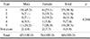
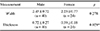
The plantaris muscle is a small muscle with a short belly and long thin tendon that forms part of the posterior superficial compartment of the calf. The purpose of this study was to classify the insertion type of plantaris tendon of Korean population, to measure anthropological characteristics by measuring the width and thickness, and to obtain clinically applicable anatomical basis data. The dissection was performed on 68 lower limbs (34 right, 34 left) fixed in formalin mixture. The types of insertion of plantaris tendon was classified according to the area and location of insertion into calcaneal tuberosity and calcaneal bone. In this study, all five types were identified. 25 limbs (36.8%) were classified into type 1, 8 limbs (11.8%) into types 2 and 3 respectively, 5 limbs (7.4%) into type 4, 18 lower extremities (26.5%) into type 5. The plantaris tendon was found to be absent in 4 lower limbs (5.9%). No differences in body side or gender were found in the insertion type of plantaris tendon. The thickness of the plantaris tendon was 0.72±0.27 mm for males and 0.59±0.18 mm for females, and males were thicker than females(p=0.029). However, there was no difference between the two genders in the width of the plantaris tendon. In conclusion, this study not only examined the morphological characteristics of plantaris tendon in Korean population, but also presented basic anatomical data that doctors can apply to clinical practice.
Figures and Tables
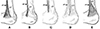 | Fig. 1Schematic representation of the major types of the plantaris tendon insertion. Medial view of the right leg with the plantaris tendon. Olewnik et al. (2017) classified into 5 types of PT insertion by location and area inserted in calcaneal tuberosity. (A) Type 1, fan-shaped insertion to the calcaneal tuberosity on the medial side of the calcaneal tendon; (B) Type 2, insertion to the calcaneal tuberosity, along with the calcaneal tendon (Fan shape is not taken); (C) Type 3, insertion at the calcaneal bone, anterior to the calcaneal tendon; (D) Type 4, insertion not being located on the calcaneal tuberosity but rather in the deep crural fascia; (E) Type 5, very wide insertion encircling the posterior and medial surfaces of the calcaneal tendon. PT: plantaris tendon, CT: calcaneal tendon. Black arrow indicates plantaris tendon.
|
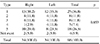
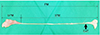 | Fig. 2Location of extension points(ExP) for measuring width and thickness in plantaris muscles. The plantaris muscle was extracted from the right leg. extension point (black arrow) is the point at which the distal tendon begins to expand before its insertion. PM: plantaris muscle, bPM: belly of plantaris muscle, tPM: tendon of plantaris muscle, ExP: extension point.
|
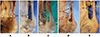 | Fig. 3Types of insertion of the plantaris tendon. Medial view of the right leg with the plantaris tendon. (A) Type 1 (36.8%); (B) Type 2 (11.8%); (C) Type 3 (11.8%); (D) Type 4 (7.4%); (E) Type 5 (26.5%). PT: plantaris tendon, CT: calcaneal tendon, ExP: extension point. Black arrow indicates plantaris tendon.
|
References
1. Aragão JA, Reis FP, Guerra DR, Cabral RH. The occurrence of the plantaris muscle and its muscle-tendon relationship in adult human cadavers. Int J Morphol. 2010; 28:255–258.

2. Olewnik L, Wysiadecki G, Polguj M, Topol M. Anatomic study suggests that the morphology of the plantaris tendon may be related to achilles tendonitis. Surg Radiol Anat. 2017; 39:69–75.
3. Olewnik L, Wysiadecki G, Podgórski M, Polguj M, Topol M. The plantaris muscle tendon and its relationship with the achilles tendinopathy. Biomed Res Int. 2018; DOI: 10.1155/2018/9623579.
4. Allard JC, Bancroft J, Porter G. Imaging of plantaris muscle rupture. Clin Imaging. 1992; 16:55–58.
5. Spang C, Alfredson H, Docking SI, Masci L, Andersson G. The plantaris tendon: a narrative review focusing on anatomical features and clinical importance. Bone Joint J. 2016; 98-B:1312–1319.
6. White WL. The unique, accessible and useful plantaris tendon. Plast Reconstr Surg Transplant Bull. 1960; 25:133–141.
7. Richard LD, Wayne V, Adam WM. Gray's anatomy for students. 3th ed. Philadelphia: Elsevier/Churchill Livingstone;2013. p. 621–623.
8. Harvey FJ, Chu G, Harvey PM. Surgical availability of the plantaris tendon. J Hand Surg Am. 1983; 8:243–247.
9. Van Sterkenburg MN, Van Dijk CN. Mid-portion Achilles tendinopathy: why painful? An evidence-based philosophy. Knee Surg Sports Traumatol Arthrosc. 2011; 19:1367–1375.
10. Lintz F, Higgs A, Millett M, Barton T, Raghuvanshi M, Adams MA, et al. The role of plantaris longus in Achilles tendinopathy: a biomechanical study. Foot Ankle Surg. 2011; 17:252–255.
11. Van Sterkenburg MN, Kerkhoffs GM, Van Dijk CN. Good outcome after stripping the plantaris tendon in patients with chronic mid-portion Achilles tendinopathy. Knee Surg Sports Traumatol Arthrosc. 2011; 19:1362–1366.
12. Jackson JB III, Philippi MT, Kolz CW, Suter T, Henninger HB. Characterization of plantaris tendon constructs for ankle ligament reconstruction. Foot Ankle Int. 2014; 35:922–928.
13. Alfredson H. Midportion Achilles tendinosis and the plantaris tendon. Br J Sports Med. 2011; 45:1023–1025.
14. Yammine K, Saghie S, Assi C. A meta-analysis of the surgical availability and morphology of the plantaris tendon. J Hand Surg Asian Pac Vol. 2019; 24:208–218.
15. Landis JR, Koch GG. The measurement of observer agreement for categorical data. Biometrics. 1977; 33:159–174.
16. Dos Santos MA, Bertelli JA, Kechele PR, Duarte H. Anatomical study of the plantaris tendon: reliability as a tendo-osseous graft. Surg Radiol Anat. 2009; 31:59–61.
17. Cummins EJ, Anson BJ. The structure of the calcaneal tendon (of achilles) in relation to orthopedic surgery with additional observation on the plantaris muscle. Surg Gynecol Obstet. 1946; 83:107–116.
18. Li Q, Xu J, Zhang D. Vascularized plantaris tendon graft: anatomic study of the donor. J Reconstr Microsurg. 2000; 16:287–290.
19. Nayak SR, Krishnamurthy A, Ramanathan L, Ranade AV, Prabhu LV, Jiji PJ, et al. Anatomy of plantaris muscle: a study in adult Indians. Clin Ter. 2010; 161:249–252.
20. Alagoz MS, Uysal AC, Tuccar E, Tekdemir I. Morphologic assessment of the tendon graft donor sites: palmaris longus, plantaris, tensor fascia lata. J Craniofac Surg. 2008; 19:246–250.
21. Ahmed N, Sarwari KN. Morphological variations and surgical importance of the plantaris muscle in humans. Indian J Fundam Appl Life Sci. 2013; 3:342–346.
22. Wehbe MA. Tendon graft donor sites. J Hand Surg Am. 1992; 17:1130–1132.
 XML Download