

 PDF
PDF ePub
ePub Citation
 Print
Print


Introduction
Afferent sensory neuron fibers, the majority of which contain calcitonin gene-related peptide (CGRP), display a broad innervation throughout central and peripheral nervous systems[1]. In kidneys, they are located in adventitia of renal vessels and renal pelvis[2]. Calcitonin gene-related peptide (CGRP) is a 37-amino acid neuropeptide primarily synthesized in and secreted from sensory neuron fibers among several mammalian species [1]. The sequence of CGRP is highly conserved among vertebrates from fish to mammals [3]. Generally, CGRP contributes to pain transmission and inflammation in the nervous system, resulting in migraine attacks [1]. However, CGRP plays other roles in extraneural sites. The concentration of CGRP in the blood of hypertensive patients is decreased because its concentration in plasma is implicated in nitric oxide-mediated vasodilation [4]. Our previous study has shown that CGRP proteins are expressed at apical or basolateral membranes of epithelial cells in dilated tubules as well as in fibers during unilateral ureteral obstruction. They can induce apoptotic cell death in tubular cells derived from mouse kidney cortex [5]. However, how CGRP contributes to the induction of cell death in kidney tubular cells remains unclear.
Reactive oxygen species (ROS) and associated oxidative stress play a pivotal role in apoptotic cell death during various pathogenesis [6]. Oxidative damage to kidneys is causally associated with acute kidney injury such as ischemia-reperfusion injury (IRI) and cisplatin nephrotoxicity [78]. Furthermore, kidney proximal tubules are more susceptible to cell death induced by IRI and cisplatin than kidney distal tubules [9]. During kidney fibrosis induced by unilateral ureteral obstruction and IRI, exogenous CGRP is detected selectively in proximal tubule epithelial cells and is linked to the induction of apoptotic cell death [510]. Therefore, it has been speculated that extraneural CGRP-induced cell death in kidney proximal tubules might be implicated in increased oxidative stress. To investigate this hypothesis, the aim of the present study was to determine whether exogenous CGRP could increase ROS production in kidney proximal tubule epithelial cells derived from human, pig, and mouse and whether pharmacological inhibitions of CGRP receptor and ROS could attenuate extraneural CGRP-induced death of kidney proximal tubule epithelial cells.
Materials and Methods
1. Cell culture and treatment
Kidney proximal tubule epithelial cell lines derived from human (HK-2), pig (LLC-PK1), and mouse (TCMK-1) were purchased from the American Type Culture Collection (ATCC; Rockville, MD, USA). HK-2 cells were maintained in Roswell Park Memorial Institute (RPMI) 1640 supplemented with 10% fetal bovine serum containing 100 units/mL penicillin and 0.1 mg/mL streptomycin at 37℃ with 5% CO2, as described previously [11]. LLC-PK1 and TCMK-1 cells were maintained in Dulbecco's modified Eagle's medium (DMEM)/high-glucose supplemented with 10% fetal bovine serum containing 100 units/mL penicillin and 0.1 mg/mL streptomycin at 37℃ with 5% CO2, as described previously [1213]. These cells were grown until 70% confluence on culture plates. After changing into serum-free medium, cells were immediately treated with 0.1, 1, or 10 nM of CGRP for 0, 6, 24, or 48 hours. Some cells were treated with either CGRP8-37 (a CGRP antagonist; 1, 3, or 10 nM), MnTMPyP(a ROS scavenger; 10, 30, or 100 µM), or phosphate buffered saline (PBS, vehicle) at 1 hour before treatment with CGRP. CGRP8-37 (human) was used to treat HK-2 and LLC-PK1 cells. CGRP8-37 (rat) was used to treat TCMK-1 cells. All chemicals were obtained from R&D Systems(Minneapolis, MN, USA).
2. Cell viability
A yellow water soluble tetrazolium dye thiazolyl blue tetrazolium bromide (MTT; Biosesang, Seongnam, Korea) was used to measure viabilities of HK-2, LLC-PK1, and TCMK-1 cells on a 24-well plate, as previously described [5]. Briefly, after removing the culture medium, cells were incubated at 37℃ with 5 mg/mL MTT in PBS for 30 minutes. After removing the MTT solution, 300 µL of dimethyl sulfoxide (DMSO, Biosesang) was added to each well and incubated at 37℃ for 5 minutes. To quantify purple-colored formazan product, the absorbance of 100 µL solution from each well after incubating with DMSO was measured on a 96-well plate (SPL Life Science, Daejeon, Korea) at 595 nm with reference wavelength of 620 nm using a VERSA max plate leader (Molecular Devices, Sunnyvale, CA, USA).
3. ROS production
An oxidative sensitive dye 2′,7′-dichlorodihydrofluorescein diacetate (DCFDA) was used to measure ROS production as described previously [14]. Briefly, 105 of HK-2 cells per well were seeded onto a 24-well plate and treated with chemicals as indicated. After that, cells were incubated with 20 µM DCFDA for 45 minutes at 37℃. After washing twice with PBS, cells were incubated with 1% Triton X-100. The intensity of 2′,7′-dichlorofluorescein (DCF) from 200 µL of cell lysate on a Nunc 96-well black plate (Thermo Fisher Scientific) was quantified with a SpectraMax i3 plate reader (Molecular Devices) using 485 nm for excitation and 535 nm for emission.
4. Statistical analysis
Analysis of variance (ANOVA) was used to compare data between groups using SigmaPlot (Systat Software, San Jose, CA, USA). Significant differences between two groups were estimated using two-tailed unpaired Student's t-tests. Pearson's correlation analysis was performed for Fig. 4. P values<0.05 were considered statistically significant.
Results
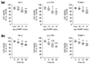
1. Extraneural CGRP decreases viability of kidney proximal tubule epithelial cells.
To determine whether extraneural CGRP could alter viability of kidney proximal tubule epithelial cells, cell viability was measured using MTT assay at 24 hours after treatment with various doses of CGRP in various species-derived kidney proximal tubule epithelial cells(HK-2, LLC-PK1 and TCMK-1 cells). After treatment with CGRP, the percentage of cell viability was decreased with increasing dose (Fig. 1a). Especially, treatment with intermediate and high doses of CGRP significantly decreased viabilities of HK-2, LLC-PK1 and TCMK-1 cells(Fig. 1a). Next, we determined the time course of the decrease in cell viability caused by exogenous CGRP. Viabilities of HK-2, LLC-PK1 and TCMK-1 cells were monitored at 0, 6, 24 and 48 hours after treatment with an intermediate dose of CGRP by using MTT assay. As shown in Fig. 1b, significant decreases in viabilities of HK-2, LLC-PK1, and TCMK-1 occurred at 6 hours and persisted to 48 hours after treatment with CGRP. These data suggest that extraneural CGRP can increase cell death of kidney proximal tubule epithelial cells in dose- and time-dependent manners.
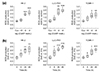
2. Extraneural CGRP increases ROS production in kidney proximal tubule epithelial cells.
To determine whether extraneural CGRP could induce ROS production in kidney proximal tubule epithelial cells, ROS levels in HK-2, LLC-PK1 and TCMK-1 cells were measured using oxidative sensitive dye DCFDA at 24 hours after treatment with various doses of CGRP. Consistent with data of cell viability, ROS production was increased at 24 hours after treatment with CGRP (Fig. 2a). Especially, treatment with intermediate and high doses of CGRP markedly increased ROS levels in all three kidney proximal tubule epithelial cell lines(Fig. 2a). Additionally, ROS levels in HK-2, LLC-PK1, and TCMK-1 cells at 0, 6, 24, and 48 hours after treatment with intermediate dose of CGRP were determined using DCFDA dye. These cells showed significant increases in ROS production at 6 hours after treatment with CGRP. The increases persisted to 48 hours after treatment with CGRP. These data indicate that extraneural CGRP can increase ROS production in kidney proximal tubule epithelial cells in dose- and time-dependent manners.
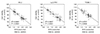
3. Extraneural CGRP induces ROS production and cell death through CGRP receptor in kidney proximal tubule epithelial cells.
CGRP can stimulate various intracellular signaling pathways via binding to its receptor consisting of two subunits, receptor activity modifying protein 1 (RAMP1) and calcitonin receptor-like receptor(CRLR)[1]. Expression levels of these subunits are not altered by exogenous CGRP in human kidney proximal tubule epithelial cells [15]. To investigate a role of CGRP receptor in kidney proximal tubule epithelial cells, a cleaved form of CGRP (CGRP8-37) as a potent antagonist was used to treat HK-2, LLC-PK1 and TCMK-1 cells. Cells were treated with various doses of CGRP8-37 at 1 hour before treatment with intermediate dose of CGRP for 24 hours. The addition of CGRP8-37 reduced the increase in the level of ROS production in CGRP-treated cells in a dose-dependent manner (Fig. 3a). Especially, the addition of low dose of CGRP8-37 significantly attenuated CGRP-induced ROS production only in HK-2 cells, but not in LLC-PK1 or TCMK-1 cells (Fig. 3a), indicating more susceptibility of human kidney proximal tubule epithelial cells to CGRP receptor antagonism. The addition of intermediate and high doses of CGRP8-37 significantly attenuated the increase in the production of ROS in all three kidney proximal tubule epithelial cell lines(Fig. 3a). The decrease of cell viability in CGRP-treated kidney proximal tubule epithelial cells was also ameliorated by the addition of CGRP8-37 (Fig. 3b). However, the addition of low dose of CGRP8-37 did not significantly alter viabilities of CGRP-treated HK-2, LLC-PK1, or TCMK-1 cells(Fig. 3b). The addition of intermediate dose of CGRP8-37 did not significantly alter viabilities of CGRP-treated LLC-PK1 or TCMK-1 cells either (Fig. 3b). These data suggest that extraneural CGRP can induce ROS production and cell death through its receptor signaling pathway in kidney proximal tubule epithelial cells.
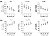
4. Extraneural CGRP-induced ROS production decreases viability of kidney proximal tubule epithelial cells.
Excessive ROS production can lead to cell death [6]. As shown in Fig. 4, the level of ROS production was significantly correlated with viability of all three kidney proximal tubule epithelial cell lines after exposure to exogenous CGRP. To test whether exogenous CGRP-induced ROS production could alter viability of kidney proximal tubule epithelial cells, a ROS scavenger MnTMPyP was added to HK-2, LLC-PK1 and TCMK-1 cells during exposure to CGRP. In all three kidney proximal tubule epithelial cell lines, the addition of MnTMPyP reduced the increase of ROS production induced by treatment with CGRP in a dose-dependent manner(Fig. 5a). Furthermore, the addition of MnTMPyP dose-dependently attenuated exogenous CGRP-induced cell death, as demonstrated by the percentage of cell viability (Fig. 5b). These data suggest that excessive ROS production induced by extraneural CGRP can cause cell death of kidney proximal tubule epithelial cells.
Discussion
Results of the present study demonstrate the following novel findings: (1) extraneural CGRP can induce ROS production through CGRP receptor in kidney proximal tubule epithelial cells; and (2) excessive ROS production during exposure to extraneural CGRP is implicated in cell death.
Primary sensory nerve-derived CGRP can induce cell death in kidney tubular cells[5]. Consistent with the previous study, the present study also demonstrated that exogenous CGRP could decrease cell viability of kidney proximal tubule epithelial cells. Our data also revealed that cell viability following treatment with CGRP was significantly higher when kidney proximal tubule epithelial cells were pretreated with a ROS scavenger. This is in contrast to previous reports that exogenous CGRP can protect vascular smooth muscle cells against apoptotic cell death induced by exposure to free radical agents[16]. In cardiomyocytes, exogenous CGRP has an antiapoptotic effect on oxidative stress-induced injury through the CGRP receptor [17]. It is possible that vascular smooth muscle cells and cardiomyocytes are more susceptible to CGRP-related immunoreactivity and hemodynamics in the cardiovascular system compared to other epithelial cells including kidney tubular cells[1819]. In addition, the altered effects in cell viability by exogenous CGRP might be dependent on cell or tissue context.
While kidney denervation dramatically prevents the development of kidney fibrosis induced by ischemia-reperfusion injury and ureteral obstruction, a local injection of CGRP into the denervated kidney mimics kidney fibrosis, suggesting CGRP is an inducer of kidney fibrosis [510]. Our previous study has shown that the upregulation of profibrogenic proteins induced by exogenous CGRP in HK-2 cells is mediated by protein kinase C (PKC) and c-Jun N-terminal protein kinase (JNK) [15]. In diabetic vascular tissues, PKC activation is dependent on NADPH oxidase activation which is responsible for increased oxidative stress [20]. The JNK signaling cascade may induce mitochondrial ROS production and is critical for initiating apoptosis in disease conditions [21]. As suggested by previous studies [2021], whether CGRP-dependent PKC and JNK activations contributes to ROS production in the fibrotic kidney remains to be explored.
ROS plays an important role in cell death induction under both physiological and pathological conditions. Intriguingly, distinct from the effects of CGRP in vascular smooth muscle cells and cardiomyocytes in the cardiovascular system [1617], kidney proximal tubule epithelial cells produced excessive ROS during exposure to exogenous CGRP in the present study, suggesting CGRP-induced oxidative stress could cause the development of cell death in kidney proximal tubule epithelial cells. Consistent with this result, neurotransmitters including norepinephrine and serotonin can also induce excessive ROS production in rat hearts and rat ventricular myocytes, respectively, leading to cell death [2223]. Kidney tubular cell death, possibly induced by oxidative stress, is a crucial early step in the development of acute kidney injury and during its long-term sequelae resulting in fibrogenesis and chronic kidney disease. Thus, inhibiting the action of CGRP may represent a novel effective therapeutic strategy to limit oxidative stress and subsequent cell death at the onset of kidney fibrosis.
In summary, the present study demonstrates that kidney sensory nerve-derived CGRP can induce cell death through ROS production in kidney proximal tubule epithelial cells. Induced expression and activation of CGRP signaling pathway maybe trigger tubular injury in acute kidney injury and subsequent tubulointerstitial fibrogenesis. Although CGRP induced oxidative injury can be a key mechanism, further investigation to delve into the possibility of additional signaling mechanisms to cause cell death in kidney proximal tubule epithelial cells is warranted.

 XML Download