Abstract
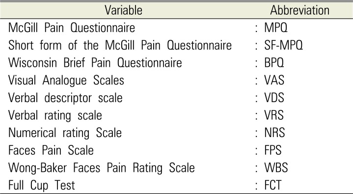
Regardless of whether it is acute or chronic, the assessment of pain should be simple and practical. Since the intensity of pain is thought to be one of the primary factors that determine its effect on a human's overall function and sense, there are many scales to assess pain. The aim of the current article was to review pain intensity scales that are commonly used in dental and oral and maxillofacial surgery (OMFS). Previous studies demonstrated that multidimensional scales, such as the McGill Pain Questionnaire, Short form of the McGill Pain Questionnaire, and Wisconsin Brief Pain Questionnaire were suitable for assessing chronic pain, while unidimensional scales, like the Visual Analogue Scales (VAS), Verbal descriptor scale, Verbal rating scale, Numerical rating Scale, Faces Pain Scale, Wong-Baker Faces Pain Rating Scale (WBS), and Full Cup Test, were used to evaluate acute pain. The WBS is widely used to assess pain in children and elderly because other scales are often difficult to understand, which could consequently lead to an overestimation of the pain intensity. In dental or OMFS research, the use of the VAS is more common because it is more reliable, valid, sensitive, and appropriate. However, some researchers use NRS to evaluate OMFS pain in adults because this scale is easier to use than VAS and yields relatively similar pain scores. This review only assessed pain scales used for post-operative OMFS or dental pain.
Pain is one of the most common complaints that patients present with at hospital [1]. Pain has several definitions that depend on the aim or scope. Mersky et al. [2] defined pain as “an unpleasant sensory and emotional experience with actual or potential tissue damage or described in terms of such damage.”. McAloon et al. [3] described pain as a subjective feeling or experience interpreted by oneself, while Gloss et al. [4] reported that pain is a complex and private experience. Briggs and Closs [5] highlighted that pain was also influenced by numerous intrinsic and extrinsic factors, and that multiple aspects of pain was assessed in different ways. Although no one interprets pain in exactly the same way, the intensity of pain depending on the patients' perception, should be assessed for effective pain management [3]. Pain perception depends on the pain threshold of each person, which might be used as a baseline to compare pain intensity.
The assessment of the intensity of pain and locating it is a routine procedure in clinical practice. Various tools have been developed for different types and subtypes of chronic pain conditions so that the effect of chronic pain on quality of life and the patient's function can be gaged [6]. The accuracy of pain assessment is very important to evaluate the appropriate treatment.
Pain intensity is the initial factor that point to its sensation and function. Therefore, pain measurement tools are used to help assess pain intensity, and monitor the effectiveness of and response to treatment decisions [7].
Multidimensional pain scales assess the following:
The aim of the current article was to provide an overview of pain measurements that are commonly used in OMFS research.
There are multidimensional and unidimensional scales of pain. The following are multidimensional scales:
Unidimensional scales to assess pain are as follows:
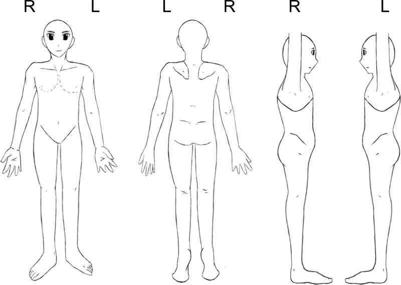
The intensity, nature, and location of pain can demonstrate its impact on the patient's activity or mood, and is useful to assess complex or persistent acute or chronic pain.
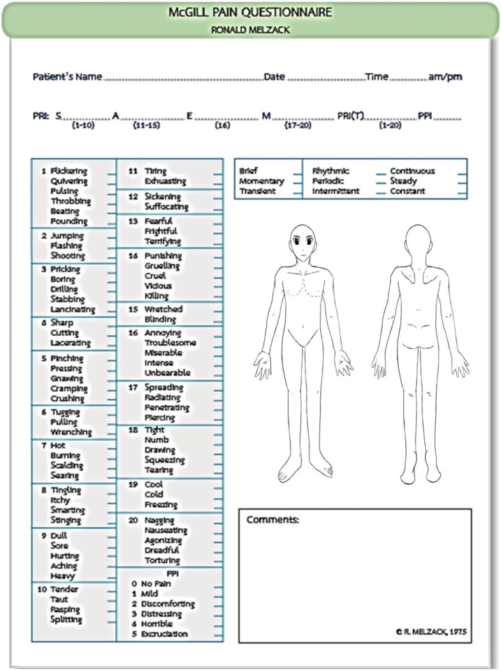
The MPQ, which was formulated in 1971 by Melzack and Torgerson., was first published in its complete form in 1975. As seen in Fig. 1 it measures several dimensions of the experience and intensity of pain experience and pain intensity [9]. The advantages of the MPQ are its reliability, validity, and its access to the multidimensionality of pain [9].
The major disadvantages of the MPQ include the following: very complex, requires tremendous patient compliance and endurance to fill the form, and takes up to 30 min, which can be tedious and time-consuming [3].
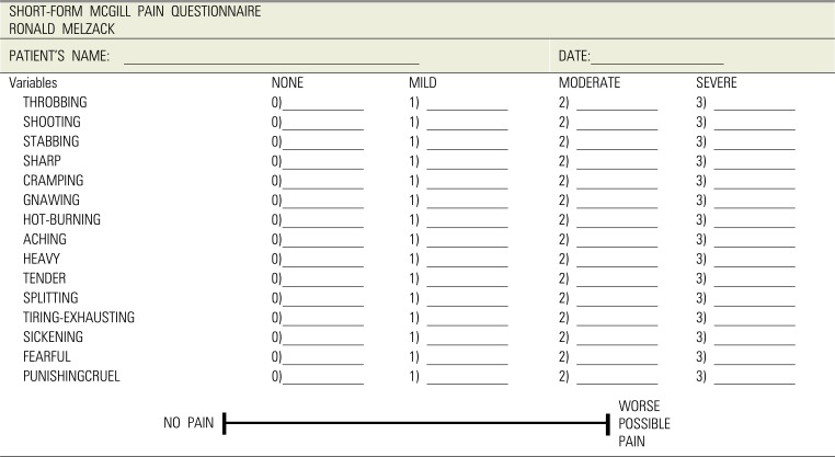
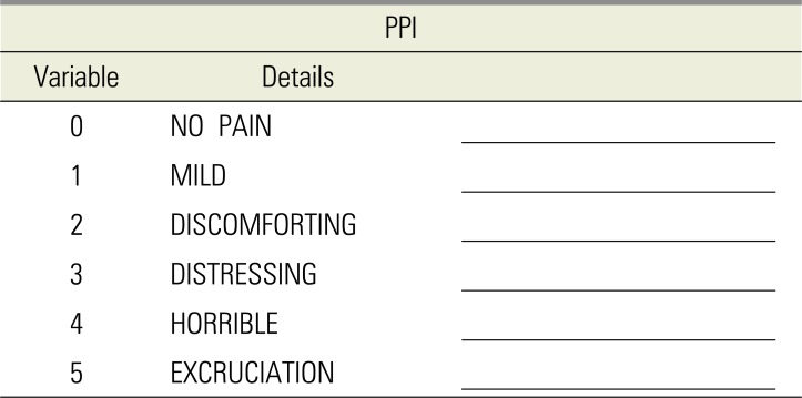
SF-MPQ is the alternative scale of the Present Pain Intensity (PPI) tool (Table 3). This scale only assesses the intensity of pain, and not its other qualities [10]. It was developed in 1980, when Melzack realized that the MPQ was too long [10]. This pain assessment scale turned out to be relatively easy, since it consisted of choosing the most commonly used sensory and affective descriptors in all clinical studies. Given its simplicity, it can be applied to assess mild, moderate, and severe cases. The SF-MPQ incorporates both the PPI and VAS, and thus, has the following five sub-scores: sensory, affective, total scores from the MPQ descriptors, overall intensity scores from the PPI, and the VAS [8]. The scales were ranked as follows:
The PPI of the standard LF-MPQ and the VAS are also included to provide overall intensity scores.
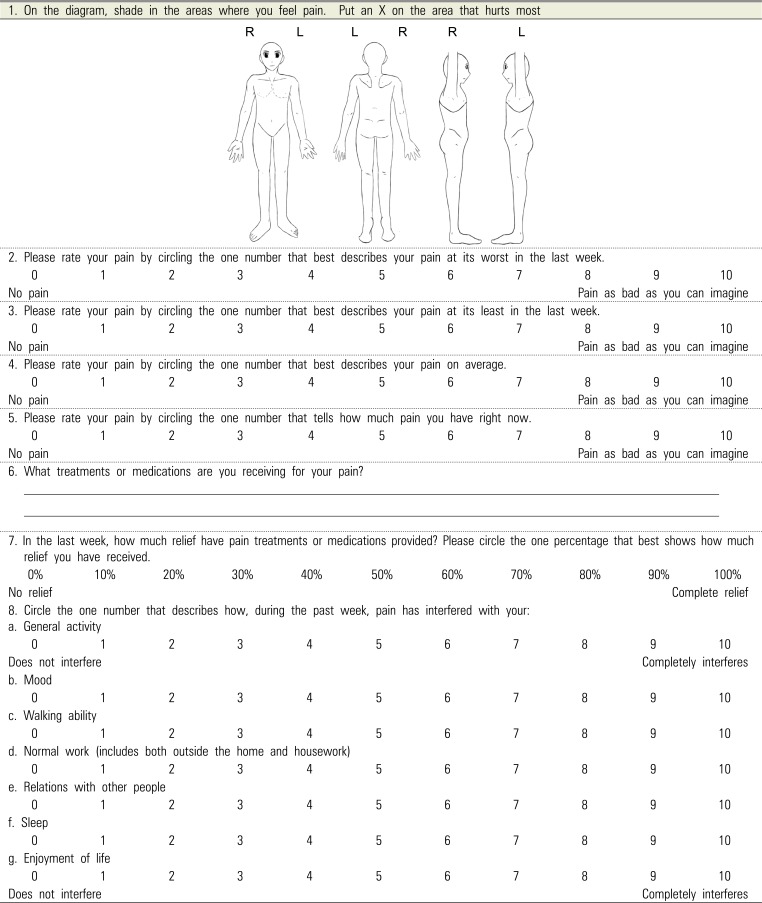
The BPQ is used to estimate the prevalence and severity of pain. It is easy to administer to a large numbers of patients, since it brief, can be self-administered, and easily determines the history, intensity, location, cause, quality, and interference with activities. However, the disadvantage is that given its brevity, it does not allow a comprehensive assessment of the pain course [11].
Most previous acute pain researches [5] used these pain measurement tool for only the sensory pain experience.
In OMFS research the VAS is commonly used to investigate many kinds of subjective experience, including pain [5]. The VAS (Fig. 2) is a straight, 100-mm line (10 cm), which can either be vertical or horizontal. It represents continuous pain intensity, where the left end of the line indicates “no pain,” while the other end denotes “pain as bad as it could possibly be.” A patients indicates their level of pain (in mm), by marking a single point on the line [9].
The VRS or VDS was created by Keele [13]. It was developed to assess patient responses to analgesics [9], with statistical validity and reliability. The VRS lists adjectives describing different levels of pain intensity [5], which consists of five numerically ranked words representing intensity of pain including the following [9]:
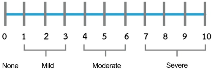
The NRS, which was developed by Downie [14] in 1978, consists of a vertical or horizontal line, with a total of 11 numbers, ranging from 0 to 10, denoting no pain to the worst possible pain, respectively [7]. Previous studies using the NRS offer great accuracy [9]. The NRS is a simple assessment tool that is easy to score. It can be administered in either the written or verbal form [9], but is not suitable for the elderly or very young child as it does not differentiate words and numbers [9].
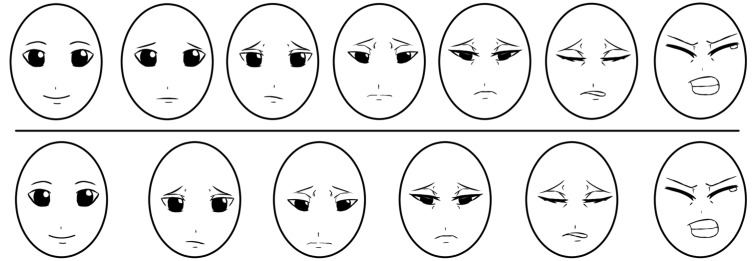
The original FPS was a 7-point number scales, which was more difficult to complete since it did not follow the usual 0-5 or 0-10 metric [15]. Thus, Hicks et al. revised the FPS scale, changing it to a more suitable 0-10 scale. Bieri et al. however, developed this measurement as seven line-drawn faces in a horizontal format, ranging from no pain to worst pain [15]. Instead of a happy face, a neutral face, with the absence of smiles and tears, was used to depict no pain [7], with increasing levels of pain intensity along the remaining six-face continuum. Patients were asked to point to the face that best represented the intensity of their pain. The scores 0, 2, 4, 6, 8, and 10 were assigned to each face consecutively from no pain to more painful faces [7].
Pain that occurs during or post-OMFS can be difficult to assess. Since patient self-reports are accepted as the gold standard for pain assessment [19], pain assessment tools should be simple and straight-forward to use [6]. These tools should ideally measure baseline discomfort and the response to remedy [17].
Several previous articles in biomedical studies suggested the use of multidimensional scales to assess chronic pain, such as in cancer and lower back pain [681011], since it can be more difficult to assess than acute pain [6].
Sensory and affective pain can be relatively easily described using the simple intensity sub-scales of the SF-MPQ, which includes the PPI and VAS. The SF-MPQ is more commonly used instead of the MPQ because the latter measures several dimensions of the experience and intensity of pain, making it more complicated to use [9]. The BPQ for estimating the prevalence and severity of pain can be easily administered to a large number of patients. The BPQ can be a quantitative or qualitative measure of pain, and is thus, the best clinical pain measurement tool.
Several previous articles suggested the use of unidimensional scales to measure acute pain [56202122232425], caused by trauma, surgery, childbirth, or an acute medical disease. Previous OMFS articles mostly used the VAS for pain assessment in acute pain. Bijur et al. [26] found that the VAS was a highly reliable tool for assessing acute pain in adults. Garra et al. [17] demonstrated that the VAS was also more informative and relatively sensitive to changes in pain, compare to other ordinal scales. The VAS tool is recommended for measuring pre- and post-operative pain. Additionally, although the VAS is similar to the NRS, it is more sensitive. Although the NRS can both underestimate and overestimate scores than the VAS, however, it is easy to administer verbally in a clinical unit and is familiar as a clinical tool [27]. Polly et al. [27] suggested that the verbally administered the NRS can be related to the VAS in acute pain measurement. Unfortunately, the VAS has also been mentioned to have more practical difficulties than the VRS and the NRS [20].
The VRS can be applied by mail or telephone [21] and it separates the pain experience into distinct categories. However, since the intervals of the VRS are not equally spaced, it cannot be assumed to be equal to ordinal data. Another weak point of the VRS scale is that application of statistical analysis to nonparametric scales is limited [5].
Previous studies suggest the use of FPS for children and older people [691516172829], since children find it easier to comprehend than non-graphics-based pain assessment scales [20]. The larger number of faces and consequent finer distinctions, however, pose difficulty for children aged 3–5 years [29].
Stanford et al. [17] demonstrated that age is a significant predictor of a child's ability to accurately apply the FPS. The WBS is one of several faces scales that has been used in multiple pediatric pain assessment settings [17]. Given the lack of tools for older people, some researchers have also suggested the use of the WBS for this population. The FPS is self-reporting scale that uses facial expressions to assess pain intensity [30]. This has appropriate psychometric properties when used with the elderly [30]. The FPS-R, however, also has strong validity and reliability properties, and has been recommended to measure pain intensity in elderly adults [30].
The full cup test is useful for pain assessment of patients with low-education patients because it does not need any numerical or verbal skills and is easy to understand and complete the scale [20].
The common intervention sequelae in lower third molar intervention (LTMI) is associated with post-operative pain, swelling, infection, and limited mouth opening. All of these may be decreased with antibiotic regimens and anti-inflammatory drugs. Previous reviews of analgesic efficacy for control pain after all types of third molar surgery contain a significant number of clinical trials of the third molar intervention study [31], and several contain pain assessment after analgesic drug usage following LTMI. The studies were shown less analgesic drug usage relate to less post-operative pain or more analgesic drug usage correlate with more painful after surgery [32].
The VAS is the most common scale to evaluate postoperative pain (especially after LTMI) [33]. Apart from due to the surgery itself, post-operative acute pain can occur due to insufficient administration of local anesthesia. In 2005, Babatunde et al. [34] compared the effect of co-administered dexamethasone and diclofenac potassium (diclofenac K) with diclofenac K alone, on postoperative pain, swelling, and limited mouth opening following LTMI. They evaluated the pain score through a four-point Category Rating Scale as follows:
In 2013, Marko et al. [35] also assessed the intensity postoperative pain during the first seven days after LTMI using the VAS. Fenlon et al. [36] determined the effect of pre-operative paracetamol administration on pain following LTMI, using the VAS, 1 hour after the intervention [36]. In 2017, Raiesian et al. [37] assessed the effect of low-level laser therapy (LLLT) on pain in LTMI, by complete a questionnaire with the VAS for pain intensity measurement. Further, Nicoli et al. [38] studied the efficacy of two different anti-inflammatory agents, i.e., Diclofenac (Deltaflogin) and Lumiracoxib (Prexige), in postoperative pain control resulting from LTMI, and also assessed pain using the VAS, as did Huskisson in 1983 [39]. The pain measurement scale also consists of a 100-mm straight line, which allows quantification of the post-operative pain.
In 2016, de Oliveira et al. [40] compared the efficacy of ibuprofen and etodolac for controlling pain after extraction of lower third molars and assessed pain using an 11-point VAS scale. They used a line with 11 identical boxes, where the extremes represented the pain limitation of patients, from absent to severe, respectively. At 6, 12, and 24 h after the LTMI, patients were instructed to mark an X in the box on the scale to indicate their pain intensity. This study demonstrated that successful use of this tool, which was adapted so that the patient had an increased understanding of the scale. Baxendale et al. in 1993 [41] evaluated the effect of a single prophylactic dose of oral dexamethasone 8 mg on these complications, in a randomized double-blind study of 50 adult patients. They also used the VAS for post-operative pain assessment in all patients of this study. In 2017, Boonsiriseth et al. [42] showed the effects of 8 mg dexamethasone injection into the pterygomandibular space on the postoperative sequelae of lower third molar surgery. They analyzed the consumption of analgesics and assesses pain using the VAS.
In 2012, Aznar-Arasa et al. [43] performed the comparative study of the analgesic and anti-inflammatory effects of preoperative and postoperative administration of ibuprofen following LTMI. They measured the intensity of postoperative pain using a 100-mm VAS scale. The intensity of pain was assessed every 2–4 h for the first 14 h, and then every 8 h between, 24 and 64 h post-surgery.
In 2012, de Santana-Santos et al. [44] investigated the relationship between preoperative findings and short-term outcome following LTMI. In this study, pain intensity was evaluated using a 10-level VAS scale. In 2010, Barreiro-Torres et al. [45] studied the difficulty and technique LTMI, assessing the ability level of fourteen dental practitioners with varying levels of experience. They also used a 100-mm VAS scale and a modified version of a surgical difficulty scale.
A study conducted by Ozveri Koyuncu et al. [46], in 2013, on postoperative pain, also used a 10-cm VAS scale. In 2015, Nedal et al. [47] studied the effects of primary versus secondary wound closure on postoperative pain, swelling, and acute alveolar osteitis after surgical extraction of partially LTMI. They also used the VAS for pain intensity assessment in all volunteers.
In contrast, in 2013, Anighoro et al. [48] assessed baseline and postoperative pain (following LTMI), using a four-point VRS verbal rating scale from 0 to 3. In 2017, Gulüen et al. [49] evaluated the efficacy of platelet-rich fibrin (PRF) on postoperative edema and pain after impacted mandibular third molar surgery using both the VAS and VRS. All patients completed the VAS and VRS to assess their pain, and there was no significant differences between the two pain scores. It must be noted, however, that the NRS is easier to use to evaluate pain in adults [33]. Further, the WBS is widely applied to assess pain in children and older adults.
In conclusion, multidimensional scales such as MPQ, SF-MPQ, and BPQ are suitable to assess chronic pain, while unidimensional scales, like the VAS, VDS, VRS, NRS, FPS, WBS, and FCT are more suitable for acute pain evaluation occurring after LTMI.
ACKNOWLEDGEMENTS
The authors would like to thank the staff and dental assistants, including co-workers at the Department of Oral and Maxillofacial Surgery, Faculty of Dentistry, Mahidol University.
References
1. Todd KH, Ducharme J, Choiniere M, Crandall CS, Fosnocht DE, Homel P, et al. Pain in the emergency department: results of the pain and emergency medicine initiative (PEMI) multicenter study. J Pain. 2007; 8:460–466. PMID: 17306626.
2. Merskey H. Classification of chronic pain: description of chronic pain syndromes and definition of pain terms. Pain. 1986; (Suppl 3):S1–S225. PMID: 3461421.
3. McAloon C, O'Connor PC, Boyer M. Patient's perception of pain on admission and discharge from the emergency department. N J Nurse. 2003; 33:7. PMID: 14692389.
4. Closs SJ, Gardiner E, Briggs M. Outcomes of a nursing intervention to improve postoperative pain at night. Acute Pain: Int J Acute Pain Manag. 1998; 1:22–31.
5. Briggs M, Closs JS. A descriptive study of the use of visual analogue scales and verbal rating scales for the assessment of postoperative pain in orthopedic patients. J Pain Symptom Manage. 1999; 18:438–446. PMID: 10641470.
6. Breivik H, Borchgrevink PC, Allen SM, Rosseland LA, Romundstad L, Breivik Hals EK, et al. Assessment of pain. Br J Anaesth. 2008; 101:17–24. PMID: 18487245.
7. Li L, Liu X, Herr K. Postoperative pain intensity assessment: a comparison of four scales in Chinese adults. Pain Med. 2007; 8:223–234. PMID: 17371409.
9. Flaherty SA. Pain measurement tools for clinical practice and research. AANA J. 1996; 64:133–140. PMID: 9095685.
11. Daut RL, Cleeland CS, Flanery RC. Development of the Wisconsin Brief Pain Questionnaire to assess pain in cancer and other diseases. Pain. 1983; 17:197–210. PMID: 6646795.
12. Heft MW, Parker SR. An experimental basis for revising the graphic rating scale for pain. Pain. 1984; 19:153–161. PMID: 6462727.
14. Downie WW, Leatham PA, Rhind VM, Wright V, Branco JA, Anderson JA. Studies with pain rating scales. Ann Rheum Dis. 1978; 37:378–381. PMID: 686873.
15. Bieri D, Reeve RA, Champion GD, Addicoat L, Ziegler JB. The Faces Pain Scale for the self-assessment of the severity of pain experienced by children: Development, initial validation, and preliminary investigation for ratio scale properties. Pain. 1990; 41:139–150. PMID: 2367140.
16. Cote CJ, Lerman J, Anderson B. A practice of anesthesia for infants and children. 5th ed. Elsevier Health Sciences: 2013;940.
17. Garra G, Singer AJ, Taira BR, Chohan J, Cardoz H, Chisena E, et al. Validation of the Wong-Baker FACES Pain Rating Scale in Pediatric Emergency Department Patients. Acad Emerg Med. 2010; 17:50–54. PMID: 20003121.
18. Ergun U, Say B, Ozer G, Yildirim O, Kocaturk O, Konar D, et al. Trial of a new pain assessment tool in patients with low education: the full cup test. Int J Clin Pract. 2007; 61:1692–1696. PMID: 17877654.
19. McCahon S, Strong J, Sharry R, Cramond T. Self-report and pain behavior among patients with chronic pain. Clin J Pain. 2005; 21:223–231. PMID: 15818074.
20. Odai ED, Ehizele AO, Enabulele JE. Assessment of pain among a group of Nigerian dental patients. BMC Res Notes. 2015; 8:251. PMID: 26087661.
21. Isik K, Unsal A, Kalayci A, Durmus E. Comparison of three pain scales after impacted third molar surgery. Oral Surg Oral Med Oral Pathol Oral Radiol Endod. 2011; 112:715–718. PMID: 21441046.
22. Lara-Munoz C, De Leon SP, Feinstein AR, Puente A, Wells CK. Comparison of three rating scales for measuring subjective phenomena in clinical research. I. Use of experimentally controlled auditory stimuli. Arch Med Res. 2004; 35:43–48. PMID: 15036799.
23. Gagliese L, Weizblit N, Ellis W, Chan VW. The measurement of postoperative pain: A comparison of intensity scales in younger and older surgical patients. Pain. 2005; 117:412–420. PMID: 16153776.
24. Sloman R, Wruble AW, Rosen G, Rom M. Determination of clinically meaningful levels of pain reduction in patients experiencing acute postoperative pain. Pain Manag Nurs. 2006; 7:153–158. PMID: 17145489.
25. Bech RD, Lauritsen J, Ovesen O, Overgaard S. The Verbal Rating Scale Is Reliable for Assessment of Postoperative Pain in Hip Fracture Patients. Pain Res Treat. 2015; 2015:676212. PMID: 26078880.
26. Bijur PE, Silver W, Gallagher EJ. Reliability of the visual analog scale for measurement of acute pain. Acad Emerg Med. 2001; 8:1153–1157. PMID: 11733293.
27. Bijur PE, Latimer CT, Gallagher EJ. Validation of a verbally administered numerical rating scale of acute pain for use in the emergency department. Acad Emerg Med. 2003; 10:390–392. PMID: 12670856.
28. Goodenough B, Champion GD, von Baeyer C, Ziegler JB. Facial expression scales for self-report of pain by children: rationale, validity and utility. In : Fourth International Symposium on Pediatric Pain; 1997.
29. Shih AR, von Baeyer CL. Preschool children's seriation of pain faces and happy faces in the Affective Facial Scale. Psychol Rep. 1994; 74:659–665. PMID: 8197303.
30. Miro J, Huguet A, Nieto R, Paredes S, Baos J. Evaluation of reliability, validity, and preference for a pain intensity scale for use with the elderly. J Pain. 2005; 6:727–735. PMID: 16275596.
31. Coulthard P, Patel N, Bailey E, Coulthard MB. Measuring pain after oral surgery. Oral Surg. 2014; 7:203–208.
32. Seymour RA. The use of pain scales in assessing the efficacy of analgesics in post-operative dental pain. Eur J Clin Pharmacol. 1982; 23:441–444. PMID: 7151849.
33. Ferreira-Valente MA, Pais-Ribeiro JL, Jensen MP. Validity of four pain intensity rating scales. Pain. 2011; 152:2399–2404. PMID: 21856077.
34. Bamgbose BO, Akinwande JA, Adeyemo WL, Ladeinde AL, Arotiba GT, Ogunlewe MO. Effects of co-administered dexamethasone and diclofenac potassium on pain, swelling and trismus following third molar surgery. Head Face Med. 2005; 1:11. PMID: 16274480.
35. Matijević M, Uzarević Z, Gvozdić V, Mikelić VM, Leović D, Macan D. The influence of surgical experience, type of instructions given to patients and patient sex on postoperative pain intensity following lower wisdom tooth surgery. Acta Clin Croat. 2013; 52:23–28. PMID: 23837269.
36. Fenlon S, Collyer J, Giles J, Bidd H, Lees M, Nicholson J, et al. Oral vs intravenous paracetamol for lower third molar extractions under general anaesthesia: is oral administration inferior? Br J Anaesth. 2013; 110:432–437. PMID: 23220855.
37. Raiesian S, Khani M, Khiabani K, Hemmati E, Pouretezad M. Assessment of low-level laser therapy effects after extraction of impacted lower third molar surgery. J Lasers Med Sci. 2017; 8:42–45. PMID: 28912943.
38. Nicoli GA, Conte-Neto N, Campos JADB, Cabrini-Gabrielli MA, Pereira-Filho VA. Efficacy of lumiracoxib versus diclofenac sodium in pain control following extraction of impacted lower third molar. Int J Odontostomat. 2017; 11:47–51.
39. Huskisson EC. Visual analogue scales. In : Melzack R, editor. Pain measurement and assessment. New York: Raven Press;1983. p. 33–37.
40. Silva de Oliveira JC, Grossi de Oliveira GA, Bassi AP. Comparative Assessment of the Effect of Ibuprofen and Etodolac on Edema, Limited mouth opening, and Pain in Lower Third Molar Surgery: A Randomized Clinical Trial. J Oral Maxillofac Surg. 2016; 74:1524–1530. PMID: 27160363.
41. Baxendale BR, Vater M, Lavery KM. Dexamethasone reduces pain and swelling following extraction of third molar teeth. Anaesthesia. 1993; 48:961–964. PMID: 8250191.
42. Boonsiriseth K, Latt MM, Kiattavorncharoen S, Pairuchvej V, Wongsirichat N. Dexamethasone injection into the pterygomandibular space in lower third molar surgery. Int J Oral Maxillofac Surg. 2017; 46:899–904. PMID: 28318872.
43. Aznar-Arasa L, Harutunian K, Figueiredo R, Valmaseda-Castellon E, Gay-Escoda C. Effect of preoperative ibuprofen on pain and swelling after lower third molar removal: a randomized controlled trial. Int J Oral Maxillofac Surg. 2012; 41:1005–1009. PMID: 22521671.
44. de Santana-Santos T, de Souza-Santos JA, Martins-Filho PR, da Silva LC, de Oliveira e Silva ED, Gomes AC. Prediction of postoperative facial swelling, pain and limited mouth opening following third molar surgery based on preoperative variables. Med Oral Patol Oral Cir Bucal. 2013; 18:e65–e70. PMID: 23229245.
45. Barreiro-Torres J, Diniz-Freitas M, Lago-Mendez L, Gude-Sampedro F, Gandara-Rey JM, Garcia-Garcia A. Evaluation of the surgical difficulty in lower third molar extraction. Med Oral Patol Oral Cir Bucal. 2010; 15:e869–e874. PMID: 20526272.
46. Koyuncu BÖ, Zeytinoğlu M, Çetingül E. Comparison of 2 different flap techniques in the surgical removal of bilateral impacted mandibular third molars. Turk J Med Sci. 2013; 43:891–898.
47. Nedal AM. The effects of primary and secondary wound closure following surgical extraction of lower third molars on post-operative morbidity: A prospective randomized clinical trial. J Dent Oral Hyg. 2015; 7:168–174.
48. Anighoro EO, Gbotolorun OM, Adewole RA, Arotiba GT, Effiom OA. Assessment of the effect of wound closure technique on postoperative sequaele and complications after impacted mandibular third molar extraction. Open J Stomatol. 2013; 3:527–532.
49. Gülşen U, Şentürk MF. Effect of platelet rich fibrin on edema and pain following third molar surgery: a split mouth control study. BMC Oral Health. 2017; 17:79. PMID: 28438151.
Table 1
Table 2
Table 3
Table 4
Detail:
Brief Pain Inventory Scoring Instructions
1. Pain Severity Score: This is calculated by adding the scores for questions 2, 3, 4 and 5 and then dividing by 4.This gives a severity score out of 10.
2. Pain Interference Score: This is calculated by adding the scores for questions 8a, b, c, d, e, f and g and then dividing by 7. This gives an interference score out of 10.
Brief Pain Inventory from Hunter Integrated Pain Service Dec 2006
Reproduced with acknowledgement of the Pain Research Group the University of Texas MD Anderson Cancer Center, USA