

 PDF
PDF Citation
 Print
Print


INTRODUCTION
Peyronie's disease (PD) is a connective tissue disorder characterized by inelastic fibrous plaques on the tunica albuginea of the penis. It induces penile pain, erectile dysfunction (ED), and penile deformity including curvature, shortening, narrowing, and hinging. Penile curvature is the most common penile deformity caused by PD [1]; it inhibits vaginal penetration and leads to a loss of self-esteem and depression [2]. Although myriad medical treatments and nonsurgical therapies have been proposed [3], few are effective; surgical treatment remains the mainstay of treatment as suggested in the current International Society for Sexual Medicine (ISSM) guidelines [4].
The surgical approach is determined by the degree of the patient's curvature, the presence of a hinge effect, and the presence of concurrent ED. Penile plication is a widely accepted option that is applied for men with curvatures <60°. Although the advantages of plication include the relative ease of operation and fewer effects on potency compared with grafting, the major concern is the associated loss of penile length [5]. Penile shortening after plication is inevitable; the procedure shortens the longer side of the penis.
In this context, there are no established surgical techniques for compensation for the loss of penile length during penile plication. Our technique is to simply detach the scrotal septum from the penile base during plication; this facilitates penile elongation. Here, we describe our technique and evaluate the efficacy and safety of the technique compared with conventional penile plication.
MATERIALS AND METHODS
1. Patients
We retrospectively reviewed the records of men with PD who underwent penile plication combined with or without penile elongation using our novel technique from January 2009 to May 2018. During this period, 38 patients were treated by a single surgeon (D.G.M.) in our center. Penile plication was indicated in those with disease that had been stable for 6 months, who had painless curvatures, and who found it either difficult or impossible to engage in coitus because of the deformity.
Preoperative curvature severity and the direction thereof were obtained during the initial history-taking and/or from photographs taken at home. Men with penile curvatures >60° or hourglass deformities creating hinge effects were offered grafting and were excluded from the study. Additionally, patients with accompanying webbed or concealed penis were excluded. Oral phosphodiesterase-5 inhibitors were prescribed to men with mild or moderate ED to confirm that penile rigidity was adequate to allow for penetration prior to penile plication. Those with refractory ED (thus, those who did not respond to pharmacologic therapy) were offered penile prostheses and excluded from the study. The study protocol was reviewed and approved by the Institutional Review Board (IRB) of Korea University Guro Hospital (approval number: 2019GR0244). Informed consent was waived because of its retrospective nature.
2. Surgical technique
The procedure was performed with the patient under general anesthesia in the lithotomy position. A 16-Fr Foley catheter was routinely placed to identify the urethra and avoid any damage thereto during dissection. An artificial erection was induced via intracorporal injection of 10 to 20 µg alprostadil to identify the extent and direction of curvature. A circumferential incision was created proximal to the corona and the penis was degloved up to the base. We have previously described our penile plication technique [6]. Sixteen or 24 dots were routinely placed on the convex side of the penis and additional sutures were placed until curvature (circumferential asymmetry) was completely corrected.
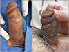
After penile plication, patients who underwent penile elongation were deeply dissected along the Buck's fascial plane to the penoscrotal junction to gain access to the scrotalseptum. At the level of the penoscrotal junction, we exposed and identified the dartos fascia that was ventrally attached to the penile base. During this step, inferior traction of the scrotal skin using the fingers, with counter-traction of the penis, facilitated exposure of the scrotal septum (Fig. 1A). The scrotal septum was ventrally detached from the penile base, and the areolar scrotal tissue was identified (Fig. 1B). An additional circumferential dissection along the Buck's fascia freed the penis from the deep dartos attachments. After complete dissection of the scrotal septum, an intraoperative photograph was taken and the gain in penile length recorded (Fig. 1C, D). Intraoperative penile length gaining was defined as the newly acquired length after detaching from the point of the penile shaft to which the existing scrotal septum was attached. The wound was closed in a three-layer manner and a mildly compressive penile wrap applied (3-inch Coban; 3M, St. Paul, MN, USA). Patients were discharged after a dressing change on the second postoperative day.
3. Outcome assessment
All patients were instructed to return for follow-up at 4 and 12 weeks. We objectively measured the preoperative, stretched penile length (SPL) at the initial physical examination and compared this with the SPL at 12 weeks postoperatively. At each follow-up visit, we assessed subjective perception of increased penile length. Patients were directly questioned about change in penile length after surgery and whether they were satisfied with the surgical outcome. All complications during the postoperative hospital stay or evident during follow-up were recorded.
4. Statistical analysis
All statistical analyses were performed with the aid of IBM SPSS Statistics ver. 22.0 for Windows (IBM Corp., Armonk, NY, USA). Normally distributed variables were expressed as means±standard deviations (SDs) and non-normally distributed variables were expressed as medians (with minima to maxima). The variables were compared according to groups using independent sample t-tests and Mann–Whitney U-test for normally distributed continuous and non-normally distributed continuous variables, respectively. Nominal data were presented as number or percentage and compared by the chi-square test. If the cells had counts of less than 5, they were re-examined with Fisher exact test. A p-value <0.05 was considered statistically significant.
RESULTS
The mean patient age was 51.6±11.5 years. Of the 38 patients, 22 patients underwent penile plication without any further procedure (conventional group) and 16 patients underwent penile plication combined with scrotal septum detachment (elongation group). The preoperative characteristics of the patients are presented in Table 1. There were no significant differences in preoperative characteristics, including age, direction of curvature, or degree of curvature between groups.
The operative time was 72.4±14.4 minutes (mean±SD) for penile plication; the penile elongation procedure required 20.0±5.2 minutes (mean±SD). There were no intra-procedural complications in either group. Most of the patients (31, 81.6%) were discharged from the hospital on the second postoperative day; two patients in the elongation group (2/16, 12.5%) were discharged 1 day later because of anxiety caused by postoperative pain.
Comparison of outcomes is presented in Table 2. There were significant differences in mean change in SPL and patient perceived penile length, respectively. Postoperative SPL increased by 1.3 cm in the elongation group, whereas it decreased by 0.5 cm in the conventional group (p<0.001). Two patients (12.5%) in the elongation group lost SPL (0.5 and 1.0 cm, respectively), whereas 19 patients (86.4%) in the conventional group lost SPL (range, 0.4 to 1.0 cm). In the elongation group, the median intraoperative gain in penile length was 3.0 cm (range, 2.0 to 4.0 cm) and postoperative SPL was correlated with intraoperative penile length gaining (p<0.001, R2=0.613). Other factors including patient's preoperative curvature degree and the plaque location showed no significant correlation (p=0.791 and p=0.538, respectively).
In the elongation group, 87.5% of patients reported perceived postoperative length increases. Of two patients who perceived postoperative length losses, the objective losses were, respectively, 1.0 cm and effectively nil. On the other hand, 77.3% of patients in the conventional group reported a perceived reduction in penile length, even though all patients had minimal SPL loss (<1.5 cm).
Early postoperative complications were rare and minor in both groups. Distal, penile skin edema and mild pain on erection were common immediately after surgery but then subsided, thus being self-limited. We encountered no delayed complications and no late failure in either group.
DISCUSSION
Numerous literature has described techniques for penile plication and their efficacy and safety profiles [78]. Recently, work has focused on minimally invasive approaches [910] and correction of more extensive curvatures (>60°) [511]. Unfortunately, surgical techniques minimizing or compensating for penile shortening have not been described previously. To the best of our knowledge, our procedure is the first technique to combine plication with compensation for loss of penile length.
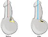
We achieved penile elongation via simple detachment of the scrotal septum from the penile base. The scrotal septum is a layer of areolar dartos fascia, or Colles fascia, which is formed by a pair of pouches fusing together in the midline of the scrotum [12]. Superficially, the scrotal septum extends above and closely adheres to the skin of the scrotal wall to form the median raphe. Deeply, the upper and anterior part of the scrotal septum are attached to the penile base ventrally and, even more deeply, to the root of the penis, forming the ligament of Luschka (the fundiform ligament) [13]. At the base of the penis, the upper and anterior parts of the scrotal septum (with the dartos fascia) form a well-defined penoscrotal angle, as does the skin [14]. Based on the pediatric procedure performed to correct a webbed penis, ventral phalloplasty looks similar with our technique. However, ventral phalloplasty is limited to excision of redundant scrotal skin. By contrast, scrotal septal detachment allows the penile base to be released from tethering the scrotal septum. As a result, the penile base is freed, resulting in not only the scrotum dropping downward but also penile lengthening (Fig. 2). The principle of penile elongation via our technique is illustrated in Fig. 3.
In this study, we achieved a mean gain of 1.2±1.3 cm in penile length postoperatively. This result is similar to the outcomes previously described for elongation surgery [1516]. However, combination of conventional penile elongation surgery with the plication procedure is limited by several aspects. Division of the suspensory ligament (with or without V–Y plasty) is the most widely accepted penile elongation technique [16]. Although the procedure is simple and effective, morbidity can be serious [17]. Paradoxically, the main adverse effect of this procedure is penile shortening; thus, placing a buffer in the place of the ligament is recommended [18]. However, buffer placement leads to the loss of simplicity of the surgery. Critically, the lack of penile support during erection after suspensory ligament release renders sexual intercourse (penetration) difficult [19]; such release is not indicated in combination with plication surgery in PD patients. Other techniques, termed “sliding elongation” and “penile disassembly” are too invasive for combination with plication surgery. “Suprapubic lipectomy” should be considered for selected patients only, particularly those with buried penis.
Encouragingly, we achieved favorable patient-reported outcomes postoperatively. Traditionally, penile plication is referred to as a “shortening procedure”; straightening of the corpora is achieved by shortening or tightening of the convex side of the tunica albuginea. The reported rate of penile length loss in the previous literature is variable at 5% to 80% [20212223]. Notably, some authors considered that SPL loss after plication surgery was negligible [1124]. However, they prioritized the physician's perspective, which differs from the patient's perspective. Actually, there was discordance between objective length changes and patient-subjective perception of length changes [2425]. This was consistent with our study results that patient dissatisfaction regarding postoperative penile length was high even though the actual SPL loss was minimal after penile plication.
The extent of postoperative patient satisfaction is associated with penile straightness but, psychosocially, penile length predicts improvements in sexual and general relationships, confidence, and libido after curvature surgery [26]. Additionally, the fear of perceived loss during preoperative counseling may prevent patients from undergoing plication surgery [7]. Thus, our technique, which results in a high rate of perceived penile length increase, would be helpful in these patients and would eliminate the unnecessary fear of penile length loss.
The most common complication of our technique was penile skin edema, apparently associated with circumcision, not scrotal septum detachment. However, circumcision is often used to deeply approach the penile base when performing penile plication. Additionally, unlike what was previously reported [9], we encountered no severe distal ischemic or lymphatic complications.
The limitations of our work include the fact that we did not measure SPL increases over the 12 postoperative weeks. We have performed the procedure for 4 years; this pilot study seeks to demonstrate the feasibility of our minimally invasive technique for penile elongation and the safety thereof. All patients are under follow-up; we will report long-term outcomes when we have the data. Also, we did not use validated questionnaires exploring erectile function (such as the International Index of Erectile Function). However, our procedure was focused on detachment of the scrotal septum from the penile base, which was not related to the damage to the dorsal neurovascular bundle.
The major strength of our technique is that can be performed with congenital curvature, webbed penis, concealed penis, and other penoscrotal approached penile surgery. Especially, simple detachment of the scrotal septum for penile elongation and penoscrotal angle reconstruction is amenable to a variety of patients with any type of concealed penis. It also provides an excellent cosmetic appearance, a significant increase in penile length, and high parent satisfaction, with minimal complications (Fig. 4).
CONCLUSIONS
In conclusion, a simple dissection detaching the scrotal septum from the penile base afforded both objective and subjective penile elongation without complications compared with conventional penile plication. This method can be used to minimize loss of penile length in PD patients undergoing plication surgery.


 XML Download