

 PDF
PDF Citation
 Print
Print


INTRODUCTION
Hydronephrosis without ureter and bladder abnormalities, or isolated hydronephrosis (IH), is a condition commonly diagnosed in antenatal screening. The rate of detection is rising as a result of the widespread use of antenatal ultrasonography [1]. These infants are at risk of urinary tract infection (UTI) [23], and the risk depends on the severity of IH and underlying uropathy [34]. A recent study has shown that mild to moderate IH, defined as an anteroposterior renal pelvis diameter (APRPD) 5 to 15 mm and the Society for Fetal Urology (SFU) grade 1 to 3 on neonatal renal and bladder ultrasonography (RBUS) performed at the age of 7 to 30 days, usually represents a self-limited condition. By contrast, severe IH, characterized by an APRPD ≥16 mm or SFU grade 4, is predictive of uropathy [5].
Continuous antibiotic prophylaxis (CAP) is considered a part of postnatal treatment in patients with IH. In the multidisciplinary consensus on the classification of prenatal and postnatal urinary tract dilation (UTD), CAP is recommended in UTD P3, where hydronephrosis is severe. The use of CAP in UTD P1 and P2, or mild to moderate hydronephrosis, is based on physician discretion [1]. A national survey of practice patterns of the general pediatricians listed in the American Medical Association has shown that 56% would prescribe CAP for infants with any degree of hydronephrosis [6]. Of 400 pediatric urologists who were members of the American Academy of Pediatrics and the European Society for Paediatric Urology, 70% chose to prescribe CAP when APRPD was larger than 10 mm and 30% of Canadian pediatric nephrologists, chose to prescribe CAP in SFU 1 to 2 [78]. Thus, variation in CAP utilization in mild to moderate IH is evidenced among providers across specialty and geography [6789].
This pilot study was conducted to determine whether CAP could reduce the incidence of UTI in mild to moderate IH in the first year of life where UTI is difficult to diagnose with accuracy and the risk of renal damage is high [10]. Our hypothesis was that infants with mild to moderate IH who received CAP would have a lower rate of UTI than those without CAP.
MATERIALS AND METHODS
1. Study design and settings and locations
This is a randomized clinical trial registered at the Clinical Trials Registry (www.clinicaltrials.in.th), number TCTR20150803001. All of the individual participant data collected during the trial will be available after deidentification immediately following publication indefinitely. To gain access, data requestors will need to sign data access agreement and direct the proposals to the corresponding author.
A two-arm parallel, unblinded, randomized controlled trial was conducted at a tertiary hospital. The present study protocol was reviewed and approved by the Institutional Review Board of Faculty of Medicine, Chulalongkorn University (approval number: 407/2015). Written informed consent was obtained from the parents or guardians of the children when they were enrolled.
2. Participants
Eligible participants were neonates aged 7 to 30 days who were referred to the nephrology unit for the evaluation of antenatal IH. Inclusion criteria were mild to moderate IH or UTD P0 to P2 in neonatal RBUS performed at the age of 7 to 30 days at our institution. Mild IH was defined as an APRPD >5 to <10 mm and SFU grade 1 to 2. Moderate IH was defined as APRPD 10 to 15 mm or SFU grade 3. Exclusion criteria were severe IH, defined as APRPD ≥16 mm or SFU grade 4 or UTD P3 [15].
3. Randomization
The randomization sequence was generated by the computer by variable block size design with an allocation ratio of 1:1. The allocation sequence was concealed in sequentially numbered sealed envelopes. A corresponding envelope was opened at the time to allocate the intervention to each participant.
4. Intervention
All subjects continued in the study until the age of 12 months. Each participant visited the hospital at enrollment and at the age of 2, 3, 6, 9, and 12 months. Follow-up telephone calls with the parents were scheduled at the age of 4, 5, 7, 8, 10, and 11 months. RBUS was performed at enrollment and at the age of 3, 6, 9, and 12 months. Hydronephrosis resolution was characterized by APRPD ≤5 mm and SFU grade ≤1 in both kidneys [5]. All RBUS was performed after adequate oral hydration in the supine position by radiologists who were blinded to the treatment allocation. Severity of hydronephrosis was graded based on APRPD and SFU grading in the midrenal transverse plane at cortical-pelvic margins within the confines of the renal cortex.
Infants in the control group did not receive CAP. Infants in the CAP group were given once daily oral CAP with 10 to 15 mg/kg of cephalexin from enrollment and followed by 2 to 3 mg/kg of cotrimoxazole after two months of age until hydronephrosis resolution or until the age of 12 months [11]. Any drug administration issue, adverse event and adherence were recorded at each clinic visit and by telephone call with the parents. Adherence to CAP was evaluated using parental self-report.
5. Trial outcomes
The parents were informed to contact when subjects developed a fever or when changes in urine characters were observed. These patients underwent clinical evaluation and urine specimens were collected by catheterization for urinalysis and cultures. The primary outcome was UTI, defined as fever ≥38℃ and urine culture of ≥50,000 colony-forming units per mL of single bacteria [12]. Bacterial sensitivity was determined based on antibiograms provided by an accredited laboratory in microbiology. Antibiotic resistance to ≥1 agent in ≥3 antimicrobial categories is considered multidrug resistance [13]. Voiding cystourethrogram (VCUG) was not universally performed. Only patients who developed UTI underwent VCUG. Patients who did not adhere to CAP were excluded from the analysis.
6. Sample size calculation and statistical analysis
Data analysis was conducted using Stata 13 software (StataCorp LP, College Station, TX, USA). All statistical tests were 2-sided. A p-value <0.05 was considered as statistically significant. Descriptive statistics included counts (percentages) and means±standard deviation. Continuous data were compared using Student's t-test. Categorical variables were compared using a chi-square or Fisher's exact test as appropriate. We assessed UTI up to 12 months in all control arm participants, and in CAP arm participants who received at least one dose of study medication.
The Kaplan–Meier method and the log-rank test were used to estimate and compare the probability of UTI between groups. Thereafter, we used cox proportional hazards regression to estimate the hazard ratio for UTI in the CAP group versus the control group.
Sample size was calculated based on the incidence of UTI of 15% in the control group and 10% in the CAP group, extrapolated from previous reports [411141516171819]. A number of 100 patients per group were required to give an 80% power with a two-sided type 1 error of 5%. The predetermined interim point was set at the completion of the 12-month study in a minimum of 80 recruited participants.
RESULTS
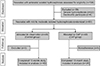
A total of 108 neonates were screened from August 2015 to December 2016. Twenty-two neonates were excluded due to severe IH. Parents of 6/86 (7.0%) eligible patients did not give consent. Eighty neonates with mild to moderate IH were randomized, 40 each to the CAP and observation arms. Six patients in the CAP arm were nonadherence and never received any antibiotic doses because the parents rejected the randomization allocation. These patients were therefore excluded from the analysis population (Fig. 1). Of 74 subjects, 64 (86.5%) were male and all of male patients were uncircumcised. Table 1 describes baseline characteristics of patients which did not significantly differ between groups. Although there was a higher proportion of patients with SFU grade 3 and moderate IH in the CAP group, the differences were not statistically significant. No major adverse effect from CAP was reported.
During the 12-month study, UTI occurred in 9/74 patients (12.2%). UTI occurred in 5/34 (14.7%) patients in the CAP group and in 4/40 (10.0%) controls. All nine patients with UTI (100.0%) had a fever ≥38℃. Although the relative incidence of UTI was 38% higher in the CAP group than in the control group, the difference was not statistically significant (hazard ratio, 1.38; 95% confidence interval, 0.37–5.16; p=0.63). As the risk of UTI could be related to grade of IH, we conducted a sensitivity analysis adjusting for severity of IH in our Cox regression model. The hazard ratio for developing UTI was similar in magnitude and significance between mild and moderate IH.
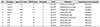
Table 2 demonstrates clinical characteristics of the nine patients with UTI, which were exclusively male. These patients underwent VCUG and did not have vesicoureteral reflux (VUR). In the control group, 1/4 (25.0%) UTI were caused by cotrimoxazole resistant bacteria. The isolate of Enterococcus faecalis from patient 1 was classified as resistant based on the intrinsic metabolism of bacteria [14]. In the CAP group, 5/5 (100.0%) UTI were caused by cotrimoxazole resistant bacteria. The chance of having UTI from cotrimoxazole resistant bacteria was four times higher in the CAP group than in the control group (relative risk, 4.0; 95% confidence interval, 1.2–13.5; p=0.02). Isolates from patient number 5, 6 and 7 were classified as multidrug resistant bacteria.
Along the study period, spontaneous resolution of IH occurred in 20/74 patients (27.0%). Spontaneous resolution occurred in 9/34 patients who received CAP (26.5%) and 11/40 untreated patients (27.5%). The resolution of IH did not significantly differ between groups (p=1.00).
Based on the interim analysis, no significant difference of UTI rates was observed between groups and there could be a potential harm to infants in the CAP group due to the increased risk of multidrug resistant UTI. The study was prematurely terminated as it deemed unjustified to continue recruitment.
DISCUSSION
Our study showed that the risk of UTI in infants with mild to moderate IH in the control group of 10% was comparable to the UTI prevalence of 7% commonly measured in the general infant population [20]. Moreover, there were no proven benefits of CAP for preventing UTI in infants with mild to moderate IH. The reported rates of UTI are influenced by study patients, study design, and diagnostic criteria of mild to moderate IH. The rate of UTI in our study was within the range of 8%–14% reported in previous prospective studies [414212223]. Male gender and uncircumcised status are risk factors for UTI in IH [14]. A high rate of UTI could be explained by the fact that male infants contributed to a large portion of the study patients as there is male predilection in IH [51014192124]. Moreover, all male infants in our study were uncircumcised.
In the present study, APRPD and SFU grading, the two commonly used measurements of hydronephrosis, were taken into account [25]. The strict diagnostic criteria of mild to moderate IH was used to create a homogeneous study population as the rates of UTI and the benefits of CAP are influenced by the severity of IH [31617]. Recent studies have shown an excellent inter-rater reliability of the interpretation of APRPD and SFU grading in neonatal RBUS [25]. Although all the ultrasounds were done according to a standard protocol at a single institution, the applicability of our findings can be reasonably extrapolated to other infants with APRPD <16 mm and SFU grade <4 on neonatal RBUS.
Our protocol is based on the presumption supported by recent studies that incidental findings of VUR detected in screening VCUG in mild to moderate IH is of little clinical significance [1824]. Thus, the VCUG result was not a prerequisite for CAP placement and VCUG was performed only in patients after UTI was confirmed. Our trial reflected the current clinical practice as a paradigm shift that supports selective VCUG in mild to moderate IH has been adopted in order to reduce excess resource utilization [22].
We focused on patients within the first year of life where the risk of UTI and renal scarring are the greatest [10]. Infants with hydronephrosis were 5 to 12 times more likely to be hospitalized for UTI compared with infants without hydronephrosis [1022]. Thus, UTI prevention is indicated in infants with IH. An accepted measure for UTI prevention is the administration of CAP [2]. However, variation in CAP utilization is evidenced in mild to moderate IH, and the benefits of CAP in mild to moderate IH is conflicting [6789].
A systematic review in patients with antenatal hydronephrosis conducted in Canada has shown a slight but not significant, protective effect of CAP against UTI whereas the benefits of CAP were not confirmed in the systematic review from the European Association of Urology [1617]. A meta-analysis showed variable preventive effects of CAP in different degree of IH. The rates of UTI were significantly decreased in children with SFU grade 3 to 4 treated with CAP but no benefits of CAP was observed in SFU grade 1 to 2 [3]. In our study, the probability of UTI in infants with APRPD <16 mm and SFU grade <4 was similar between infants receiving CAP and controls.
A potential harm related to the use of CAP is the emergence of resistant bacteria. Antibiotics including cephalexin, nitrofurantoin, trimethoprim, and cotrimoxazole are used as CAP, with cotrimoxazole being the most commonly prescribed CAP [2627]. The use of cephalosporin prophylaxis increases the risk of UTI secondary to extended spectrum β-lactamase producing bacteria [28]. A systematic review showed that nitrofurantoin had a lower risk of antibiotic resistance than cotrimoxazole [29]. We used cotrimoxazole in the present study as nitrofurantoin is locally available only in pills. Cephalexin was used during the first two months of life when cotrimoxazole is contraindicated [11].
Bacterial drug resistance is a growing problem and has become a major public health concern [2728]. The Randomized Intervention for Children with Vesicoureteral Reflux (RIVUR) trial reported a bacterial resistant rate of 60% within the group receiving cotrimoxazole [27]. The trial has shown that antibiotic resistance related to CAP is of transient nature and has no impact on recurrence of UTI [27]. In our study, infants treated with CAP were at risk of UTI due to cotrimoxazole resistant and multidrug resistance bacteria. The difference in the probability of UTI between the CAP and the control group should be interpreted with caution. A slight increase in the probability of UTI in infants receiving CAP could be the result of microbiome dysbiosis and may be clinically meaningful. Nevertheless, the probability of UTI did not statistically differ between groups.
Our study has several limitations. First, the small number of subjects decreased the power of the study and impeded a firm conclusion of the impact of CAP on UTI. Moreover, there were only nine patients who developed UTI. Although all patients with UTI were male and uncircumcised, the small number of patients with UTI precluded the accurate evaluation of potential risk factors. Although collaboration between multiple centers might ensure properly powered research, the negative impact of CAP on bacterial resistance observed in the current study requires further evaluation.
Second, the study was not a placebo-controlled trial and unblinded. Parental awareness of the assignment may modify their behaviors such as practice of the perineal or foreskin care and the use of prebiotics or probiotics, which could change the risk of UTI and would thereby alter the study results [2]. Nevertheless, biased determination of the observed outcome should be minimized in our study as the diagnosis of UTI was uniform using urine culture from catheterized specimens and based on prospective documentation.
Third, parental self-report, an indirect method, was used to evaluate nonadherence in the current study. There is no gold standard method for measuring adherence. Indirect measurement is practical but has a potential bias towards overestimation of adherence [30]. Thus, the actual nonadherence rate was likely to be higher than the reported rate. Nonadherence to CAP is a common phenomenon. A recent study in children with VUR has found that the adherence rate to CAP measured by the medication possession ratio was 40% at one year [26]. Interventions to promote adherence are needed when CAP is recommended as the benefit of CAP will be achieved only if the patient adheres to the treatment and lack of adherence could interfere with clinical trial outcomes. Subtherapeutic antibiotic levels can foster the development of resistant organisms and could explain the significant increase in the resistance of cotrimoxazole in infants receiving CAP [30].
Fourth, bowel and bladder dysfunction (BBD) was not formally evaluated. Physicians may be unaware of BBD, given that the diagnosis depends on parental report and that symptoms frequently go unrecognized in infants. The presence of BBD could increase the probability of UTI. Fifth, the effectiveness of CAP in prevention of renal scars was unknown. The ultimate goal of CAP is to reduce the renal scars, which are detected by dimercaptosuccinic acid (DMSA) scans. However, DMSA scans were not performed in our study patients.
CONCLUSIONS
As a starting point, this pilot study has shown that infants with mild to moderate IH are susceptible to UTI but the benefits of CAP are inconclusive. CAP conferred a high risk of resistant bacterial organisms when UTI occurs. Differential treatment in unblinded study and nonadherence could result in biased estimates of the impact of CAP. Further study is required to better identify which infants with mild to moderate IH are most likely to benefit from CAP. Our results are in agreement with recent observational studies that conservative management with a low threshold for investigation and treatment of UTI is appropriate in IH when APRPD is 5 to 15 mm and SFU grade <4.

 XML Download