

 PDF
PDF Citation
 Print
Print


INTRODUCTION
Since the introduction of targeted therapy (TT), immunotherapy (IT) with multiple cytokine therapies has been substituted for diverse targeted agents, which were the standard systemic therapy for metastatic renal cell carcinoma (mRCC) [1]. The advantage of TT is that it is relatively well tolerated and targets specific angiogenetic receptors with less severe adverse effects than IT and with improved survival prognoses, with markedly extended progression-free survival (PFS) intervals and observed CSS rates of 16 to 26 months [2]. However, the weakness of TT is the insignificant difference in long-term survival, including very few cases of complete remission, and dismal 5-year survival rates of approximately 10%.
Estimating prognosis is important for planning a therapeutic strategy in patients with mRCC; however, diverse and unexpected prognostic outcomes are frequently encountered in clinical practice. Thus, many researchers have developed prognostic risk assessment to classify patients into favorable-, intermediate-, and poor-risk groups according to their survival prognoses to better predict therapeutic outcomes [34]. One of the most commonly used risk stratification systems is the International Metastatic Renal Cell Carcinoma Database Consortium (IMDC) model, also known as the Heng prognostic criteria [3]. The Heng criteria were established in the TT era and were validated with mRCC patients who were administered TT [5]; however, these criteria also have good predictability for mRCC patients treated with IT [6]. The criteria comprise six readily assessable parameters: time from diagnosis to treatment (treatment-free interval, TFI), Karnofsky performance status, hemoglobin, neutrophil count, platelet count, and serum calcium concentration.
Among the three Heng risk groups, the favorable- and poor-risk groups have uniform survival outcomes, whereas the intermediate-risk group, which has one or two risk parameters, is composed of a wide range of diverse patients with variable disease burdens and clinicopathologic characteristics, resulting in unpredictable and diverse responses to systemic therapy compared with the favorable- and poor-risk groups [789]. This was because patients were grouped with various heterogeneous people with heterotrophic and pleomorphic tumor burdens and with different clinicopathologic characteristics, including different pathophysiology and metabolic activity [1011]. Several previous studies have addressed the necessity of understanding survival outcomes according to more specific stratification of intermediate-risk patients to increase the predictability of survival outcomes. Therefore, this retrospective study analyzed PFS, caner-specific survival (CSS), and overall survival (OS) in intermediate-Heng-risk mRCC patients treated with TT compared with IT in the first-line setting and evaluated the significant risk factors for the three survival outcomes.
MATERIALS AND METHODS
1. Ethics statement
Following approval of this retrospective study by the Institutional Review Board (IRB) of the National Cancer Center (approval number: NCC2016-0263), the IRB waived the requirement for written informed consent. All patient data were anonymized and deidentified before our analysis. All study protocols were performed in accordance with the ethical tenets of the Declaration of Helsinki.
2. Patient criteria and evaluation tools
The medical records of 186 mRCC patients with intermediate Heng risk and treated with IT or TT between January 2000 and December 2017 were retrospectively reviewed after the exclusion of those with incomplete medical records, aged <20 years, or unavailable for follow-up. All the included mRCC patients were of intermediate Heng risk [12] and had undergone a complete evaluation after every 1 to 4 cycles (6–12 weeks) of IT and every 2 cycles of TT (12 weeks). The follow-up protocol, which included laboratory and imaging evaluations, was described in detail previously [6].
Treatment continued until disease progression was detected. Patients were further stratified into groups with a TFI <1 year or ≥1 year and into metastatic types of either synchronous or metachronous. Other ages, gender, Eastern Cooperative Oncology Group Performance Status (ECOG PS), anemia, hypercalcemia, neutrophilia, thrombocytosis, histology, clinical TN stage [13], Fuhrman nuclear grade [14], treatment duration, and survival outcomes including PFS, CSS, and OS were evaluated as baseline characteristics of each IT and TT group to analyze the predictive risk factors of survival outcomes. The Response Evaluation Criteria in Solid Tumors v1.1 (RECISTv1.1) was used to determine the therapeutic response to systemic therapy [15].
3. Treatment regimens
The choice of first-line systemic agent (IT or TT) was at the discretion of the treating urologist (J.C.) according to each patient's pathology and coverage by the National Health Insurance System, as described previously [6]. Combination IT comprised subcutaneous recombinant human interleukin (IL)-2 (Proleukin; Chiron Italia s.r.l., Milan, Italy) and recombinant human interferon (IFN)-α (IFN-alpha-2a, Roferon-A; Hoffmann-La Roche Inc., Nutley, NJ, USA).
All TTs were administered either orally or intravenously with the recommended regimen of the National Comprehensive Cancer Network (NCCN) guidelines, from 2005 until 2017 (available at https://www.nccn.org/professionals/physician_gls/pdf/kidney.pdf). First-line TT comprised sunitinib, sorafenib, pazopanib, or temsirolimus. Of the 122 patients who received TT, 73 patients (61.9%) received sunitinib, 15 patients (12.7%) received sorafenib, and 30 patients (25.4%) received pazopanib. The target agent regimens were described previously [611].
An overlapping period of 10 years between 2007 and 2017 existed. During the overlapping period, the National Health Insurance System in Korea changed; IT was considered the first-line therapy for mRCC before 2007, and then subsequently, TT became the first-line therapy since 2007 while IT became the second-line therapy. However, a small portion of patients still received an interleukin therapy as the first-line therapy until 2010. In addition, interferon therapy was at times also given as a second-line and third-line therapy after TT.
4. Statistical analysis
Baseline characteristics of Heng intermediate-risk patients were expressed as frequency with percentage for categorical variables and median with range or mean with standard deviation for continuous variables. Differences between the IT and TT groups were compared by using t-test, Wilcoxon rank sum test, chi-square test, and Fisher's exact test as appropriate. The Kaplan–Meier method was used to compute the probabilities of survival, and comparison of survival curves was performed by use of log-rank tests. Cox proportional hazard models were used to identify the prognostic factors. The multivariable model was performed with the backward variable selection method with an elimination criterion of 0.1. All statistical analyses were considered statistically significant at a p-value <0.05 and were performed by using SAS (version 9.4, SAS Institute Inc., Cary, NC, USA) and R Foundation for Statistical Computing (version 3.5.2).
RESULTS
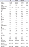
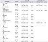
The median age of the 186 patients was 58 years (range, 22–83 years) and the ratios of histology, clinical T stages, and Fuhrman nuclear grades were 55.9%/4.3% for clear cell/non-clear-cell, 44.1%/33.9%/10.8% for T1–2/T3–4/Tx. stages, and 6.5%/39.8%/53.8% for low/high/unknown grades. During a median of 5.1 months of systemic treatment and 92.22 months of follow-up, the median PFS, OS, and CSS were 5.16, 18.44, and 19.04 months, respectively, and the RECISTv1.1 responses were 1.6%, 18.8%, 42.5%, 19.9%, and 17.2% for complete response, partial response, stable disease, progressive disease, and unknown, respectively (Table 1). The comparison of baseline characteristics between the two groups showed a significantly higher rate of T3–4 stages, a lower rate of high nuclear grades, a shorter duration of follow-up, longer treatment durations, lesser rates of cytoreductive nephrectomy, a lower objective response rate, and no cases of complete response in the TT group compared with the IT group (p<0.05, Table 1).
Of the 186 patients included in the analysis, 88 (47.3%) underwent secondary treatment and 73 patients (83.0%) had progression since the start of the second treatment. In addition, 72 patients (81.8%) received TT as a second treatment and 16 patients (18.2%) received IT. The median PFS, OS, and CSS were 4.50, 10.65, and 12.0 months, respectively, for the second-line therapy (Table 2).
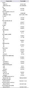
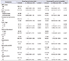
The multivariable analyses using metastatic types of either synchronous or metachronous mRCC, systemic therapeutic agents of either IT or TT, TFI of less than or greater than 1 year, cytoreductive nephrectomy, clinical T stages, ECOG PS, and presence of anemia, thrombocytosis, hypercalcemia, and neutrophilia showed that synchronous metastatic type (hazard ratio [HR], 2.285; 95% confidence interval [CI], 1.154–4.523), IT (HR, 1.746; 95% CI, 1.257–2.426), and TFI of less than 1 year (HR, 1.926; 95% CI, 0.997–3.720) were significant factors for PFS (p<0.05, Table 3), whereas none of risk factors were significantly left in the model for OS and CSS (p>0.05, Table 4).
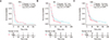
The Kaplan–Meier curve of each survival shown for a comparison between IT and TT in Fig. 1A that only PFS was significantly different (IT, 4.1 months vs. TT, 7.0 months; p<0.05). The comparison of OS and CSS showed that IT (18.7/19.0 months) and TT (18.4/19.0 months) had approximately similar survival results (p>0.05; Fig. 1B, C).
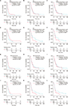
The Kaplan–Meier survival analysis according to the predictive risk factors including TFI of 1 year, systemic agents, and metastatic types for PFS showed that TT (7.0/5.5/8.0/4.8 months) had significantly longer PFS than IT (4.6/4.0/4.5/3.8 months), respectively (p<0.05) (Fig. 2).
DISCUSSION
Since the introduction of TT, improvement in prognostic survival of mRCC has been demonstrated, especially in PFS, whereas long-term gains in OS or CSS were not reached until the recent introduction of immune checkpoint inhibitors (nivolumab, ipilimumab) and the newly introduced targeted agents (axitinib, cabozantinib) [1617]. The results of the present study also support the prognostic outcomes of TT compared with IT similar to previous studies in that significant differences were not shown in OS or CSS, but only in first-line PFS in intermediate-risk mRCC (Table 1, Fig. 1). This advantage of prolonged PFS in TT was expected because TT is commonly known to have less severe adverse effects and higher tolerability of therapy than IT. The insignificant differences in OS and CSS between the two groups can be explained by various reasons.
First, because PFS is a short-term terminology of survival and OS and CSS are long-term survival terminology, OS and CSS might be affected by various lines of sequential applications of multiple targeted agents after the failure of first-, second-, and third-line TT. Second, heterotrophically and pleomorphically diverse tumor cell types are encountered in mRCC, leading to different metabolic and pathophysiologic activities within the tumor microenvironment. Thus, different metastatic tumors are influenced by different therapies in different organs, resulting in newly acquired therapeutic resistance and decreased therapeutic resistance [18]. Last, the different mechanism of action needed for therapeutic action between IT and TT might be another influencing factor, as discussed later in this discussion section.
The diverse heterogeneity of the intermediate-risk mRCC group has been an important issue of debate since this group of patients led to unpredictable clinical outcomes after systemic treatment compared with other favorable and poor-risk mRCC groups [45678910]. Many researchers have tried to find factors to classify the intermediate-risk group into more thoroughly divided prognostic risk subgroups [789]. This study proved two significant risk factors, TFI <1 year (HR, 0.894) and metastatic type (synchronous vs. metachronous; HR, 1.444) in the multivariate analysis and Kaplan–Meir curve with log-rank comparison (p<0.05; Table 2, Fig. 1). In a study by Tanaka et al. [8] of 245 patients with mRCC, approximately one-quarter of the patients were reclassified into different risk groups of the IMDC model after TT administration in the first-line and second-line settings; the reclassification included 15.7% of favorable-risk patients reclassified as intermediate risk, 21.6% of intermediate-risk patients reclassified as poor risk, and 65.5% of poor-risk patients reclassified as intermediate risk. Our research team also previously suggested that the neutrophil-to-lymphocyte ratio and other risk factors significantly potentiated the subgroup classifications of the current intermediate-risk group with better predictors of prognosis than the Heng risk criteria [19].
TFI with a 1-year cut-off came from the time from diagnosis to treatment from the Heng risk criteria [5], which is a well-known prognostic risk factor for mRCC [20212223]. It indirectly depicted the growth rate and aggressiveness of the tumor. A tumor with a TFI of <1 year might suggest a rapidly growing, aggressively invading, or metastasizing tumor with hypermetabolic states. In contrast, a TFI ≥1 year might indicate a slowly progressing tumor with low metabolic activity and less aggressiveness [22]. This study also supported the statements that IT might be more suitable for patients with slow-growing mRCC with a TFI ≥1 year and that TT might be adequate for fast-growing mRCC because an interesting finding was observed when intermediate-risk patients were stratified by TFI. In terms of PFS, the TT group was associated with superior PFS (7.0/5.5 months) regardless of TFI compared with the IT group (4.6/4.0 months, p<0.05). However, the IT group had insignificantly better OS/CSS than the TT group (25.2 vs. 20.1 months) among patients with a TFI ≥1 year (p>0.05, Fig. 2D–F), whereas the TT group had insignificantly better OS/CSS (TT, 18.3/18.9 vs. IT, 14.9/19.0 months, p>0.05; Fig. 2G–L).
These results might be explained by the mechanism of action of each systemic therapy in the tumor environment and corporal immune system. IT is suitable for slow-glowing tumors with low metabolic activity because it needs time for antigen presentation and boosting of the cellular and acquired immune system with delayed sequential therapeutic responses to attack the tumor and prevent tumor growth, resulting in long-term, durable responses in mRCC patients [2425]. On the contrary, TT directly attacks multiple specific vascular-related target receptors of tumor cells and peri-tumoral vessels for antiangiogenesis in the tumor microenvironment quickly enough to induce rapid tumor necrosis and inhibition without neovascularization [17262728]. Accordingly, these therapeutic mechanisms might induce the combination of TT with IT to improve prognostic survival and to increase the long-term curable state in mRCC. Recent immune checkpoint inhibitors and other immune therapies have shown an increased rate of long-term durable states in recent clinical trials [24262728], changing first-line therapeutic settings in the international European Association of Urology (EAU) [16] and NCCN guidelines v2019 [17].
Another significant prognostic factor found for PFS in this study was the metastatic type of either metachronous or synchronous mRCC. The metastatic type also implied other significant prognostic factors, such as nephrectomy and the aforementioned time from diagnosis to treatment [2930]. Metachronous mRCC treated by nephrectomy to remove the primary kidney tumor had better prognostic HRs than synchronous mRCC. Bozkurt et al. [31] demonstrated a potential prognostic value of late recurrence in terms of PFS, OS, and objective response rate: among 86 patients with mRCC who received TT, those 56 metachronous mRCC patients had recurrence within 5 postoperative years and had significantly worse survival. Kroeger et al. [21] investigated 10 mRCC patients treated with TT after surgery, and the 26% of patients who relapsed after 5 postoperative years had a more favorable prognosis.
This study had some inherent limitations related to its retrospective design, small number of intermediate-risk patients, and treatment with different therapeutic modalities that have different mechanisms of action. The effect of second-line agents was somewhat limited owing to the small number included in our study. We were therefore unable to evaluate the efficacy of IT and TT on PFS, OS, and CSS individually. The short follow-up period was another limitation. The 16 patients who received IT and 72 patients who received TT as second-line therapy were compared in terms of survival. Overall, an insignificant difference was found for OS and CSS (p>0.05); however, there was a significant difference observed in PFS (IT, 2.8 months vs. TT, 4.8 months; p=0.0469) (Supplementary Fig. 1). In addition, metastatic lesions diagnosed pathologically might not represent the entire disease burden of mRCC, especially for metachronous mRCC because of differences between primary and metastatic lesions. However, the results of this study suggest the necessity of future studies to investigate multiple additional genetic, imaging, and inflammatory markers; incorporated together, these markers could improve prognostic models for intermediate-risk patients with mRCC based on different therapeutic modalities.
CONCLUSIONS
This study reported prognostic results of IT and TT in intermediate-Heng-risk mRCC and suggested TFI and metastatic type as significant risk factors for PFS as well as potential factors for categorizing intermediate-risk patients into subgroup classifications. Additional large studies are warranted to investigate the influence of these prognostic parameters on survival.

 XML Download