

 PDF
PDF ePub
ePub Citation
 Print
Print


The important role of angiogenesis in tumor development and progression is well known [1]. Additionally, the prognostic significance of increased angiogenesis has been demonstrated in a wide range of solid tumors [234] and hematologic malignancies [567]. Multiple myeloma (MM) is the first hematological malignancy, in which the prognostic relevance of increased bone marrow (BM) microvessels was demonstrated [8]; since then, many studies have reported the prognostic significance of BM microvessel density (MVD) in MM patients [910]. To estimate angiogenesis grade, MVD is usually defined as the microvessel count per field in “hot spots” of anti-CD34 stained trephine biopsies, as endothelial cell proliferation is particularly active in highly vascularized regions [11].
However, the evaluation of MVD by manual counting is highly labor-intensive and can thus become a burden on hematology laboratories. Moreover, the subjectivity of manual counting can result in inter-observer variability. To provide a more objective and less labor-intensive evaluation, we developed an automated image analyzer to assess MVD in BM biopsies of MM patients. This study protocol was approved by the Institutional Review Board of the National Cancer Center, Korea (IRB no. NCC2015-0078). To the best of our knowledge, this is the first software developed that can automatically evaluate MVD using anti-CD34 staining of BM biopsies.
Two color models were used to assess MVD using images of BM biopsies stained with anti-CD34 antibodies: an RGB (red, green, and blue) model and an HSV (hue, saturation, value) model. The red and hue channels were merged, and a bilateral filter was applied to classify the microvessels. Next, histogram and GLCM (gray level co-occurrence matrix) texture analyses were performed on the labeled microvessels. The feature values of each label were used to distinguish microvessels from non-microvessels by applying a regression equation that was derived by statistical analysis. The final MVD was determined after removing the signal from the non-microvessels. We provided the program files as a Google Drive link (https://drive.google.com/file/d/19HPPKC0NDfEL2JHqwlu-YjtYnKKcyEfQ/view?usp=sharing). A self-extractable file, MVD Analyzer.tar, which contains NCC_MVDTool.exe, and all other related files are available. The program works only with the Windows operating system.
To evaluate the automated image analyzer, BM biopsy samples from 84 MM patients (median age: 62 years, range: 38–84 years) were used for MVD quantification. The patients included in the study were initially diagnosed as having MM through a comprehensive diagnostic workup at the National Cancer Center, Goyang, Korea, between March 2009 and March 2014. Informed consent was exempted as no personal identification information was collected or used for this study. Paraffin-embedded BM biopsy samples were decalcified in 10% neutral-buffered formalin (Australian Biostain, Pty. Ltd., Traralgon, Australia), according to standard procedures. Thin-layer sections were prepared and stained with hematoxylin and eosin, and anti-CD34 antibodies. Immunohistochemistry (IHC) staining for CD34 was performed using the ultraView Universal DAB Detection Kit (Ventana Medical Systems Inc., Tucson, AZ, USA) on a Ventana Benchmark XT platform (Ventana Medical Systems, Tucson, USA), according to the manufacturer's instructions. The slides were first immersed in citrate buffer and boiled for 30 minutes in a microwave for antigen retrieval. The slides were then dewaxed, pretreated with a mild cell-conditioning buffer (CC1, Ventana Medical Systems Inc.), incubated with a 1:500 dilution of a primary antibody against CD34 (clone QBEnd10; Novocastra, Leica Biosystems, Newcastle upon Tyne, UK) for 32 minutess, counterstained by hematoxylin and eosin, and mounted.
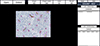
The MVD results obtained using the automated image analyzer were compared with manual counting results. For manual counting, MVD was evaluated by two independent hematopathologists in a blinded manner using a microscope (Zeiss, Jena, Germany), as described previously, with some modifications [12]. First, the slides were scanned at 100×magnification to identify areas showing conspicuously increased MVD (hot spots). Three hot spots were identified per slide and stained vessels, including arterioles and venules, were counted in each hot spot at 400×magnification (0.24 mm2 covered per spot). Round CD34-positive cells showing distinct nuclei were considered as hematopoietic precursors and were excluded from the analysis. Stained cells in the trabecular bone and periosteum were also excluded from the analysis. Finally, the numbers of vessels in the three hot spots were averaged. The hot spot image at 400×magnification (0.24 mm2 covered per spot) was also assessed using the automated image analyzer, and the results from three hot spots per slide were averaged (Fig. 1).
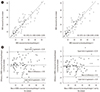
The agreement between the MVD results determined by the hematopathologists and by the automated image analyzer was evaluated by generating an intraclass correlation coefficient (ICC) and Bland-Altman plots. The difference between the MVD results was evaluated by a paired t-test. Statistical analyses were performed using the R software version 3.5.2 (R Foundation for Statistical Computing, Vienna, Austria). P<0.05 was considered statistically significant.
Manual assessment of the MVDs by the two hematopathologists from the 84 BM biopsy samples resulted in a mean±SD of 19.4±11.8 and 20.0±11.8. The MVD results obtained by the hematopathologists demonstrated very good agreement (ICC [95% confidence interval (CI)], 0.984 [0.974–0.990]). However, there was a statistically significant difference between the results obtained by hematopathologists according to the paired t-test (P<0.001). The automated image analyzer resulted in a mean±SD of 19.5±11.2. The MVD results by the analyzer exhibited very good agreement with results by both hematopathologists, with few outliers, based on the Bland-Altman plot (ICC=0.893 [0.840–0.929] and ICC=0.906 [0.859–0.938]) (Fig. 2). No statistically significant difference was observed between the results by the analyzer and the hematopathologists based on the paired t-test.
We developed an automated image analyzer and evaluated its utility for assessing the MVD in BM biopsy samples from MM patients. The MVD measurement showed a very good agreement between the automated image analyzer and hematopathologists. As the two hematopathologists had over 10 years of experience in their specialty, their results showed a very high correlation. One of the hematopathologists tended to consistently count more microvessels than the other, highlighting the need for a more objective evaluation of MVD, especially in hematology laboratories, where experienced pathologists are not present.
As many studies have provided persuasive evidence that MVD has a significant impact on the clinical outcome of MM patients, ongoing studies are examining novel drugs targeting angiogenesis as combination regimens [13141516]. The routine measurement of MVD in MM patients at initial diagnosis can provide additional information for patient care. Methodologies for the automated analysis of IHC images have been developed recently owing to advances in image processing software, especially for cancer diagnosis [171819]. Automated methods can provide rapid and accurate results and eliminate any human-related bias. The automated image analyzer we have developed may provide time- and labor-saving benefits and more objective results in hematology laboratories that evaluate the MVD of MM patients.
 XML Download