

 PDF
PDF ePub
ePub Citation
 Print
Print


INTRODUCTION
To date, the known sources of foreign bodies found in intravenous (IV) lines are drug ampoules, syringes, or the IV set.12 Therefore, if some foreign body is found in IV line, we usually assume that it came from one of these sources.
If we fortunately find a foreign body in IV line before connecting it to the patient, we can retrieve it. However, if the foreign body is found after IV line is connected to the patient, this would cause some anxiety for the patient.
Here we report a case of a worm-like foreign body found in the patient-controlled analgesia (PCA) line, which was later identified as a fibrin clot by pathologic examination.
CASE REPORT
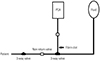
A 22-year-old man (45 kg, 171 cm) with a recurring spontaneous pneumothorax presented for elective surgery. Video-assisted thoracoscopic wedge resection was performed under general anesthesia. General anesthesia was induced with propofol that was administered through an intravenous line inserted in the right antecubital vein. A non-filter intravenous line, a non-return valve, and a 25-cm extension line were used (Fig. 1). An automated blood pressure cuff was placed on the patient's right arm, along with an IV line. The patient was in the right lateral decubitus position with his right arm abducted 90 degrees alongside his body. The operation ended without any incident. Since the patient wanted PCA, we connected PCA to intravenous line in the post-anesthesia care unit (PACU). Two 3-way stopcocks were used near the patient's body, and PCA was connected to the one on the distal side (Fig. 1). We used 100 mL of PCA (AUTOMED® 3200 module, ACE Medical. Co., Seoul, Korea) with a regimen of fentanyl 2500 mcg, nefopam 60 mg, ramosetron 0.6 mg, and normal saline 40 mL at settings of 0 mL continuous flow, bolus of 1 mL, and lockout time of 5 minutes. After PCA connection, no further drugs were administered to the patient via IV line. After confirming full recovery from anesthesia and adequate pain control, the patient was sent to the ward.
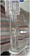
The next day, the patient complained that there was a worm in PCA line (Fig. 1). A white material about 5 mm in length was seen in the patient's PCA line (Fig. 2). No other foreign material was found in IV line. PCA was disconnected immediately, and the material was retrieved. The foreign body was sent to the pathology department of our institute for examination. Pathological examination of the foreign material revealed that it was a fibrin clot. The patient agreed and signed informed consents for the case report.
DISCUSSION
Recently, there have been several reports of foreign bodies being discovered in the IV line. The cases reported finding glass, plastic, rubber, and dissolved solids.12 If even a small particle contained in IV line or fluid enters the body through the patient's vessels, the patient may experience complications, such as phlebitis or pulmonary embolization, depending on the type of foreign body.345
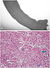
In this case, a foreign body was found in the PCA line connected to the main IV line. It was a long, white deposit that looked just like a worm. However, pathology test result confirmed that it was actually a fibrin deposit (Fig. 3). There was a non-return valve on the IV line connected to the patient's body. Non-return valve is designed to prevent the backflow of blood from the patient's body. Therefore, no one thought that this would be a fibrin clot from the patient's blood. However, even with a non-return valve, regurgitation is not always prevented.678 It is possible that the non-return valve did not work when air was lodged in the disc.8 In this situation, if there is excessive backflow pressure on IV line, such as measuring blood pressure in an arm with IV line, or moving the patient excessively or changing the elevation of IV fluid bag lower than the patient, the patient's blood can be regurgitated, even if there is a non-return valve.89 In this case, a fibrin deposit formed 10 cm proximal to the non-return valve, which means that the blood regurgitated by more than 10 cm. If a significant amount of RBCs remained in the clot to make it look more like a blood clot, the patient and caregivers would have been less upset. However, since there were few RBCs, it was not suspected to be a blood clot. We attempted to reproduce this phenomenon using discarded PCA set and blood, and confirmed that white fibrin clot could be made under certain conditions. Usually, when something is seen in the IV set, it is easy to think that it is a foreign body that came from the fluid, IV line, drugs, and so on. However, it may not be a foreign body, as in this case. Therefore, this case demonstrates two important points. First, blood clots can be formed in IV line due to unexpected blood regurgitation, even when a non-return valve is present; and second, RBCs in the blood clot can be almost washed out, leaving only the fibrin network, which may resemble a worm or a larva. To our knowledge, this case was the first to report the possibility of a white fibrin clot. If caregivers keep this possibility in mind, the patients can be reassured until pathological findings are reported.
 XML Download