Journal List > J Dent Anesth Pain Med > v.15(2) > 1143234

Ping, Kiattavorncharoen, Saengsirinavin, Im, Durward, and Wongsirichat: The efficacy of an elevated concentration of lidocaine HCl in impacted lower third molar surgery

Abstract

Background

There have been few studies on the effect of an elevated concentration of lidocaine hydrochloride in the surgical removal of an impacted lower third molar. This study aimed to examine the efficacy of 4% lidocaine along with 1:100,000 epinephrine compared to 2% lidocaine along with 1:100,000 epinephrine as inferior alveolar nerve block for the removal of an impacted lower third molar.

Methods

This single-blind study involved 31 healthy patients (mean age: 23 y; range: 19-33 y) with symmetrically impacted lower third molars as observed on panoramic radiographs. Volunteers required 2 surgical interventions by the same surgeon with a 3-week washout period. The volunteers were assigned either 4% lidocaine with 1:100,000 epinephrine or 2% lidocaine with 1:100,000 epinephrine as local anesthetic during each operation.

Results

We recorded the time of administration, need for additional anesthetic administration, total volume of anesthetic used. We found that the patient's preference for either of the 2 types of local anesthetic were significantly different (P < 0.05). However, the extent of pulpal anesthesia, surgical duration, and duration of soft tissue anesthesia were not significantly different.

Conclusions

Our study suggested that inferior alveolar nerve block using 4% lidocaine HCl with 1:100,000 epinephrine as a local anesthetic was clinically more effective than that using 2% lidocaine HCl with 1:100,000 epinephrine; the surgical duration was not affected, and no clinically adverse effects were encountered.

INTRODUCTION

An impacted lower third molar is a common abnormality observed in the eruption pattern of human teeth, which requires surgical removal to prevent further complications and to reduce the susceptibility to disease, including bone pathology [1]. Such surgery requires an effective local anesthetic to achieve sufficient nerve block. Inferior alveolar nerve block (IANB) does not always result in successful pulpal anesthesia during the operation, even when the correct injection technique is followed [23]. Researchers have manipulated a range of variables to improve the effectiveness of the local anesthetic used in IANB, such as increasing its volume, modifying its concentration, adjusting its pH, or adding a buffering agent to the local anesthetic cartridge [3456789]. Rood and colleagues have reported the efficacy of 5% lidocaine HCl in routine dental treatment [67810]. Besides 2% lidocaine with epinephrine, which is readily available commercially, 3% lidocaine with norepinephrine has become available in dental cartridge form [11]. However, very few studies report the effect of increasing lidocaine HCl concentration for surgical removal of impacted lower third molars. Thus, the purpose of this study was to examine the efficacy of 4% and 2% lidocaine, with an identical concentration of epinephrine (1:100,000), in IANB for removal of an impacted lower third molar.

MATERIALS AND METHODS

This single blind study included 31 healthy patients (mean age: 23 y, range: 19-33 y) with bilaterally symmetrical impacted lower third molars, as observed on a panoramic radiograph. Table 1 shows the eligibility criteria for this study. The volunteers were injected with either of 2 types of local anesthetics: 4% lidocaine or 2% lidocaine (both with 1:100,000 epinephrine) as nerve block for surgical removal of impacted lower third molars. The operations were performed within a washout period of 3 weeks. Before injection of the local anesthetic, all volunteers were tested for pain perception and pulpal sensitivity on a healthy canine and first or second molar ipsilateral to the third molar scheduled for removal.
The study protocol was approved by the Committee in the Ethics of Research in Human Dentistry and Pharmacy of Mahidol University, Institutional Review Board (Protocol No. MU-DT/PY-IRB 2014/036.0509). The procedure was explained to patients and written consent was obtained from each patient prior to the operation.
The local anesthetic was prepared under sterile conditions immediately before injection by the operator.
  • A. 2% solution-1.8 ml of the local anesthetic in cartridges containing 2% lidocaine with 1:100,000 epinephrine (Novocol Pharmaceutical Inc, Cambridge, Canada) was withdrawn into a 3-ml plastic syringe.

  • B. 4% solution (72 mg lidocaine solution)- 0.02 ml of the local anesthetic in a 2-ml ampule containing 40 mg/ml lidocaine (Jayson Pharmaceuticals Ltd; Dhaka, Bangladesh) was withdrawn using a micropipette. Then, 0.02 ml of 1:1000 epinephrine bitartrate (1 mg/ml) was withdrawn from a 1-ml ampule into the lidocaine ampule.
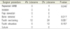
The following formula was used to calculate the volume of the active ingredient:
C(i) × V(i) = C(f) × V(f)
where C(i) = initial concentration, V(i) = initial volume, C(f) = final concentration, V(f) = final volume. Therefore, for 1:100,000 epinephrine, the following volume was required (1:1000) (X) = (1:100, 000) (2 ml); so that X = 0.02 ml of 1:1000 epinephrine 1 mg/ml needed to be added to the is lidocaine solution.
Then, after combining 1.98 ml of 40 mg/ml lidocaine with 0.02 ml of 1:1000 epinephrine, a final 2-ml volume of a solution containing 39.6 mg/ml lidocaine with 1:100,000 epinephrine was obtained. The final solution was drawn into a 3-ml plastic syringe with a 27 G needle attached. Then, 0.2 ml of the solution was flashed out, leaving 1.8 ml in the syringe for injecting into the soft tissue. This solution was prepared for another 3 syringes, for additional anesthesia if required.
IANB was administered using 1.8 ml of anesthetic solution. Following evaluation of anesthesia, another 0.3 ml of the same anesthetic solution was dispensed into the mucosa of the retromolar pad to anesthetize the long buccal nerve in order to reduce intra-operative bleeding, and 0.2 ml was dispensed into the vestibule of the second molar region. Immediately thereafter, removal of the impacted lower third molar was initiated following standard surgical techniques.
Postoperatively, all volunteers were instructed to fill a Patient Record Form (PRF) for evaluating the duration of anesthesia, as indicated by the regain of sensation on the lower lip, and also for recording any unfavorable event that may have occurred postoperatively. Postoperative medications were 500 mg paracetamol (1 tablet, prn q4-6 h for pain), 400 mg ibuprofen (1 tablet, three times a day, after a meal), and 500 mg amoxicillin (1 capsule, 4 times a day, before a meal and before bed), administered orally for 5 days.
The following parameters were assessed:
  1. Type of impaction

  2. Total volume of local anesthetic used during the operation (in ml).

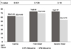
  3. Subjective onset: Vincent sign (in seconds)

  4. Objective onset: Absence of pain to the pinprick test on the canine vestibule and molar lingual vestibule mucosa (in seconds) If the volunteer still responded to a pinprick after 15 min post-injection, the anesthesia was judged to have failed. Then, the operator injected 1.5 ml of the same local anesthetic solution for a second IANB. If local anesthesia was not achieved even after this second injection, the subject was withdrawn from the study.

  5. Pulpal anesthesia: After administration of the local anesthetic, an electric pulp testing (EPT) value of 80 µA indicated profound pulpal anesthetic [1213141516]. Ten minutes post-injection, EPT was performed at the healthy canine and lower first or second molar, on the side of the injection and the obtained EPT values were compared to the base line values.

  6. Pain assessment: The 100-mm visual analogue scale (VAS) was used for measuring pain during the procedure. This scale uses 4 levels of pain, which is counted from the extreme left side and is categorized as: no pain (0-4 mm), mild pain (5-44 mm), moderate pain (45-74 mm), and unbearable pain (75-100 mm) [17]. The VAS was measured when the drug was dispensed into the tissue for the first time, during the operation, and 1-h post operation.

  7. Duration of local anesthesia: This was recorded by each patient in the patient report chart and signified the time lapsed from the subjective onset of anesthesia to the time when the numbness at the lower lip wore off (in minutes).

  8. Incidence of postoperative severe adverse effects: This was observed by the surgeon and also reported by the patient during the operation and in the first few hours after the operation (e.g., dizziness, anxiety, any neuron dysfunction, etc.).

  9. pH of local anesthetic solutions: This was measured using a millivolt pH meter (Orion 3 Star pH Benchtop; Thermo Scientific, Waltham, MA, USA)

    a. 4% lidocaine without epinephrine
    b. 4% lidocaine with epinephrine 1:100,000
    c. 2% lidocaine with epinephrine 1:100,000
Paired t-test or Wilcoxon's signed rank test was used to compare the difference in outcome variables between the 2 treatments in the same patient. Outcomes of interest were: VAS during disposal of local anesthetic solution, hemodynamic changes, subjective and objective onset of anesthesia, distribution of the volunteers according to the occurrence of pulpal sensibility after 10 min, duration of anesthesia, and intensity of intra-operative pain.
McNemar's test was used to calculate the distribution of patients in each group in terms of the need for repeated anesthesia and the total amount of anesthetic solution administered.

RESULTS

The control group had a pH = 3.96, which was lower than that of the test group (pH = 5.79), after mixing of 4% lidocaine (pH = 5.83) with epinephrine (pH = 3). The type of impaction was accessed using a panoramic radiograph. All cases had bilaterally symmetrical impaction; mesial angulation was observed in 41.9% of the cases, followed by horizontal (38.7%), and vertical angulation (19.4%).
Table 2 shows that the onset of local anesthesia was significantly different for the 2 types of IANB, both subjectively and objectively. The onset of anesthesia was significantly more rapid with 4% lidocaine than 2% lidocaine (subjective onset: 113 ± 63 and 159 ± 57 min, respectively, after administration; objective onset: 5.46 ± 2.02 and 8.19 ± 4.24 min, respectively). Moreover, the duration of anesthesia with 4% lidocaine was longer than that with 2% (228 ± 55 min vs. 207 ± 54 min), although this was not significantly different. Additionally, there was no significant difference between the solutions in the surgical time required for the interventions.
Table 3 shows the intensity of intra-operative pain that volunteers rated in the 0-100 mm VAS. In terms of pain during the administration of the drug into the soft tissue (Table 3), the 4% lidocaine group experienced more pain (20.77 ± 23.3 mm) than the 2% group (16.51 ± 14.21 mm), but the difference was not statistically significant. During tooth-sectioning, however, the difference between the 2 types of anesthetic solutions was nearly significant (P = 0.052; Table 3 and 4). There was no significant difference between the 2 types of local anesthetics in terms of intensity of intra-operative pain.
Table 4 shows the degree of intra-operative pain, from the absence of pain to severe pain, based on the VAS value as rated by the volunteers.
In this study, the EPT device was used to confirm pulpal anesthesia. Based on the status of healthy teeth, 62 canine, 58 first molars, and 4 second molars were tested. The results showed no significant differences between the solutions in terms of baseline values of pulpal anesthesia. The distribution of the volunteers according to the occurrence of pulpal sensibility after 10 min was not significantly different between the groups (Fig. 1). Among all cases, we found that the 4% lidocaine solution was superior to the 2% lidocaine solution in terms of pulpal anesthesia, but the difference was not statistically significant. With 4% lidocaine, 10/31 canines (32.3%), and with 2% lidocaine, 9/31 canines (29%), showed EPT values of 80 µA. Among the molars examined, 19/31 (61.3%) in the 4% lidocaine group and 16/31 (51.6%) in the 2% lidocaine group showed EPT values of 80 µA; however, none of the differences were statistically significant.
The proportion of cases requiring additional anesthetic was significantly greater in the 2% lidocaine group than in the 4% group (26/36 vs. 16/31; P = 0.006). The requirement for repeated IANB (P = 0.046) and additional anesthetics during the tooth-sectioning period (P = 0.001) are shown in Table 5. The 2% lidocaine group required a significantly greater total volume of anesthetic to control intra-operative pain than the 4% group (2.91 ± 0.58 ml (range 2.3-4.7 ml) vs. 2.49 ± 0.21 ml (range 2.3-2.9 ml), P = 0.001).
Postoperatively, there were no signs of nerve dysfunction, paresthesia, or infection reported or assessed. Only a few volunteers reported a longer-acting anesthesia in the 4% lidocaine group, as shown in Table 2, but these values were not significantly different.
In terms of overall intra-operative comfort, volunteers had a significantly higher preference for 4% lidocaine (77.4%) than for 2% (22.6%) (P = 0.004).

DISCUSSION

Volunteers with bilaterally symmetrical impaction provide us with the opportunity to conduct a blind split-mouth clinical study; in this way, the individual bias based on the difference in the degree of discomfort and the intensity of the pain response can be reduced. Some previous studies have addressed the effects of anesthetic solutions in third molar surgery without a split-mouth design, which confounds the comparison of the results [18]. We found that onset of anesthesia in the 4% lidocaine group was significantly faster, both subjectively and objectively, than in the 2% group. Our results were similar to those of a study by Chaney et al. [2]. The onset of local anesthesia can be influenced by the pH of the solution. We found that the test solution had a higher pH than the standard solution (5.79 vs. 3.96). A literature review indicated that on the injection side, the local anesthetic is converted to a quaternary salt (BH+) and a tertiary base (B), which is one of the core factors that enhances the onset of anesthesia, via the pKa and the pH of physiological tissue [1920]. It has been suggested that a rapid onset is promoted by a high pH [321]. Recently, Whitcomb et al. [3] found that adjustment of the pH of the lidocaine to 7.5 did not have any negative effects. Acidic solutions have been reported to produce a burning sensation in the tissue during drug administration [20]; however, in our research, the incidence of pain during drug administration was not significantly different between high and low pH solutions. These results corroborated those of other previous studies on pH-adjusted local anesthetics [23922]. The rating of moderate (3%-16%) and severe pain (3.22%; Table 4) in our groups was lower than that reported in a study by Ridenour (2001), which rated moderate pain as 13%-23% and severe pain as 7% [22]. In terms of the VAS, the test and control groups rated pain as 20.77 ± 23.3 and 16.51 ± 14.21 mm, respectively, but these results were not significantly different.
The duration of soft tissue numbness is shown in Table 2. Our study showed that the 2% lidocaine group had approximately 207 ± 54 min of soft tissue anesthesia. This finding was similar to that of Becker and Reed, Haas, and Thomson et al, which found that soft tissue anesthesia with 2% lidocaine with 1:100,000 epinephrine ranged from 3 to 5 h [1923]. On the other hand, in the 4% lidocaine group, anesthesia lasted 228 ± 55 min, which was superior to that in the 2% lidocaine group, even though the differences between the 2 groups were not significantly different. In the test group, the longer duration of soft tissue anesthesia may have been due to the higher concentration of lidocaine, as both solutions contained the same concentration of epinephrine.
Tables 3 and 4 show the degree of intra-operative pain that the volunteers rated on the 0-100 mm VAS. In both groups, it was noted that the surgical removal of an impacted lower third molar was a painful operation, especially during the tooth-sectioning period, and required supplementary local anesthetic. The pain scale showed that the pain sensation remained between mild to moderate in both groups, but that the 4% lidocaine group experienced less intra-operative pain, although the difference was not significant. We also found that patients significantly preferred 4% lidocaine to 2% for surgical removal of impacted lower third molars (P = 0.004), because of reduced intra-operative pain. This result is in agreement with that of Rood's pilot study [6]. Another double-blind trial conducted by the same author showed that the success rate of IANB for the extraction of pulpally inflamed teeth was higher with 5% lidocaine (93%) with 1:80,000 epinephrine than with the standard 2% solution (22%).
Complications and adverse effects of anesthesia were other important variables that were evaluated in our research. Postoperatively, the volunteers were placed in a comfortable room for 1 h and were observed for any immediate side effects of the drug. None of the volunteers reported any adverse complications or paresthesia postoperatively or on the 7th-day follow-up visit. These findings were consistent with those of previous reviews and studies that have stated that lidocaine is the gold standard local anesthetic with low toxicity [2024]. However, a high concentration of lidocaine has been reported to produce neurotoxicity in some in vitro studies [25]. Controversially, there have been no reports of neurotoxicity in humans [24]. Additionally, a study by Eldridge and Rood reported that the use of 50 mg/ml lidocaine with 1:80,000 epinephrine for routine dental treatment did not show any adverse clinical events, following which, this local anesthetic solution was used in a pediatric patient clinical trial, in which it was considered to be safe and in which no adverse reactions were observed [58]. In 1978, Rood evaluated the plasma levels of lidocaine after injection of equal doses of different concentrations of lidocaine (1 ml of 5% lidocaine versus 5 ml of 1% lidocaine). The solution was infiltrated into the maxillary buccal sulcus of the second premolar at separate appointments. After 2 h of evaluation, the results showed that the lidocaine plasma level was between <0.1 and 0.2 µg/ml in the 5% lidocaine group. However, it may have reached a toxicity level in the 1% lidocaine group (0.1-5 µg/ml). The paper finally concluded that injection of a higher concentration of lidocaine did not produce a notable leveling of the lidocaine plasma concentration in healthy adults [10]. Evidence of lidocaine plasma leveling toxicity may commence at concentrations > 5 µg/ml, but convulsive seizures generally require concentrations > 10 µg/ml [19]. Although adverse clinical reactions were not observed in our study, the plasma level associated with 4% lidocaine HCl should be investigated further in future.
EPT has become the standard research method to identify the extent of pulpal anesthesia after endodontic administration of local anesthetic [1213141516]. In our study, there was no statistical difference in the percentage of teeth that achieved the maximum EPT output of 80 µA after 10 min post-injection with either solution (Fig. 1), although we found a higher percentage of maximum EPT output in the 4% lidocaine group than in the 2% lidocaine group. Interestingly, even when the EPT reached the maximum output of 80 µA, some patients still complained of pulpal sensitivity during tooth sectioning. It may have been because our procedure was more traumatizing to the pulp nerve bundle. Based on these findings, we suggest that a maximum output of 80 µA by the EPT device may not represent complete pulpal anesthesia in impacted lower third molar surgery during tooth sectioning. Moreover, the total volume of local anesthetic used in each group was significantly higher in the 2% lidocaine group than in the 4% lidocaine group.
In our study, four cases in the 2% lidocaine group required repeated IANB, although those were subjective in nature and had reported complete lower lip numbness at the injection site. It is possible that failure of the injection or the effectiveness of the standard drug solution was inadequate to produce a sufficient objective onset of anesthesia. Moreover, there was a greater requirement for additional local anesthetic in the 2% lidocaine group; this need was significantly greater during the tooth-sectioning period, which may have been due to an insufficient volume of anesthetic to control pain during the operation.
Based on our results, we suggest that 4% lidocaine with 1:100,000 epinephrine can be used as a standard local anesthetic in dentistry, especially during the surgical removal of an impacted lower third molar. This concentration of lidocaine did not produce any adverse reaction in healthy adult patients in our study. Patients also significantly preferred it to 2% lidocaine for reducing operative pain.

Figures and Tables

Fig. 1

Distribution of the volunteers according to the occurrence of pulpal sensitivity at 10 min after inferior alveolar nerve block administration, based on electric pulp testing.

jdapm-15-69-g001
Table 1

Eligibility criteria used in this study

jdapm-15-69-i001
Table 2

Subjective onset, objective onset, and duration of local anesthesia and surgery

jdapm-15-69-i002
Table 3

Intensity of intra-operative pain

jdapm-15-69-i003
Table 4

Intensity of intra-operative pain

jdapm-15-69-i004
Table 5

Number of additional anesthetics administration

jdapm-15-69-i005

*IANB: inferior alveolar nerve block

Acknowledgment

We would like to express our gratitude to the patients who participated in our study. We also thank Dr. M. A. Wadud Sarker and Dr. Md. Asikul Wadud who provided us with the 4% lidocaine solution (Jayson Pharmaceuticals) and thank Dental Siam Co., Ltd. that provided us with the 2% lidocaine solution (Novocol Pharmaceutical) for our research. We would like to thank all the staff of the Department of Oral and Maxillofacial Surgery, Faculty of Dentistry, Mahidol University. for their help in providing the facilities and support during our study.

Notes

Funding none

Competing Interests none

Ethic Approval Committee in the Ethics of Research in Human Being of Dentistry and Pharmacy Mahidol University, Institutional Review Board with Protocol No. MU-DT/PY-IRB 2014/036.0509.

References

1. Rakprasitkul S. Pathologic changes in the pericoronal tissues of unerupted third molars. Quintessence Int. 2001; 32:633–638.
2. Chaney MA, Kerby R, Reader A, Beck FM, Meyers WJ, Weaver J. An evaluation of lidocaine hydrocarbonate compared with lidocaine hydrochloride for inferior alveolar nerve block. Anesth Prog. 1991; 38:212–216.
3. Whitcomb M, Drum M, Reader A, Nusstein J, Beck M. A prospective, randomized, double-blind study of the anesthetic efficacy of sodium bicarbonate buffered 2% lidocaine with 1:100,000 epinephrine in inferior alveolar nerve blocks. Anesth Prog. 2010; 57:59–66.
crossref
4. Nusstein J, Reader A, Beck FM. Anesthetic efficacy of different volumes of lidocaine with epinephrine for inferior alveolar nerve blocks. Gen Dent. 2002; 50:372–375.
5. Eldridge DJ, Rood JP. A double-blind trial of 5 per cent lignocaine solution. Br Dent J. 1977; 142:129–130.
crossref
6. Rood JP. Inferior alveolar nerve blocks. The use of 5 per cent lignocaine. Br Dent J. 1976; 140:413–414.
crossref
7. Rood JP, Sowray JH. Clinical experience with 5 per cent lignocaine solution. Journal of Dentistry. 1980; 8:128–131.
crossref
8. Sandy J, Rood JP. Five per cent lignocaine solution in children's dentistry. Journal of Dentistry. 1980; 8:312–314.
crossref
9. Vreeland DL, Reader A, Beck M, Meyers W, Weaver J. An evaluation of volumes and concentrations of lidocaine in human inferior alveolar nerve block. J Endod. 1989; 15:6–12.
crossref
10. Kanaa MD, Meechan JG, Corbett IP, Whitworth JM. Speed of injection influences efficacy of inferior alveolar nerve blocks: a double-blind randomized controlled trial in volunteers. J Endod. 2006; 32:919–923.
crossref
11. Hannan LRA, Nist R, Beck M, Meyers WJ. The use of ultrasound for guiding needle placement for inferior alveolar nerve blocks. Oral Surg Oral Med Oral Pathol Oral Radiol Endod. 1999; 87:658–665.
crossref
12. Dagher FB, Yared GM, Machtou P. An evaluation of 2% lidocaine with different concentrations of epinephrine for inferior alveolar nerve block. J Endod. 1997; 23:178–180.
crossref
13. Yared GM, Dagher FB. Evaluation of lidocaine in human inferior alveolar nerve block. J Endod. 1997; 23:575–578.
crossref
14. Hannan L, Reader A, Nist R, Beck M, Meyers WJ. The use of ultrasound for guiding needle placement for inferior alveolar nerve blocks. Oral Surg Oral Med Oral Pathol Oral Radiol Endod. 1999; 87:658–665.
crossref
15. Jensen MP, Chen C, Brugger AM. Interpretation of visual analog scale ratings and change scores: a reanalysis of two clinical trials of postoperative pain. J Pain. 2003; 4:407–414.
crossref
16. Moore PA, Boynes SG, Hersh EV, DeRossi SS, Sollecito TP, Goodson JM, et al. The anesthetic efficacy of 4 percent articaine 1:200,000 epinephrine: two controlled clinical trials. J Am Dent Assoc. 2006; 137:1572–1581.
17. Becker DE, Reed KL. Local anesthetics: review of pharmacological considerations. Anesth Prog. 2012; 59:90–101.
crossref
18. Malamed SF. Handbook of Local Anesthetisia. 6 ed. St. Louis, Missouri: Mosby;2014.
19. Hobeich P, Simon S, Schneiderman E, He J. A prospective, randomized, double-blind comparison of the injection pain and anesthetic onset of 2% lidocaine with 1:100,000 epinephrine buffered with 5% and 10% sodium bicarbonate in maxillary infiltrations. J Endod. 2013; 39:597–599.
crossref
20. Ridenour S, Reader A, Beck M, Weaver J. Anesthetic efficacy of a combination of hyaluronidase and lidocaine with epinephrine in inferior alveolar nerve blocks. Anesth Prog. 2001; 48:9–15.
21. Haas DA. An update on local anesthetics in dentistry. J Can Dent Assoc. 2002; 68:546–551.
22. Ping B, Kiattavorncharoen S, Saengsirinavin C, Durward CS, Im P, Wongsirichat N. Effect of high concentration lidocaine for mandibular teeth anesthesia: Review literature. M Dent J. 2014; 34:364–372.
23. Kanai Y, Katsuki H, Takasaki M. Lidocaine disrupts axonal membrane of rat sciatic nerve in vitro. Anesth Analg. 2000; 91:944–948.
crossref
24. Hiller A, Karjalainen K, Balk M, Rosenberg PH. Transient neurological symptoms after spinal anaesthesia with hyperbaric 5% lidocaine or general anaesthesia. Br J Anaesth. 1999; 82:575–579.
crossref
25. Rood JP, Cannell H. Plasma levels of lignocaine after peri-oral injections of two different concentrations. Pharmacol Ther Dent. 1978; 3:45–47.
TOOLS
Similar articles