

 PDF
PDF ePub
ePub Citation
 Print
Print


Beckwith-Wiedemann syndrome is mainly characterized by macroglossia, omphalocele, and gigantism. It was first described by Beckwith in 1963 and later substantiated by Wiedemann in 1964 [123]. Macroglossia is a condition observed in patients with this syndrome, and is associated with difficult perioperative anesthetic management [456]. It has been reported that perioperative anesthetic management might be complicated by anatomical airway abnormalities [123456]. Here, we report the perioperative airway management of a 14-month-old girl with Beckwith-Wiedemann syndrome who underwent a procedure for the reduction of macroglossia.
Informed consent was obtained from the patient for this case report.
CASE REPORT
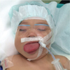
The patient was a 14-month-old girl, 78.2 cm in height and 9.8 kg in weight. She was diagnosed with Beckwith-Wiedemann syndrome based on a chromosomal analysis. Her clinical features included macroglossia, exomphalos, and earlobe grooves in both sides. Although the patient's tongue was remarkably large and protrusive, she was able to eat and drink without any problem. No apparent history of apnea and seizure was observed. Auscultation of heart and lungs was within the normal limits, although the clinical assessment of her airway was Mallampati's grade IV, with a mouth opening of 2 cm. Blood oxygen saturation level (SpO2) was 100% in room air. No abnormal findings were observed on chest X-ray and laboratory data, and her cardiac function was normal.
In this case, we had scheduled tracheal intubation management after the tongue reduction procedure and transferred the patient to the intensive care unit (ICU) until the tongue edema decreased.
At admission for a tongue reduction procedure under general anesthesia, her heart rate (HR) was 156 bpm and SpO2 was 100% on room air. On the day of the surgery, no premedication was administered before transferring her to the operating room. Anesthesia was induced with administration of sevoflurane (1-8%) in oxygen after the start of the noninvasive monitoring for SpO2 (100%). After the loss of consciousness, mask ventilation was provided for the oral airway. Subsequently, monitoring of electrocardiogram (ECG), blood pressure (BP), and HR was initiated. Fentanyl (20 mcg), atropine (50 mcg), and rocuronium (6 mg) were administered after achieving peripheral intravenous access. Intubation was easily performed with a 4.0-mm uncuffed tracheal tube using the Macintosh laryngoscope blade (size 2; Fig. 1). The Cormack-Lehane classification was grade I. Anesthesia was maintained with sevoflurane (1-3%) in air and oxygen. BP was maintained at 80–108/40–48 mmHg; HR was 130-140 bpm and end-tidal carbon dioxide (EtCO2) was 35-40 mmHg. The tongue reduction procedure was completed in 71 minutes without any surgical and/or other anesthetic problems. The blood loss was minimal during the surgery, and she received a total of 213 mL lactated Ringer's solution with 1% glucose. Urine volume was 35 mL. At the end of the surgery, we administered a bolus of midazolam (1 mg), and a combination of dexmedetomidine (0.6 mcg/kg/h) and midazolam (1 mg/h). Tracheal intubation was continued; she was observed in the ICU under controlled ventilation (synchronized intermittent mandatory ventilation [SIMV]). As tongue edema was resolved on postoperative day 1, we discontinued the administration of sedatives, and she gained consciousness in about 10 minutes. Then we assessed for air leak (pressure < 30 mm Hg) and signs of sufficient respiratory condition (body movement, eye opening, regular breathing pattern [> 15], gag reflex, and SpO2 was 100%). Subsequently, we removed the tracheal tube. After extubation, her respiratory and hemodynamic conditions were stable. No remarkable changes and/or complications were observed since the surgery, and the patient was discharged from the hospital 5 days later.
DISCUSSION
Beckwith-Wiedemann syndrome is caused by mutations in the genes; the prevalence of this condition is 1 per 13,700-15,000 births, with equal sex distribution [3456]. Clinical features of the syndrome include macroglossia, omphalocele, umbilical hernia, and neonatal hypoglycemia [4567]. Macroglossia is especially associated with Beckwith-Wiedemann syndrome in 95% of the patients [8]. Early treatment with surgical reduction of the tongue might often be required to prevent respiratory complications and to improve feeding.
Anesthetic administration is difficult because of the airway obstruction caused by macroglossia. Although awake intubation in pediatric patients might be difficult, inhalational induction with sevoflurane in 100% O2 can be used as an alternative method because it is safe [5]. The administration of high concentrations of sevoflurane or muscle relaxants to the patients lying in the supine position might cause the tongue to fall into the retrolingual space, which could lead to severe airway obstruction during the induction of anesthesia [1]. Thus, the following strategy was followed for the anesthetic administration. Ventilation via a mask was performed using gradually increased sevoflurane (1-8%) with an oral airway. Sevoflurane can be safely used in the induction of general anesthesia, because of low fat:blood solubility [9]. After achieving mask ventilation, rocuronium was administered for neuromuscular blockade. It has been reported that maintaining spontaneous breathing is important during anesthesia induction in patients with upper airway problem [10]. Additionally, the adequate depth of sevoflurane anesthesia provides muscle relaxation. However, recent studies have showed that the rates of difficult intubation increase without muscle relaxants. Muscle relaxants might be effective for suppressing laryngospasm induced by stimulation using the laryngoscope [11]. Therefore, we administered muscle relaxant, with a preparation of sugammadex for the reversal of muscle relaxation, in case the intubation was unsuccessful [10].
Preparation before the induction of anesthesia is especially important in cases that may show potential for difficult ventilation and intubation. In the operating room, different-sized masks, tracheal tubes, nasal and/or oral airways, a stylet, laryngeal masks, fiberscope, and a tracheostomy set should be kept available [35].
Airway obstruction due to postoperative tongue edema is an important problem [1213]. Postoperative edema can potentially cause airway obstruction. The criteria for extubation after a tongue reduction procedure are unclear. The surgical procedure to be followed would depend on the severity of the tongue edema. In this case, we performed intubation to protect the airway from post-surgical edema. At extubation on postoperative day 1, we assessed for air leak and signs of sufficient spontaneous respiration. Then we removed the tracheal tube.
In summary, we reported a case of Beckwith-Wiedemann syndrome in which perioperative upper airway obstruction posed a challenge for anesthetic management. Prior preparation and strategies to manage the airway obstruction is essential for the successful administration of anesthesia.
 XML Download