

 PDF
PDF ePub
ePub Citation
 Print
Print


Nasotracheal intubation (NTI) is a common procedure performed in the operating room, especially in patients undergoing oral and maxillofacial surgeries. NTI improves the efficiency of surgical procedures for intra-oral pathological anomalies, structural abnormalities, and maxillofacial fracture and orthognathic surgery [1]. However, NTI may cause complications if the tube comes in contact with the surrounding structures when it passes through the nasopharyx and oropharynx. The incidence of cleft lip and palate (CLP) was 1 per 554 births in the Republic of Korea. Up to 20% of patients with cleft palate will develop velopharyngeal dysfunction after primary palatoplasty, requiring further treatment with pharyngoplasty [2]. Many patients will undergo further orthognathic surgeries, and NTI is performed under general anesthesia during these surgeries. If a pharyngeal flap is present, NTI can lead to tearing of the flap, which may result in a traumatic massive hemorrhage. In this case report, we describe a case of NTI-related pharyngeal flap damage in a patient who underwent palatoplasty and pharygoplasty previously.
CASE REPORT
A 20-year-old man (height, 177 cm; weight, 60 kg) was hospitalized for an orthognathic surgery. He was diagnosed with bilateral CLP and mandibular prognathism associated with midface deficiency, and was scheduled to undergo LeFort I osteotomy, right iliac block bone graft procedure, and bilateral sagittal split osteotomy. He had previously undergone cheiloplasty, palatoplasty, cleft maxillary repair, and pharyngoplasty to correct the CLP. Preoperative laboratory findings, chest X-ray image, and electrocardiogram were normal. He had no other diseases, including upper airway obstruction or obstructive sleep apnea symptoms.
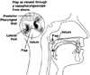
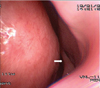
He had maxillary deficiency with malocclusion, mandibular prognathism, a scar due to a pharyngeal flap, and cheiloplasty. As a premedication, glycopyrolate (0.2 mg) was injected intramuscularly. Standard monitoring was performed in the operating room. After preoxygenation with 100% oxygen, general anesthesia was induced with remifentanil (1 µg/kg), propofol (120 mg), and rocuronium (50 mg). After providing adequate muscle relaxation, flexible fiberoptic bronchoscope (FFB)-guided NTI was attempted using a nasotracheal tube (NTT) of 6.5 mm internal diameter; this procedure was performed instead of a direct rigid laryngoscope because FFB technique and smaller sized NTT are more suitable for adults who have intact pharyngeal flaps. The FFB was inserted into the both nostrils for examination and passed through the posterior pharynx; we observed that the velopharyngeal port (between the nasopharynx and oropharynx) was divided into two small-sized pharyngeal ports by the pharyngeal flap (Fig. 1). Because right side of pharyngeal port was a little wider, a scope was passed carefully through the right side pharyngeal port and vocal cords to the carina; NTT was passed over the FFB smoothly. There was no specific resistance during NTT insertion, but minor bleeding appeared on the nasopharynx. After the intubation, the patient was mechanically ventilated to administer desflurane (6 vol%), with a fresh gas flow rate of 3 L/min and 50% oxygen in air. After the induction of anesthesia, active bleeding was observed in the mouth immediately. To assess the bleeding focus, we examined the oral cavity with a dental mirror, and bleeding was seen at the pharyngeal flap. The flap was probably torn at the time of the NTI. Subsequently, hemostasis was achieved using a 4 × 4 gauze moistened with 1:1000 epinephrine solution. After the bleeding was controlled, cotton swabs soaked in 2% lidocaine and 1:1000 epinephrine were inserted into the torn flap and nostril. Subsequently, the pharyngeal bleeding stopped, and the surgery was completed without further complications. After the surgery, the patient recovered without any problems. A follow-up examination using a nasendoscopic view that was performed two years later showed that the pharyngeal flap remained intact after the recovery (Fig. 2). For this case presentation, we received consent from the patient.
DISCUSSION
Patients with congenital CLP must undergo a number of corrective surgical procedures during their infancy and early childhood. The scars that result from these procedures have been shown to affect the growth of the maxilla, and this often leads to maxillary deficiency [3]. Many patients with CLP require orthognathic surgery during their childhood for the correction of the skeletal asymmetry and often pharyngoplasty is performed with pharyngeal flap (in 20% of patients) to improve the quality of speech and velopharyngeal function [4]. This procedure causes a scar that obstructs the velopharyngeal port. Thus, the pharyngeal flap can be damaged during NTI, and it can lead to significant bleeding, thereby causing damage to the airway.
Bell et al. suggested that preoperative nasendoscopy must be performed in patients who underwent pharyngoplasty previously and were scheduled to undergo NTI [5]. Nasendoscopy is a minor procedure that is usually performed in the ear-nose-throat clinic to assess the structures of the nose and throat. This is useful for evaluating the position, shape, and size of the velopharyngeal port. However, this procedure requires the cooperation of the patient; hence, performing this procedure in patients below 6 years of age can be difficult [6].
A few guidelines for performing NTI in patients with a pharyngeal flap have been provided in the literature. For minimizing the damage to a pharyngeal flap, it is essential to establish some strategies for performing NTI in these patients. First, a suitable vasoconstrictor should be used prior to intubation. If pharyngoplasty is performed in patients without a nasendoscopic examination, a vasoconstrictor preparation should be applied to the velopharyngeal port to prevent bleeding [7]. In our case, hemostasis was achieved with 2% lidocaine and 1:1000 epinephrine solutions. However, prior to the insertion of FFB and NTT through the nose, it would be better to pre-treat the velopharyngeal port with a vasoconstrictor. The bleeding due to turbinates and septum damage also interferes with visualization; thus, it is necessary to pre-treat the nose with a vasoconstrictor. Second, after checking the pharyngeal flap with nasendoscopy, FFB must be used to visualize the pharyngeal flap ostium [8]. Furthermore, FFB-guided NTI must be performed with utmost care because NTI can injure the flap. Epistaxis is the most frequently observed complication, with an incidence ranging from 10% to 80% [9]. Other complications include impingement of the endotracheal tube (ETT) in the subglottic region, thereby further hindering the advancement of the tube after passing the glottis; sinusitis; bacteremia; and dislodgement of the adenoids [101112]. In addition, pharyngoesophageal perforation and inadvertent intracranial placement of NTT have also been reported [13]. Third, small-sized NTT should be used, and the tube may also be softened by soaking it in warm saline and well lubricated with lidocaine jelly to decrease the incidence of epistaxis and pharyngeal flap damage [14].
This case report emphasizes that thorough history taking of the patient is essential. If a patient who has a possibility of velopharyngeal obstruction undergoes oral maxillofacial surgery, the aforementioned strategies must be followed to prevent the damage of pharyngeal flap.
 XML Download