

 PDF
PDF ePub
ePub Citation
 Print
Print


INTRODUCTION
Sedation during dental treatment can minimize the patient's anxiety and fear and increase the approachability of dental treatment, thereby improving the quality of care. Sedation can be classified as minimal, moderate, or deep sedation, according to its depth. In clinical practice, the depth of sedation can be varied according to the anxiety level of the patient, as well as the degree of stimulation. Drugs used for sedation typically suppress respiratory function and decrease respiratory rate. Since unintended entry into deep sedation can cause respiratory distress and resulting hypoxemia, respiration monitoring is necessary [1]. If respiratory depression is not detected early, serious complications, including death, can occur [23].
The traditional method of respiration monitoring is visual observation of thoracic and abdominal movements to assess respiratory rate and the depth of respiration. Continuous monitoring of respiration sound, by fixing an auscultation bell on the patient's anterior chest or trachea, can also be applied. Many dental sedation guidelines recommend respiratory monitoring [45]. However, when the attending physician is focused on the procedure, auscultation or observation of chest wall movement may be difficult. Therefore, respiratory monitoring by using capnography is considered the standard of care for respiration monitoring; the use of capnography monitoring is especially important when moderate and deep sedation are in place [56]. This is a non-invasive method that measures the partial pressure of CO2 in inspired and expired gas, then expresses it in waveforms that indirectly evaluate the respiratory rate, respiration pattern, and alveolar gas exchange.
Recently, a device that measures acoustic respiratory rate (RRa) for respiration monitoring was introduced in clinical practice. A sensor is attached in the cervical area near the trachea, which measures respiratory sounds. RRa has been reported to have higher specificity than capnography in monitoring apnea [7], but it may be affected by noise, since the device uses acoustics. An ultrasonic scaler, a commonly used device in dental practice, causes vibration and noise that may affect the accuracy of the acoustic respiration monitoring device. This study aimed to evaluate the effect of noise from the ultrasonic scaler on RRa respiration monitoring.
MATERIALS AND METHODS
This study used data from a previous study on patient-controlled sedation, approved by the Seoul National University IRB (the clinical research information service [CRiS], Republic of Korea [https://cris.nih.go.kr/cris/index.jsp], registration number KCT0001618) [8]. Sixty adult volunteers, 20–60 years of age, with American Society of Anesthesiologists statuses of I or II, were allocated to propofol and midazolam groups.
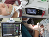
Before the administration of drugs, a nasal cannula (3469DU-00 LoFloTM CO2 sampling O2 delivery nasal cannula, Respironics Co. Inc, MN, USA) was placed in the nasal cavity and philtrum of all subjects. Subjects were administered 2 L/min of oxygen; end-tidal CO2 partial pressure and respiratory rate were measured via a capnography monitoring device (LoFloTM CO2 Sensor, Respironics Co. Inc, MN, USA) and recorded with a patient monitor (BM7, Bionet, Seoul, Korea) (Fig. 1A, 1B). An RRa sensor (Masimo Corp., Irvine, CA, USA) was attached to each subject's right cervical area near the trachea for the monitoring of respiratory rate (Fig. 1A, 1C).
The clinical monitoring in this study was divided into preparation, sedation, and scaling periods. Only monitoring was performed without sedative administration during the preparation period. During the sedation period, each subject was given sedatives using patient controlled infusion system. In the scaling period, dental scaling was performed under sedation (Fig. 1D). RRa settings were respiratory pause at 15 s, averaging time at “No Averaging,” freshness at 1 min. The “no breath detected” time-out on the capnography monitor was set to 20 s.
Respiratory rates from capnography and RRa were stored as capnogram waves on a computer. Respiratory rate over 3 min from each period were extracted from stored data for this analysis. Three sets of data, 3-min in length, were collected from the following periods: immediately before drug administration in the preparation period, at 5 min after the start of drug administration in the sedation period, and at the time of the initialization of the use of scaler in the scaling period.
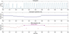
Respiratory rate was measured by using RRa at 2-s intervals, and capnography data were extracted at 2-s intervals to match. The two monitoring devices were synchronized for measured time, and times when the respiratory rate was not recorded were defined as “missing values.” (Fig. 2).
Means and standard deviations of the respiratory rates measured with the two devices over the three periods were calculated; the probabilities of missing values were compared with chi-squared test. Respiratory rates measured by RRa and capnography in each period were analyzed for correspondence by using correlation coefficient and Bland-Altman analysis. P < 0.05 was considered statistically significant.
RESULTS
After the extraction and review of data from 60 subjects, 49 subjects with confirmed respiration on capnogram wave were included in the analysis. The average age of the subjects was 26.6 ± 5.6 years, average height was 172.2 ± 7.6 cm, and average weight was 69.4 ± 13.0 kg. Thirty-five subjects were male and 14 were female.
Ninety samples were collected over 3 min from each subject and period, totaling 13,230 samples from 49 subjects. Respiratory rates by period are shown in Table 1. Missing values not recorded by RRa occurred 248 times in the preparation period, 355 times in the sedation period, and 1056 times in the scaling period; thus, there was a greater number of missing values during scaling (P < 0.001). Apnea, assessed by RRa measurement, occurred 39 times in the preparation period, 37 times in the sedation period, and three times in the scaling period. Apnea, assessed by capnography, occurred zero times in the preparation period, 14 times in the sedation period, and 11 times in the scaling period.
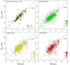
Correlation coefficients for respiratory rate, measure by two separate methods, were 0.692 (95% CI: 0.675–0.707) in the preparation period, 0.677 (95% CI: 0.659–0.693) in the sedation period, and 0.562 (95% CI: 0.546–0.591) in the scaling period. Fig. 3 shows the distribution of correlation coefficients.
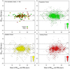
Respiratory rates of the 49 subjects during each period, measured by RRa and capnography, were compared by using Bland-Altman analysis. The mean capnography-RRa biases in Bland-Altman analyses were −0.03, −0.27, and −0.61 in each respective period (P < 0.001), and limits of agreement were −4.84–4.45, −4.89–4.15, and −6.18–4.95 (P < 0.001) (Fig. 4).
DISCUSSION
Detection of respiratory depression or apnea by respiratory rate measurement is very important for safe sedation [4]. Visual inspection of the chest wall movement or auscultation of the respiratory sounds is the traditional method of respiratory monitoring. However, various respiratory rate monitoring methodologies have been introduced, such as impedance pneumography (measures respiratory rate via electrodes on the chest wall) [9], piezoelectric plethysmography, or fiberoptic plethysmography [10]. However, in clinical practice, capnography, which measures partial pressure of CO2, is the generally accepted method for respiratory rate measurement during sedation [11].
RRa is a noninvasive and continuous monitoring method for assessing respiration rate, provided in a device developed by Masimo. Studies of RRa report that it is a reliable method for respiratory monitoring in patients undergoing extubation in the recovery room after general anesthesia [1213], patients in the intensive care unit [14], patients in the emergency department [15], and patients undergoing dental treatment under sedation [7]. Studies also report that there is no significant difference in accuracy, compared with capnography or impedance pneumography [161718].
However, as RRa uses acoustics for measurement, a noisy environment may compromise its accuracy. In their study of the accuracy of respiratory rate measurement by noise level, Yabuki et al. reported that a 76–85 dB environment can decrease accuracy, especially in patients with a low respiratory rate [19]. Ouchi et al. reported that respiratory rate measurement by RRa can exhibit reduced accuracy during the use of a high-speed hand piece [7]. Studies of noise during ultrasonic scaler use have revealed that 59.7 dB of noise occurred when the scaler was simply switched on, whereas 78.3–85.3 dB of noise occurred with the application of the scaler during the actual procedure and during the use of suction [20]. This is slightly higher than the noise that occurs with a high-speed hand piece (73.6–79.8 dB).
Missing values occurred during the preparation and sedation periods with RRa, which were likely due to movement or speaking by the subject or the researcher, especially during the preparation period. However, there were cases of missing values even without movement or speaking, which may be due to sensor malfunction or other causes. However, in periods without missing values, there was no difference in respiratory rate, as measured by capnography or RRa.
Missing values ratios were higher during the scaling period, and significantly differed from the preparatory and sedation periods. This is likely due to the noise that occurs from the use of scaler and suction, but may also have been affected by the movement of the researcher and the subject, as well as the sounds of the researcher's oral instructions and the subject's voice. It is difficult to determine whether the results of this study are purely related to the vibration and noise from the scaler; however, these results are more clinically relevant, since the setting of this study is more similar to that involved in an actual scaling procedure.
On Bland-Altman analysis, the limits of agreement, using both acoustic and CO2 partial pressure methods, were broader during the scaling period; the absolute value of bias showed a statistically significant difference during the scaling period. This indicates the possibility that scaling affected the acoustically measured respiratory rate. However, it is difficult to assess clinical significance, since this difference is < 1 respiratory rate per period. If acoustic measurement of the respiratory rate during scaling is recorded, it can be considered a clinically reliable method to measure respiratory rate, despite the difference from the respiratory rate measured by partial pressure of CO2. However, the periods of missing values can be extended when measuring respiratory rates with acoustic methods; therefore, the use of RRa in respiratory rate monitoring during scaling should be implemented with caution.
Since this study was not designed to measure respiratory rate, we could not determine which device showed higher accuracy in detecting apnea during sedation. Subjects who exhibited oral respiration were also excluded from analysis, since the accuracy of respiration could not be confirmed. Further studies are needed regarding the cause of missing values, as well as regarding accuracy during apnea or oral respiration.

 XML Download