

 PDF
PDF ePub
ePub Citation
 Print
Print


INTRODUCTION
High-resolution intravital imaging through an abdominal imaging window enables the visualization and quantification of cell dynamics in a solid abdominal organ in vivo [123]. However, unlike the human pancreas which is a well-defined solid organ, the mouse pancreas is a soft sheet-like structure constantly affected by the bowel movement and physiologic respiration [4]. Therefore, a longitudinal imaging of microscale structures such as the islets in a microscopic resolution over an extended period of time has been technically challenging. A stabilized imaging capability through multiple imaging sessions is imperative to observe cellular-level dynamics to investigate the pancreas cell biology and pathophysiology over several weeks [5]. To overcome this hurdle, previous studies have used glue on the titanium ring to adhere it to the pancreas [12]. However, using an adhesive could induce unexpected adverse effects such as a toxic response and non-physiological microenvironmental changes as well as unintended adhesion of the pancreas to an adjacent abdominal organ [6].
Herein, we developed a novel pancreatic imaging window optimized for long-term imaging of the pancreas islets. By integrating a modified metal plate for non-invasive immobilization of soft tissue at the window, the long-term stability of pancreas was achieved without using additional adhesives on the organ surface. Using the pancreatic imaging window, we successfully tracked a single islet up to 3 weeks with cellular-resolution and visualized its dumbbell-shape morphogenesis in young mice.
METHODS
Mice
All animal experiments were conducted in accordance with the standard guidelines for the care and use of laboratory animals and were approved by the Institutional Animal Care and Use Committee (IACUC) of KAIST (protocol No. KA2017-08). All mice were housed in ventilated and temperature & humidity-controlled cages under 12-hour light/12-hour dark cycle and provided with standard diet and water ad libitum. For experimental use, 4 to 12 weeks old male of C57BL/6N (OrientBio, Seongnam, Korea) and mouse insulin 1 promoter (MIP)-green fluorescent protein (GFP) (C57BL/6J strain, Tg [Ins1-EGFP] 1Hara, kindly provided by Dr. H. Kim at KAIST) mice were used in this study. All painful procedure including surgeries were conducted under anesthesia, and all efforts were made to minimize suffering.
Imaging system & image processing
To visualize islet through pancreatic imaging window, a custom-built video-rate laser-scanning confocal microscopy system was utilized. For multi-color fluorescence imaging, three continuous laser modules (Wavelength at 488 nm [MLD488; Cobolt, Solna, Sweden], 561 nm [Jive; Cobolt], and 640 nm [MLD640; Cobolt]) were used as excitation light source. The laser beam was transferred to the pancreas of mice through the commercial objective lens (UplanSApo, 10X, NA 0.4, Olympus, Tokyo, Japan; LUCPLFLN, 40X, NA 0.45, Olympus). Fluorescence signals from mouse pancreas were simultaneously detected by three bandpass filters (BPF1; FF02-525/50, BPF2; FF01-600/37, BPF3; FF01-685/40, Semrock, Rochester, NY, USA) and photomultiplier tubes (R9110; Hamamatsu Photonics, Hamamatsu, Japan). Using a custom-written imaging software based on Matrox Imaging Library (MIL9; Matrox, Dorval, QC, Canada), video-rate movies were displayed and recorded in real time at the frame rate of 30 Hz and frame size of 512×512 pixels. The real-time image frames were averaged over 30 frames to improve contrast and signal-to-noise ratio.
Pancreas imaging window implantation
Mice were anesthetized with zoletil (30 mg/kg) and xylazine (10 mg/kg). The thermal probe was carefully inserted into the rectum for continuous monitoring of body temperature of the mouse which was maintained at 37.0℃ by using the homeothermic system (RightTemp; Kent Scientific, Torrington, CT, USA). Mice were positioned in the right lateral decubitus followed dissection of the left flank. Skin and muscle were dissected until the exposure of spleen, followed by careful exteriorization of the spleen. Pancreas connected with the spleen was overlayed on the plate of pancreas imaging window, and spleen was positioned on the opening side of the imaging window. Purse string suture was carefully done surrounding the imaging window, and the cover glass was covered on the imaging window. N-butyl cyanoacrylate glue was carefully applied to the margin of imaging window to touch with cover glass instead of the tissue.
Intravital pancreas imaging
Mice anesthesia and temperature control were performed with the same method described above. After anesthesia, pancreas imaging window of the mouse was introduced to window holder prepared in the stage of intravital microscopy setup. To visualize the vessel, 25 µg of anti-CD31 (Clone 31, 553708; BD Biosciences, Franklin Lakes, NJ, USA) conjugated with Alexa 647 (A-20006; ThermoFisher Scientific, Waltham, MA, USA) was intravenously injected 2 hours before intravital imaging session.
RESULTS
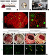
The novel pancreas imaging window was designed to effectively isolate the soft sheet-like pancreas from the neighboring bowel movement. Two-thirds of the base of the imaging window was supported with a thin metal plate, and the pancreatic tissue was overlaid on the plate (Fig. 1A and B, Supplementary Fig. 1). Applying the imaging window to an angle-tilting window holder on the stage of a custom-built laser-scanning video-rate confocal microscopic imaging setup [789], intravital imaging of the mouse pancreas was successfully achieved in vivo (Fig. 1C). From the real-time videos acquired at a speed of 30 frames per second, a significantly enhanced tissue imaging stability with the novel pancreas imaging window was clearly noticeable while the islets in the conventional abdominal imaging window were continuously wandering accompanied with complex non-uniform deformation due to peristalsis (Supplementary Video 1). Due to severe blurring induced by motion-artifacts, the quality of the averaged image using the abdominal imaging window was too impaired to recognize the microscale structure (Supplementary Fig. 2). Additionally, physiologic characteristics were measured to assess the detrimental effect of window implantation for 5 days and no significant decrease in body weight nor increase of blood glucose were observed (Supplementary Fig. 3).
Next, we used MIP-GFP mice [10] for simultaneous imaging of pancreatic islets and adjacent vasculature. By acquiring multiple overlapping images through the pancreas imaging window, a wide-area mosaic image was produced with a high enough resolution to distinguish individual islets and capillary (Fig. 1D). Subsequently, we verified the imaging stability of the islets using the window for longitudinal time-lapse imaging over several days. Compared to the conventional abdominal imaging window where tracking of the same islets was unfeasible, the pancreas imaging window showed a remarkable immovability of the pancreatic tissue in the gross view as well as in the microscopic view identifying the MIP-GFP expressing islets until post-implantation day 7 (Fig. 1E).
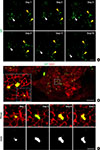
Up to post-implantation day 19, we successfully achieved the tracking of the same pancreatic islets in vivo (Fig. 2A). We next investigated the imaging of the morphogenesis of the islets in the pancreas in vivo in 4-week-old mice [1112]. With repetitive imaging from post-implantation day 1 to 5, we observed a dumbbell-shape transformation of the islets from a single islet in vivo (Fig. 2B and C).
DISCUSSION
Our study is highlighted for the specificity on pancreas as well as direct visualization of the morphogenesis of islets in situ. Technical hindrance of accessibility and stability of islets imaging has proposed low-resolution imaging or heterotopic transplantation model [1314] which has limited ability to evaluate the islet fission. In addition, their microenvironment might be different from the pancreas in situ. Our study convincingly demonstrated the finding of a previous mathematical model study [11] and ex vivo study [15], which suggested the dumbbell-shape morphogenesis was mostly derived from the fission of a single islet instead of the fusion of separate islets.
This imaging window could be a useful method for studying embryology, cell biology, and endocrinology of islets in the pancreas. Additionally, its use can be extended to observe the orthotopic pancreatic cancer model which recapitulates the tumor-microenvironment more accurately than that of the ectopic model [1617].

 XML Download