

 PDF
PDF ePub
ePub Citation
 Print
Print


INTRODUCTION
The cosmetics industry is interested in discovering materials with skin whitening properties. Recently, reactive oxygen species (ROS) were shown to have paradoxical effects on melanocytes [1]. Specifically, they increase pigmentation and cause oxidative stress-induced damage to the melanocytes. Thus, ROS scavengers and inhibitors of ROS production may suppress melanogenesis [23].
Sodium 2-mercaptoethanesulfonate (mesna) is a protective agent that is used as a chemical antidote and adjuvant in various therapeutic areas. Mesna is widely used in medicine because of its protective and antioxidant effects [45]. As an antioxidant, mesna acts by effectively maintaining the intracellular thiol status. Although mesna exists in both reduced and oxidized forms, it mainly exists in the reduced form. Mesna acts as an antioxidant by scavenging freeradicals, which inhibit melanogenesis by suppressing tyrosinase activity [6]. Therefore, mesna is predicted to reduce de novo melanogenesis.
Melanin synthesis is regulated by several signal transduction pathways with important functions in melanogenesis. Forkhead box-O3a (FoxO3a) was recently demonstrated to be an antimelanogenic factor that mediates ROS-induced skin pigmentation, suggesting a close association between melanogenesis and antioxidants, especially ascorbic acid (AA) and N-acetylcysteine (NAC) [7]. Although mesna is a powerful radical scavenger, its whitening effect and signaling mechanism have not yet been investigated.
In this study, we aimed to investigate the inhibitory effect of mesna on melanin synthesis and on the expression of melanogenesis factors, to elucidate the mechanism underlying its effective depigmentation action.
METHODS
Cell culture and cell viability assay
The human melanoma (MNT-1) cells used in this study were maintained in minimum essential medium (MEM; Gibco, Grand Island, NY, USA) containing 10% Dulbecco's MEM, 20 mM HEPES (Sigma-Aldrich Co., St. Louis, MO, USA), 20% FBS, 100 U/ml penicillin G, and 100 mg/ml streptomycin sulfate. Cells were propagated at 37℃ with 5% CO2. Cell viability was measured by 3-(4,5-dimethyl-thiazol-2-yl)2,5-diphenyl tetrazolium bromide (MTT) assay. Briefly, after incubation with test substances, the culture medium was removed and MTT solution (5 mg/ml) was added. Then, cells were maintained for 1 h 37℃, and extracted with DMSO. Absorbance was determined at 590 nm using a microplate reader.
2,2-diphenyl-1-picrylhydrazyl (DPPH) radical scavenging activity assay
The DPPH radical scavenging activity assay was performed as previously described [8], with a few modifications. Briefly, 10 µl of mesna (3–400 µg/ml, dissolved in ethanol) was added to 190 µl of ethanol solution containing DPPH radicals (100 µM) in each well of a 96-well plate. The mixture was shaken and incubated for 30 min at 37℃ in the dark, after which the absorption was measured at 517 nm with AA as the positive control. A lower absorbance represents a higher DPPH scavenging activity. The percent scavenging effect was calculated as follows: scavenging rate (%) = [1 − (A1 − A2)/A0] × 100%, where A0, A1, and A2 are the absorbance of the control (without sample), test sample, and sample without DPPH, respectively. Experiments were repeated three times independently.
Mushroom tyrosinase assay
The in vitro mushroom tyrosinase assay was performed with L-tyrosine and L-3,4-dihydroxyphenylalanine (L-DOPA) as the tyrosinase substrate. The inhibitory activity of each sample (mesna and AA) against tyrosinase-catalyzed oxidation of L-tyrosine was determined according to the methods of Chang et al . [9]. In brief, 40 µl of 1.5 mM L-tyrosine (substrate) dissolved in 0.1 M phosphate buffer (pH 6.8) and 120 µl of the same buffer were mixed with 20 µl of each sample at different concentrations. Then, 20 µl of mushroom tyrosinase (2,000 U/ml in phosphate buffer) was added to initiate the reaction, and the assay mixture was incubated at 37℃ for 15 min. Finally, the increase in absorbance at 475 nm caused by the formation of dopachrome was monitored using a microplate reader (Opsys MR; Dynex Technologies, Ltd., Frankfurt, Germany).
The inhibitory effect of each extract on L-DOPA oxidation by mushroom tyrosinase was determined according to the method of Masamoto et al . [10], with minor modifications. Briefly, 100 µl of 0.1 M phosphate buffer was mixed with 20 µl of varying concentrations of each sample. Reactions were initiated by the addition of 20 µl of mushroom tyrosinase (2,000 U/ml in phosphate buffer). The mixture was subsequently incubated at 37℃ for 5 min and then added to 40 µl of L-DOPA (4 mM in 0.1 M phosphate buffer). This reaction mixture was incubated for 10 min at 37℃, after which its absorbance at 475 nm was measured. The percentage inhibition of tyrosine or L-DOPA oxidation was calculated as follows: inhibition (%) = 100 − (B/A × 100), where A and B were the ΔOD475 values recorded at 10 min without and with the test sample, respectively. Experiments were repeated three times independently.
Measurement of melanin content
Cellular melanin content was determined by a modified version of the method reported by Hosoi et al. [11]. Briefly, MNT-1 cells were seeded onto a 24-well plate at a density of 1 × 10⁵ cells/well and incubated overnight. The medium was replaced with medium containing different concentrations of mesna, NAC, and AA, after which the mixtures were incubated for a further 72 h and the medium was removed. The cells were then washed twice with PBS, harvested by trypsinization using 0.25% trypsin/0.02% EDTA in PBS, pelleted, and solubilized in 1 N NaOH for 1 h at 80℃. After centrifugation at 3,000 × g for 10 min, the OD of the resulting supernatant was measured at 490 nm by a microplate reader. Experiments were performed in triplicate.
Quantitative real-time RT-PCR (qRT-PCR)
Total cellular RNA was extracted from MNT-1 cells using an RNeasy Mini Kit (Qiagen, Valencia, CA, USA). For cDNA synthesis, 1 µg of RNA was reverse transcribed using a Primerscript first strand cDNA synthesis kit (Takara Bio Inc., Shiga, Japan). Quantitative real-time PCR (qPCR) was performed using iQ SYBR Green Supermix (Bio-Rad, Hercules, CA, USA). qRT-PCR thermocycling parameters included an incubation at 95℃ for 10 min, followed by 45 cycles at 95℃ for 10 sec, 72℃ for 1 sec, and 40℃ for 30 sec. The reactions were performed in a Bio-Rad CFX96 Real-Time PCR Detection System. At the end of each qRT-PCR run, the results were automatically analyzed and an amplification plot was generated for each cDNA. All experiments were performed in triplicate. mRNA expression levels were quantified using the relative CT method and normalized to that of GAPDH, the housekeeping gene transcript.
Westernblot analysis
Protein extracts of MNT-1 cells were generated using cold radioimmunoprecipitation assay (RIPA) buffer (50 mM Tris-HCl, pH 7.2, 150 mM NaCl, 1% NP40, 0.1% SDS, 0.5% DOC, 1 mM PMSF, 25 mM MgCl2, and phosphatase inhibitor cocktail). The protein contents of the resultant cell lysates were quantified using a Pierce bicinchoninic acid (BCA) protein assay kit (Thermo Scientific, Waltham, MA, USA). Equal amounts of protein were resolved by SDS-PAGE (8%–12% gels) and transferred onto PVDF membranes (Millipore, Billerica, MA, USA). After blocking with 5% skim milk in Tris-buffered saline containing 0.5% Tween 20 (Sigma-Aldrich Co.) for 2 h, the membranes were probed with primary antibodies and incubated with horseradish peroxidase-conjugated secondary antibodies. Hybridized antibodies were detected using enhanced chemiluminescence solution (ATTO Co., Tokyo, Japan), and images were acquired using an LAS-1000 LuminoImage Analyzer (Fujifilm, Tokyo, Japan). All experiments were performed in triplicate.
Cellular fractionation assay
Cytoplasmic and nuclear protein fractions were generated using an NE-PER nuclear and cytoplasmic extraction reagent kit following the manufacturer's protocol (Pierce Biotechnology, Rockford, IL, USA). The extraction reagents were supplemented with phosphatase inhibitors, 10 mM sodium pyrophosphate, protease inhibitor tablets (Roche Diagnostics, Basel, Switzerland), and 100 mM sodium orthovanadate. Experiments were performed in triplicate.
FoxO3a immunofluorescence assay
MNT-1 cells were seeded onto four-chamber cell culture slides (SPL Life Sciences Co., Ltd., Pocheon, Korea) and treated with each test substance (mesna, NAC, or AA) for 1 day. Next, the slides were washed with 1 × PBS and fixed with 4% paraformaldehyde for 20 min. After washing again with 1 × PBS, the slides were incubated with 0.01% Triton X-100 (Sigma-Aldrich Co.) in 1 × PBS for 20 min, blocked with 2% BSA in 1× PBS for 2 h, and then incubated with anti-FoxO3a antibodies overnight at 4℃. After washing with 1 × PBS, the slides were incubated with FITC-conjugated goat anti-rabbit IgG (1:1,000, NB730-F; Novus Biologicals, Littleton, CO, USA) and mounted using 4′,6-diamidino-2-phenylindole (DAPI; Golden Bridge International, Inc., Mulkiteo, WA, USA). Cell morphology was examined using a DP70 fluorescence microscope using DP controller software (Olympus Optical Co., Ltd., Tokyo, Japan). Experiments were performed in triplicate.
RESULTS
Effect of mesna on DPPH scavenging activity
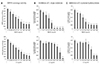
DPPH is widely used to assess the antioxidant efficacy of complex mixtures and individual compounds. The in vitro antioxidant activity of mesna was evaluated by DPPH radicalscavenging activity assays and compared to that of AA. The IC50 values were 23.49 and 21.89 µg/ml for mesna and AA, respectively, indicating that both compounds were strong radical scavengers and that their radical scavenging abilities were comparable (Fig. 1A and Table 1).
Mesna inhibits mushroom tyrosinase activity
To investigate whether mesna directly inhibits the key enzyme in melanogenesis, an in vitro mushroom tyrosinase assay was performed. Both mesna and AA inhibited mushroom tyrosinasemediated oxidation of L-DOPA and tyrosine in a dose-dependent manner (Fig. 1B, C). As expected, the positive control (AA) strongly inhibited mushroom tyrosinase, while mesna showed similarly strong inhibitory activity (Table 1). The IC50 values of mesna against L-DOPA and L-tyrosine were 22.5 and 16.3 µg/ml, respectively, while those of AA were 120.8 and 166.9 µg/ml, respectively. Taken together, our results demonstrate that mesna is a stronger inhibition of mushroom tyrosinase activity than AA.
Mesna reduces melanin synthesis in MNT-1 cells
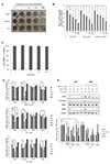
To compare the inhibitory effects of mesna, NAC, and AA on melanin contents, MNT-1 cells were treated with different concentrations of these antioxidants. All three agents significantly decreased melanin production in a dose-dependent manner compared with melanin production in untreated cells (Fig. 2A, B). The IC50 values for melanin synthesis were 230 nM, 173 nM, and 812 nM for mesna, NAC, and AA, respectively. The effective concentration of mesna was not cytotoxic to MNT-1 cells (Fig. 2C). These results suggest that the inhibitory effect of mesna on melanin synthesis correlates well with the effects of mesna on mushroom tyrosinase activity. Additionally, we evaluated changes in the expression of MITF, TYR, TRP-1, and TRP-2 in MNT-1 cells (Fig. 2D). Mesna and NAC significantly downregulated MITF, TYR, and TRP-2 expression at 12–24 h, but the levels were recovered after 48 h. Furthermore, AA downregulated MITF, TYR, TRP-1, and TRP-2 expression levels, but their levels subsequently increased after 48 h. We also analyzed the protein levels of MITF, TYR, TRP-1, and TRP-2 at 24–48 h and found that the levels were significantly decreased following treatment with each agent for 24–48 h (Fig. 2E). These data indicate that mesna, NAC, and AA all reduce melanin synthesis, likely through the downregulation of MITF, TYR, and TRP-2.
Mesna induces nuclear translocation of FoxO3a
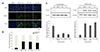
NAC and AA have been reported to induce the nuclear translocation of FoxO3a, which regulates melanogenesis and decreases melanin synthesis. They have also been reported to induce MITF, TYR, TRP-1, and TRP-2 expression in MNT-1 cells [7]. We hypothesized that FoxO3a phosphorylation in nuclear translocation is involved in the melanogenesis processes induced by mesna. To test our hypothesis, we performed an immunofluorescence assay. The results showed that mesna, NAC, and AA all induced the nuclear translocation of FoxO3a (Fig. 3A, B). In addition, the level of endogenous FoxO3a was increased in the nuclear fraction of antioxidant-treated cells (Fig. 3C). These results suggest that mesna regulates FoxO3a nuclear translocation, specifically by enhancing FoxO3a nuclear translocation, thereby reducing melanin synthesis.
Mesna induces phosphorylation of MST1 and JNK
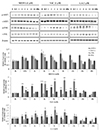
Since FoxO3a interacts with various proteins, it may bind to melanogenesis-related factors in the nucleus to form hig-hmolecular-weight complexes that accomplish its associated functions. The nuclear accumulation of FoxO3a has been reported to activate MST1, which is followed by JNK activation [1213]. To investigate the mechanisms underlying nuclear translocation of FoxO3a, Western blot analysis of a time-course experiment was used to track changes in FoxO3a-related signals induced by mesna. We confirmed that treatment with mesna, NAC, and AA significantly increased the phosphorylation of MST1 and JNK (Fig. 4). These results suggest that mesna, NAC, and AA regulate nuclear translocation of FoxO3a through the phosphorylation of MST1 and JNK.
DISCUSSION
Melanogenesis, a major function of differentiated melanocytes, play an important role in protecting skin from damage by UV-B radiation and is primarily responsible for skin color. Abnormal accumulation of melanin pigment is responsible for pigmentation disorders such as melasma and senile lentigo. Melanogenesis involves a series of tightly regulated enzymatic oxidation processes that culminate in the synthesis and assembly of melanin polymers. Therefore, many antioxidative agents have been used to treat skin hyperpigmentation. Recently, FoxO3a was reported to be involved in the regulation of melanogenesis and to exert antimelanogenic effects. Specifically, the antimelanogenic activity of antioxidants is mediated by FoxO3a activation via its nuclear translocation [7].
Mesna is used therapeutically to prevent haemorrhagic cystitis and haematuria due to cyclophosphates such as ifospamide. Mesna is a small molecule that is able to scavenge ROS by virtue of its sulfhydryl group [14]. In this study, we showed that mesna has strong radicalscavenging ability, similar to that of AA (Fig. 1A). The purpose of the present work was to investigate the inhibitory effects of mesna on melanin formation, which is intimately involved in skin pigmentation. We found that mesna decreased melanin production in and secretion by MNT-1 melanoma cells. This effect was due to a sharp increase in FoxO3a activation and nuclear translocation, resulting in downregulation of the master genes of melanogenesis (e.g., MITF, tyrosinase, TRP1, and TRP2 [DCT]) after 12 h of treatment.
MST1 activation promotes FoxO3a nuclear accumulation, which is accompanied by JNK activation. A previous study in melanoma cells showed that MST1 promotes cell death in response to oxidative stress by directly activating FoxO3a [12]. Meanwhile, another study reported that MST1 is crucial for survival of naïve T cells in response to intracellular ROS by regulating FoxO protein levels [15]. In our study, mesna induced FoxO3a nuclear translocation by activation of MST1 and JNK, respectively. However, mesna did not change the number of MNT-1 cells (Fig. 2D), suggesting that mesna does not induce cell apoptosis. Thus, MST1 appears to act according to cell conditions to modulate the FoxO function to regulate cell death and survival differently.
Melasma is an acquired pigmentary disorder occurring mainly on the face and largely affecting women of childbearing years. Depending on the degree of melanin deposition in the skin, it is classified as epidermis, skin and mixed melanin. Epidermal melanoma is the most common form of the disease, characterized by increased melanin in the epidermis. Dermal melasma is characterized by an increase in melanin in the dermis, while mixed melanin refers to a combination of epidermal and dermal melasma [16]. The currently available melasma treatment options include topical agents, chemical peels, lasers, and light therapy [17]. Of these treatments, a topical combination therapy consisting of hydroquinone, tretinoin, and fluocinolone acetonide has been shown to be the most effective, but approximately 40% of patients showed erythema and peeling. Moreover, chemical peels, laser therapy, and light therapy produced mixed therapeutic results, with increased risk of irritation and subsequent hyperpigmentation. Therefore, the current treatment modalities for melasma are unsatisfactory. AA, otherwise known as vitamin C, is a potent antioxidant and radical scavenger that interferes with melanogenesis by inhibiting oxidative reactions in the melanin production process [18]. In a 12-week randomized double-blind split-sided study, Korean women suffering from melasma reported noticeable improvement on the treated side using iontoporesis with vitamin C [19]. Thus, we hypothesize that mesna, an antioxidant and radical scavenger, also suppresses melanin production and may thus be a useful therapeutic for the clinical treatment of hyperpigmentation disorders.

 XML Download