

 PDF
PDF Citation
 Print
Print


INTRODUCTION
Cervical cancer is the fourth most common female cancer with an estimated global incidence of 527,600 new cases and 265,700 deaths each year [1]. Compared to Western countries, the prevalence of cervical cancer is much higher in Korea. In 2019, it was expected to account for 2.8% (2,856) of newly diagnosed female cancers in Korea, in contrast to 1.5% (13,170) of new cases in the United States [23].
The first step in routine cervical cancer screening is the Papanicolaou test (Pap test) performed by either conventional cervicovaginal smears or liquid-based cytology. It is a simple and cost-effective test for early detection of squamous intraepithelial lesion (SIL) or squamous cell carcinoma (SqCC) of the uterine cervix. Among the categories of the 2001 Bethesda system for reporting cervical cytology [4], women with high-grade SILs (HSILs) require special attention; 60%–90% are diagnosed with cervical intraepithelial neoplasia (CIN) grade 2 or greater by either colposcopy-directed biopsy (CDB) or loop electrosurgical excision procedures (LEEPs) [567].
For management of women with cytologic HSILs, the current clinical guideline of the American Society for Colposcopy and Cervical Pathology (ASCCP) recommends colposcopy or immediate LEEP [7]. It is undeniable that colposcopy plays an important role in early detection of CIN and cervical cancer [8]. Nevertheless, previous studies have reported discrepancies between CDB and LEEP results with overall concordance or agreement rates ranging from 43% to 86% [910111213141516]. However, study populations and designs as well as statistical methods differed among the studies. While various types of baseline Pap tests were included, few studies have focused solely on cytologic HSILs.
Therefore, this study aimed to investigate pathologic discrepancies between the CDB and LEEP in women with cytologic HSILs. Considering the LEEP as the gold standard, the diagnostic performance of CDB for identifying CIN grades 2 and 3, adenocarcinoma in situ (AIS), and cancer was evaluated.
MATERIALS AND METHODS
This single-institution retrospective study was conducted with the approval from the Institutional Review Board of Seoul National University Bundang Hospital (No. B-1903/526-103).
1. Study population
In order to include all patients who underwent the LEEP for cytologic HSILs, we retrospectively investigated 1,355 consecutive LEEP cases at Seoul National University Bundang Hospital, a high-volume tertiary institutional hospital, between January 2015 and December 2018. During the study period, only the LEEP and not cold-knife conization were performed. After excluding 54 duplicated patients, we further excluded patients with any of the following characteristics: 1) received Pap tests during pregnancy (n=19); 2) were referred after the LEEP at an outside clinic (n=16); 3) had previous histories of the LEEP before the study period (n=8); or 4) had unknown Pap test results (n=85). After reviewing the initial Pap test results, we identified 411 patients with cytologic HSILs. Among them, only 297 patients who had undergone CDB before the LEEP were included in this analysis.
2. Data collection
At our institution, the standard practice for the management of women with abnormal Pap test results is in line with the current clinical guidelines of the ASCCP [7]. Similar to the ASCCP guidelines, we recommend either immediate LEEP (unless a patient is pregnant or 21–24 years of age) or colposcopy for women with cytologic HSILs. A diagnostic excisional procedure (LEEP) is performed when the colposcopic examination is inadequate, except during pregnancy. In addition, we also consider endocervical curettage (ECC) for women with cytologic HSILs when the colposcopic examination is unsatisfactory or when the colposcopic examination is satisfactory, but no lesions are seen.
Through a comprehensive review of medical records and pathology reports, we collected information for patients' clinicopathologic characteristics, such as age at diagnosis, implementation of human papillomavirus (HPV) tests and the results, and CDB and LEEP results. At our institution, pathologists report the lesion size only in cases of cancers when examining LEEP specimens. For cancer cases, we reported the lesion size as well as the depth of invasion.
Before 2017, HPV genotyping was performed using a GG HPV-40 DNA Genotyping Chip (Goodgene Inc., Seoul, Korea) according to the manufacturer's instructions, as described previously [17], after which a peptide nucleic acid probe-based fluorescence melting curve analysis technology was used in real-time polymerase chain reaction systems (PANA RealTyperTM HPV Kit, PANAGENE Inc., Daejeon, Korea). Using these methods, high-risk, low-risk, and other HPV genotypes were detected.
To evaluate the diagnostic performance of CDB, we regarded LEEP as the gold standard method. Also, we set up a 2-tiered classification for both CDB and LEEP results: HSIL+ (CIN2,3, SqCC, AIS, and adenocarcinoma [AC]); and HSIL− (CIN1 or low-grade SIL [LSIL], and chronic cervicitis).
3. Statistical analysis
Descriptive statistics were used to describe clinicopathologic characteristics of the study population. The diagnostic performance of CDB for identifying HSIL+ was presented in the form of a 2×2 table. To evaluate the consistency of pathologic results between the CDB and LEEP, Cohen's kappa (κ) coefficient was calculated. We considered κ values <0 as no agreement, 0–0.20 as slight, 0.21–0.40 as fair, 0.41–0.60 as moderate, 0.61–0.80 as substantial, and 0.81–1.00 as almost perfect agreement. In order to evaluate whether CDB and LEEP result in different HSILs (+ and −), we performed exact McNemar's test on the samples, regarding the null hypothesis as the probability of multiple paired outcomes being equal.
Characteristics were compared between patients who were under-diagnosed from CDB and those who were correctly or over-diagnosed using Student's t and Mann-Whitney U tests for continuous variables and Pearson's χ2 and Fisher's exact test for categorical variables. Multivariate analysis was performed using a logistic regression model.
For subgroup analyses according to patients' age, patients were divided into 3 age groups <35 years, 35–50 years, and ≥50 years. The following statistical analyses were performed to identify whether each subgroup disagreed with the overall trend: we generated 1,000 replicates for each subgroup and checked how many times the balanced accuracy of the replicate was inferior to those of the overall samples (empirical p-values). Each replicate was generated by down-sampling 90% of the given dataset. We determined that there was significant disagreement between each age group and the overall trend when the empirical p-value was <0.05.
All statistical analyses were performed using SPSS statistical software (version 25.0; SPSS Inc., Chicago, IL, USA) and R statistical computing software (version 3.5.0, https://www.r-project.org/). A p-value <0.05 was considered statistically significant.
RESULTS
1. Patients' characteristics
The selection of the study population is depicted in Fig. 1. In total, 297 patients with cytologic HSILs who underwent both CDB and LEEP were included in this analysis. Table 1 presents patients' clinicopathologic characteristics. The mean age of the patients was 45.6±12.5 years (range, 21.7–81.5). The number of patients <35 years of age were 67 out of 297 (22.6%). HPV testing was performed in 63.3% (188/297) of the patients. Among the patients who received HPV testing, the proportions of negative results, other types, and high-risk genotypes were 2.7%, 1.6%, and 95.7%, respectively. Low-risk HPV genotypes were not observed in any cases.
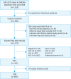 | Fig. 1Flow diagram illustrating the selection of study population.AGC, atypical glandular cells; AIS, adenocarcinoma in situ; ASC-H, atypical squamous cells, cannot exclude HSIL; ASC-US, atypical squamous cells of undetermined significance; CDB, colposcopy-directed biopsy; HSIL, high-grade squamous intraepithelial lesion; LEEP, loop electrosurgical excision procedure; LSIL, low-grade squamous intraepithelial lesion; Pap test, Papanicolaou test; SNUBH, Seoul National University Bundang Hospital; SqCC, squamous cell carcinoma.
|
Table 1
Clinicopathologic characteristics of study population
Data are shown as number (%).
AC, adenocarcinoma; AIS, adenocarcinoma in situ; HPV, human papilloma virus; HSIL, high-grade squamous intraepithelial lesion; LEEP, loop electrosurgical excision procedure; LSIL, low-grade squamous intraepithelial lesion; SD, standard deviation; SqCC, squamous cell carcinoma.
*Calculated among the patients who underwent HPV test.
![]()
The overall incidence of pathologic HSIL+ among women with cytologic HSILs was 90.9% (270/297). In terms of detailed LEEP results, the proportions of no residual tumor, chronic cervicitis, LSIL, HSIL, SqCC, AIS, and AC were 1.7%, 2.4%, 5.1%, 72.1%, 17.5%, 0.7%, and 0.7%, respectively.
2. Diagnostic performance of CDB
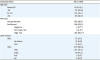
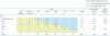
Considering LEEP as the gold standard, we evaluated the diagnostic performance of CDB for identifying HSIL+ with the following results: sensitivity, 87.8%; specificity, 59.3%; balanced accuracy, 73.6%; positive predictive value, 95.6%; and negative predictive value, 32.7% (Fig. 2). Cohen's κ coefficient for CDB and LEEP was 0.344, indicating fair agreement. However, the exact McNemar's test revealed that CDB was significantly different in the diagnosis of HSIL+ compared to the LEEP in all patients (p<0.001).
 | Fig. 2Diagnostic performance of CDB in patients with cytologic HSILs. (A) 2×2 contingency table, (B) Value for each parameter.AC, adenocarcinoma; AIS, adenocarcinoma in situ; CDB, colposcopy-directed biopsy; CIN, cervical intraepithelial neoplasia; HSIL, high-grade squamous intraepithelial lesion; LEEP, loop electrosurgical excision procedure; LSIL, low-grade squamous intraepithelial lesion; SqCC, squamous cell carcinoma.
|
Detailed characteristics of 33 patients who showed false negative CDB results are presented in Table 2. The median number of biopsies was 4 (range, 1–6), and ECCs were performed in 12 out of 33 false negative cases (36.4%). However, 5 out of 12 ECCs (41.7%) yielded inadequate results (i.e., “tissue insufficient for diagnosis”). After the LEEP, the final diagnoses of these patients included CIN2,3 (n=29) and early stage cervical cancers (n=4). While 3 cases were International Federation of Gynecology and Obstetrics stage IA1 SqCC, 1 case was diagnosed with stage IB1 endocervical AC. All 4 cervical cancer patients underwent primary surgical treatments.
Table 2
Detailed characteristics of patients who showed false negative results on CDB
AC, adenocarcinoma; CDB, colposcopy-directed biopsy; CIN, cervical intraepithelial neoplasia; DOI, depth of invasion; HSIL, high-grade squamous intraepithelial lesion; HPV, human papilloma virus; LEEP, loop electrosurgical excision procedure; LSIL, low-grade squamous intraepithelial lesion; RM, resection margin; SqCC, squamous cell carcinoma; SqCIS, squamous cell carcinoma in situ.
*According to Querleu and Morrow's classification.
![]()
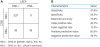
Next, we compared detailed pathologic results between the CDB and LEEP (Fig. 3). The overall concordance rate was 67.7% (201/297). Under-diagnosed cases were observed in 26.6% of cases (79/297). Among 79 under-diagnosed cases, 5 (6.3%), 29 (36.7%), 43 (54.4%), and 2 (2.5%) were finally diagnosed with LSIL, HSIL, SqCC, and AC, respectively. Specifically, among 20 cases that had been diagnosed with chronic cervicitis by CDB, 5 (25.0%) and 12 (60.0%) cases were correctly diagnosed with LSIL and HSIL by the LEEP, respectively. SqCC and AC were confirmed in 1 case (5.0%) each. Therefore, the under-diagnosis rate was 95.0% in CDB-proven chronic cervicitis. Among 29 cases that had been diagnosed with LSIL by CDB, 17 cases (58.6%) were correctly diagnosed with HSIL and 2 cases (6.9%) with SqCC. Therefore, the under-diagnosis rate was 65.5% in CDB-proven LSILs.
 | Fig. 3Comparison of pathologic results between CDB and LEEP in patients with cytologic HSILs.AC, adenocarcinoma; AIS, adenocarcinoma in situ; CDB, colposcopy-directed biopsy; HSIL, high-grade squamous intraepithelial lesion; LEEP, loop electrosurgical excision procedure; LSIL, low-grade squamous intraepithelial lesion; SqCC, squamous cell carcinoma.
|
Clinicopathologic characteristics, such as age and the presence of high-risk HPV genotypes, were not statistically different among the patients who were under-diagnosed from CDB (n=79) and those who were correctly or over-diagnosed (n=218) (Supplementary Table 1). In multivariate analysis, neither patients' ages (adjusted odds ratio [OR]=1.593; 95% confidence interval [CI]=0.798–3.180; p=0.187) nor high-risk HPV infections (adjusted OR=1.071; 95% CI=0.208–5.529; p=0.935) affected the under-diagnosis rate of CDB.
3. Subgroup analysis according to patients' age
Clinicopathologic characteristics of patients among the 3 subgroups are presented in Supplementary Table 2. HPV testing was performed in 70.1%, 64.9%, and 56.3% of the patients in age groups <35 years, 35–50 years, and ≥50 years, respectively (p=0.169). High-risk HPV infection rates were not different among the 3 subgroups (p=0.704).
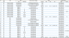
We also performed subgroup analyses according to patients' ages. In the subgroup of patients aged <35 years (n=67), the diagnostic performance of CDB for identifying HSIL+ was as follows: sensitivity, 95.3%; specificity, 66.7%; balanced accuracy 81.0%; positive predictive value, 98.4%; and negative predictive value, 40.0% (Fig. 4A and B). Cohen's κ coefficient for CDB and LEEP was 0.470, indicating moderate agreement. Overall rates for under-, correctly-, and over-diagnosed cases were 22.4%, 76.1%, and 1.5%, respectively.
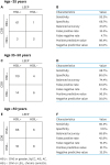 | Fig. 4Diagnostic performance of CDB in patients with cytologic HSILs aged <35 years (upper), 35–50 years (middle), and ≥50 years (lower). (A, C, E) 2×2 contingency table, (B, D, F) Value for each parameter.AC, adenocarcinoma; AIS, adenocarcinoma in situ; CDB, colposcopy-directed biopsy; CIN, cervical intraepithelial neoplasia; HSIL, high-grade squamous intraepithelial lesion; LEEP, loop electrosurgical excision procedure; LSIL, low-grade squamous intraepithelial lesion; SqCC, squamous cell carcinoma.
|
In the subgroup of patients aged 35–50 years (n=134), the diagnostic performance of CDB for identifying HSIL+ was as follows: sensitivity, 88.7%; specificity, 60.0%; balanced accuracy 74.4%; positive predictive value, 96.5%; and negative predictive value, 30.0% (Fig. 4C and D). Cohen's κ coefficient for CDB and LEEP was 0.334, indicating fair agreement. Overall rates for under-, correctly-, and over-diagnosed cases were 27.8%, 65.2%, and 7.0%, respectively.
In the last subgroup of patients aged ≥50 years (n=96), the diagnostic performance of CDB for identifying HSIL+ was as follows: sensitivity, 80.5%; specificity, 57.1%; balanced accuracy 68.8%; positive predictive value, 91.7%; and negative predictive value, 33.3% (Fig. 4E and F). Cohen's κ coefficient for CDB and LEEP was 0.290, indicating fair agreement. Overall rates for under-, correctly-, and over-diagnosed cases were 30.2%, 61.5%, and 8.3%, respectively.
Finally, when evaluating whether the samples in each subgroup agreed with the trend of the entire data set, the age groups <35 years and 35–50 years showed good agreement with the overall samples (p=0.496 and 0.406, respectively). However, the age group ≥50 years showed statistically significant disagreement with the overall samples (p=0.036), magnifying the pathologic discrepancy between CDB and LEEP results.
DISCUSSION
Considering the high incidence of pathologic HSIL+ in women with cytologic HSILs, identifying every presence of CIN2,3, SqCC, AIS, and AC is an important issue. In the current study, we evaluated the diagnostic performance of CDB in this population by comparing CDB results directly with LEEP results, and observed discrepancies between these procedures.
Such pathologic discrepancies between the CDB and LEEP have been also reported in various studies. In contrast to our study that was limited to cytologic HSILs, most studies included patients with various types on baseline Pap tests. Owing to the differences in study details, direct comparisons of the concordance rates among the studies might be difficult. While some studies reported the concordance rate as <70% [910111415], others reported the rate as ≥70% [121316]. Rates for under-diagnosis by CDB were reported as 20%–40% throughout the studies. In a prospective study conducted by a British group who assessed 170 paired CDB and LEEP specimens, poor overall agreement between CDB and LEEP results was observed and CDB results had a tendency to underestimate high-grade CINs [18].
In the current study, the overall concordance rate between CDB and LEEP results was 67.7% (201/297) and under-diagnosed CDB cases were observed in 26.6% (79/297). Statistically, we also observed a significant difference in the diagnosis of HSIL+ in all patients. Moreover, we demonstrated a high CDB false negative rate (12.2%; 33/270) for identifying HSIL+. Among false negative cases, 12.1% (4/33) were ultimately diagnosed with cancer which might have been missed if the LEEP had not been performed. Therefore, we emphasize the substantial risk of misdiagnosis of actual cancer and the risk of progression of cervical dysplasia when the LEEP was not performed or delayed based on favorable CDB results in women with cytologic HSILs.
While some researchers pointed out an inherent inaccuracy of CDB as the main cause of the discrepancy, others have also investigated the factors that might affect agreement between CDB and excisional biopsy results. Using the database of the Gardasil clinical trials, Stoler et al. [11] included a total of 594 cases and identified patients' age, number of biopsies, lesion sizes, presence of HPV 16/18 genotypes, and geographic region as factors affecting agreement between the CDB and excisional biopsy results. An Italian retrospective study reported that patients' age and invisibility of the squamocolumnar junction were associated with CDB under-diagnoses [19]. A recent Korean retrospective study suggested that baseline Pap test results and the number of vaginal deliveries as additional factors affecting the diagnostic discrepancies between the CDB and LEEP [15].
However, in this study, neither patients' age nor the presence of high-risk HPV was different between under-diagnosed cases and the other cases. The main reason for such an inconsistency might be that we chose only women with cytologic HSILs as the study population, unlike the former 3 studies which did not restrict the baseline Pap results. Although the current ASCCP guidelines do not recommend triage using reflex HPV testing for women with cytologic HSILs [7], HPV testing is commonly performed at our institution. Among the patients who underwent HPV testing, 97.3% showed positive results and 95.7% had high-risk HPV genotypes. Beside the extremely high prevalence of HPV infection, the patients’ mean age (45.6 years) was higher than that in other studies.
In subgroup analyses according to patients’ ages, we found that the diagnostic accuracy of CDB for identifying HSIL+ decreased as age increased with the balanced accuracy of CDB in age groups <35 years, 35–50 years, and ≥50 years being 81.0%, 74.4%, and 68.8%, respectively. Although the study population in the study by Stuebs et al. [16] was quite different from that in our study, the authors reported similar trends in the accuracy rates for detecting HSILs, which were 93.1% (age 0–34), 83.6% (age 34–55), and 80% (age 55 or older). Relatively poor diagnostic performances for identifying HSIL+ in patients ≥50 years of age in both studies might originate from the tendency of postmenopausal women to have unevaluable squamocolumnar junctions or cervical lesions whose limits are not well visualized with colposcopy.
Displaying discrepancies between CDB and LEEP results does not imply that we are eliminating colposcopic examinations for women with cytologic HSILs. Rather, we agree with the importance and value of colposcopy for the evaluation of abnormal or inconclusive cervical cancer screening tests [8]. However, by any means, HSIL and SqCC must not be underdiagnosed.
We believe that there were 2 possible factors that might affect the diagnostic inaccuracy of CDB in this study: first, a proficiency issue among physicians performing CDB might exist. It is well known that colposcopists’ expertise influences the accuracy of colposcopic grading [20]. Some studies also reported the tendency of junior colposcopists to overestimate rather than underestimate colposcopic findings [21]. However, this study was retrospective, and we were unable to perform quality assessments of the CDB procedures. The accuracy of CDB might be improved or optimized by implementation of well-designed training and quality assurance programs for colposcopy practice [2122]. A standardized practice guideline for colposcopic examination from ASCCP is available [23].
Secondly, pathologic examination-related issues should be considered for CDB. It is well known that discrepancies in the interpretation of cervical histology exist among pathologists; in particular, the differential diagnosis between LSIL (CIN1) and HSIL (CIN2–3) tends to be difficult [2425]. To aid objective CIN grading and identification of true HSILs, several biomarkers have been suggested. The most extensively used biomarkers are Ki-67 and p16INK4a (CDKN2A) [25]. Currently, additional biomarkers, such as cytokeratin 7 and topoisomerase IIa, have been proposed with promising results [262728]. Therefore, a combined approach using these biomarkers is expected to increase the diagnostic accuracy of CDB.
The current study has several limitations. First, although we identified consecutive patients with clearly defined eligibility criteria, inevitable issues might arise in retrospective studies such as selection bias. Secondly, as a single-institution study, sample size might be insufficient to determine the diagnostic performance of CDB with high accuracy. Thirdly, we were unable to investigate the colposcopists' expertise in preforming CDB, which might affect the diagnostic accuracy of CDB to identify HSIL+. Lastly, we did not evaluate other risk factors for cervical dysplasia and cervical cancer, such as parity, smoking, socioeconomic status, and use of oral contraceptives. Despite this study's limitations, our results, using clearly defined methods, provide additional scientific evidence for the diagnostic inaccuracy of CDB, especially for those with cytologic HSILs.
In conclusion, there were significant pathologic discrepancies between CDB and LEEP results in women with cytologic HSILs. Our results showed that the overall concordance rate between CDB and LEEP was 67.7% and under-diagnosed cases by CDB was 26.6%. Two-thirds of the cases with negative or low-grade lesions using CDB were diagnosed with pathologic HSIL+ by LEEP. Moreover, diagnostic inaccuracies of CDB were magnified in those ≥50 years of age. Further prospective studies are warranted.
 XML Download