

 PDF
PDF ePub
ePub Citation
 Print
Print


INTRODUCTION
Adjuvant endocrine therapy is an important treatment option for patients with hormone-sensitive breast cancer [1]. Selective estrogen receptor modulators (SERMs) and aromatase inhibitors (AIs) form the backbone of adjuvant endocrine therapy. The SERM tamoxifen is a competitive agonist of the estrogen receptor (ER) and has been the standard drug used for adjuvant endocrine therapy for decades, in both premenopausal and menopausal women. In contrast, AIs deplete estrogen by directly inhibiting aromatase, thereby stopping estrogen biosynthesis in the extra-gonadal organs, including the adrenal glands, liver, bones, and particularly, the adipose tissue [2]. AIs are contraindicated for stand-alone therapy in premenopausal women as they stimulate estrogen production in the ovaries. Ovarian function suppression is, therefore, a prerequisite for AI therapy in premenopausal women. However, in postmenopausal women, current guidelines recommend third-generation AIs instead of tamoxifen as the gold standard treatment, due to lower recurrence rates and better disease-free survival (DFS) [345].
Despite the notably higher efficacy of AIs, their use is associated with numerous adverse effects including musculoskeletal pain, osteoporosis, and increased cardiovascular events. These lead to gradually decreasing compliance rates and in the long-term, to non-breast cancer related deaths [56]. Consequently, guidelines recommend the use of SERMs instead of AIs in patients with contraindications or intolerance to AIs due to the concern that the adverse effects of AI may outweigh its survival benefits in those patients [4]. Therefore, there is a high demand for a novel biomarker that predicts the desired survival benefits while avoiding unwanted side effects. However, despite substantial advances in the molecular profiling of breast cancer during the last few decades, there are currently no established predictive biomarkers for individualized adjuvant endocrine regimens, other than the ER.
Phosphorylated ribosomal S6 kinase 1 (pS6K1) is known to be a key driver of the mammalian target of rapamycin (mTOR) pathway and overexpression of S6K1 has been associated with poor prognosis in breast cancer [78]. Interestingly, pS6K1 has recently been shown to play a role in early adipocyte differentiation (adipogenesis) in preclinical models [91011]. Adipocytes are closely related to hormone-sensitive breast cancer development and prognosis, by contributing to increased local estrogen production [1213]. These results provide a theoretical link between pS6K1, adipogenesis, and local estradiol levels, which are associated with the prognosis of breast cancer. This suggests that patients with tumors expressing pS6K1 could obtain greater survival benefits from estrogen-depleting AIs than from SERMs. As such, in this study, we evaluated the potential of pS6K1 as a predictive marker for adjuvant AI therapy in postmenopausal or ovarian function suppressed patients with hormone-sensitive breast cancer.
METHODS
Study design and patient selection
A total of 2,663 patients with primary breast cancer who underwent surgical treatment between March 2008 and May 2015 were included in the study. Among them, 428 women with postmenopausal (n = 401) or ovarian function-suppressed (n = 17) estrogen receptor-positive and node-positive breast cancer and who had received adjuvant endocrine therapy were identified and selected for further analysis. Premenopausal women who underwent bilateral oophorectomy during adjuvant endocrine therapy were also included in this analysis (n = 10). Gonadotropin-releasing hormone agonists were administered monthly for a minimum of 2 years for suppression of ovarian function. Letrozole, anastrozole, or exemestane had been used as components of upfront, switch, or extended adjuvant endocrine therapy regimens. For the purposes of this study, patients who were treated with AIs at any point during their course of adjuvant endocrine therapy were defined as AI-treated patients. All patients in the cohort received treatment after providing informed consent, in accordance with the principles of the Declaration of Helsinki. The need for informed consent for using patient medical records was waived owing to the retrospective nature of this study.
Medical records were retrospectively reviewed to obtain clinicopathologic information regarding age, body mass index (BMI), tumor size (≤ 2 cm vs. > 2 cm), nodal (N) stage (N1 vs. N2–3), progesterone receptor (PR) status, human epidermal growth factor receptor (HER)-2 status, histologic grade, history of previous chemotherapy, types of surgery, and pS6K1 status. The Institutional Review Board of the Korean Cancer Center Hospital approved the protocol version 3.0 (K-1707-002-015).
Immunohistochemical staining
Immunohistochemical staining of ER, PR, HER-2, and pS6K1 was used to evaluate expression status and had been routinely performed for each patient on core needle biopsy specimens obtained during the diagnosis of primary breast cancer. Surgical specimens were used in cases where core needle biopsy specimens were unavailable. ER or PR positivity was defined as the expression of the ER or PR in at least 1% of tumor cells as shown by immunohistochemistry. The pS6K1 expression status was evaluated on a scale of 0 to 3+ by immunohistochemistry using the mouse monoclonal anti-pS6K1 antibody (Cell Signaling Technology, Danvers, USA; dilution 1:50). Details of the procedures are described in our previous report, which evaluated pS6K1 status in 304 hormone receptor-positive breast cancer patients [14]. Scores of 0 and from 1+ to 3+ were regarded as negative and positive pS6K1 expression, respectively.
Statistical analysis
Statistical analyses were performed using the χ2 and Fisher's exact test to assess the correlation between pS6K1 expression and other clinicopathological parameters. The DFS of patients treated with AIs or SERMs was evaluated with respect to the pS6K1 status. DFS was defined as the time from diagnosis to the detection of any recurrence, contralateral breast cancer, secondary malignancy, or to death without recurrence. Survival curves were constructed using the Kaplan-Meier method. Differences in survival rates were assessed using the log-rank test. For univariate and multivariate analysis, the Cox proportional-hazards model was used to calculate hazard ratios (HRs) and 95% confidence intervals (CIs). A p-value of < 0.05 was considered statistically significant.
RESULTS
Patient characteristics
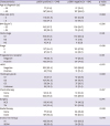
Among the 428 patients analyzed, 244 (57.1%) and 184 (42.9%) patients had pS6K1 positive and negative tumors, respectively (Table 1). The 2 groups were well balanced on a number of patient characteristics, including age, N stage, hormone receptor status, histologic factors, surgical treatment methods, and adjuvant endocrine therapy. BMI, an indicator of systemic adiposity, did not differ significantly between patients with a different pS6K1 status (p = 0.526). Analysis of patient risk factors according to pS6K1 and adjuvant endocrine therapy status are shown in Supplementary Table 1. AIs and SERMs were administered to 288 (67.3%) and 140 (32.7%) patients, respectively. Seventeen (4%) premenopausal patients underwent ovarian function suppression.
Table 1
Patients characteristics
Expression of pS6K1 and patient outcomes
During the median follow-up period of 44 months (range, 1–90), 43 cases of recurrence were observed, which included 12 and 33 locoregional recurrences and distant metastases, respectively. Secondary malignancies were observed in 4 patients. Only one death occurred in this cohort.
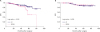
In the pS6K1 positive group, DFS was significantly longer in patients treated with AIs than in those treated with SERMs (Figure 1A, p = 0.016). The 5-year DFS rates were 83.5% and 50.7% for the AI and SERM group, respectively. In contrast, in patients with pS6K1 negative tumors, there was no significant difference in DFS between the groups treated with AIs or SERMs (Figure 1B, 5-year DFS 87.6% vs. 91.4%, p = 0.630). The five-year DFS rates were 75.7% and 88.6% for pS6K1 positive and negative patients, respectively (HR, 0.43; 95% CI, 0.23–0.82, p = 0.010).
Figure 1
Kaplan-Meier DFS curves for adjuvant AIs and SERMs in patients with (A) positive pS6K1 expression status and (B) negative pS6K1 expression status. Patients with positive pS6K1 expression status showed better DFS when treated with AIs than with SERM (p = 0.016). However, in pS6K1 negative patients, there was no difference in DFS between AIs and SERMs (p = 0.630).
DFS = disease-free survival; AI = aromatase inhibitor; SERM = selective estrogen receptor modulator; pS6K1 = phosphorylated ribosomal S6 kinase 1.
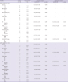
On univariate analysis, AI therapy, as well as PR and HER-2 expression, were significantly associated with better DFS in the pS6K1 positive group. In the pS6K1 negative group, tumor size, stage, as well as PR and HER-2 overexpression were significantly associated with better prognosis (Table 2). Variables that were significant in the univariate analysis were examined on the multivariate analysis according to pS6K1 status. On multivariate analysis, AI therapy remained a significant predictor of better DFS in patients with a positive pS6K1 expression status (HR, 0.37; 95% CI, 0.18–0.78, p=0.008, Table 2). In contrast, DFS was not affected by the type of endocrine therapy in pS6K1 negative patients (HR, 1.37; 95% CI, 0.38–4.91; p = 0.632). We performed a subgroup analysis according to age, as younger patients (age < 50) may have received SERMs more frequently than AIs, due to the unique reimbursement system of our country (Supplementary Table 1). The Kaplan-Meier estimates in patients with ages < 50 showed improved DFS in patients with pS6K1 positive tumors treated with AIs than in those treated with SERMs (p = 0.021, Supplementary Figure 1A). In patients with ages ≥ 50, the difference in DFS was not statistically significant. However, it seemed that patients with pS6K1 positive tumors had better outcomes when treated with AIs than with SERMs (HR, 0.48; 95% CI, 0.16–1.44; p = 0.188, Figure 1C)
Table 2
Univariate and multivariate analyses of DFS based on pS6K1 status using Cox proportional hazard regression
DISCUSSION
In this study, we found that AI therapy was associated with better DFS in patients with pS6K1 positive tumors, when compared to SERM therapy. To the best of our knowledge, this is the first study to evaluate pS6K1 expression status as a potential predictive marker for adjuvant AI therapy benefits in postmenopausal women.
The exact mechanism for the difference in prognosis between groups with different pS6K1 expression status is not yet fully understood. However, we suggest that the connection between adipogenesis, local estradiol levels, and the prognosis of hormone-sensitive breast cancer may help explain the predictive role of pS6K1 in postmenopausal women. Presumptively, the microenvironment of tumors expressing pS6K1 may have higher local estradiol levels than that of tumors not expressing pS6K1. As such, we suggest that pS6K1 may predict adjuvant AI therapy benefits in postmenopausal women. However, our results indicated that the pS6K1 expression status could be a more effective predictor in patients with ages < 50 than patients with ages ≥ 50.
The adipose tissue is a dynamic component of the endocrine system, wherein mature adipocytes occupy 90% of the volume, while adipocyte precursor cells become mature adipocytes through an interactive two-step process called adipogenesis. A precursor adipose stem cell differentiates into a preadipocyte (early adipogenesis) and then undergoes terminal differentiation to become a mature adipocyte (late adipogenesis). The mTOR pathway and its effector pS6K1, appear to be critical mediators in the development of obesity, defined as an excess of adipose tissue, and of adipogenesis [91011].
In postmenopausal patients with hormone-sensitive breast cancer, obesity remains a definite risk factor for poorer outcomes. The epidemiological link between obesity and hormone-sensitive breast cancer is well established [151617]. However, the molecular links between adipogenesis and breast cancer are not completely understood. A small number of the preclinical studies investigating the relationship between adipogenesis and local estradiol levels in the breast cancer microenvironment have suggested that the development of breast cancer might cause alterations in adipocyte differentiation in the breast [18]. Changes in adipogenesis may induce modifications of the aromatase promoters and thereby could increase local estrogen production [1920]. Others have reported that cytokines involved in adipogenesis may contribute to estradiol elevation by increasing aromatase expression and upregulating the ER in adipocytes and cancer cells [2122]. Because the oncogenic effect of estradiol on the ER is mediated via the PI3K/AKT/mTOR pathway in hormone-sensitive breast cancer [2324], increased pS6K1 levels might be representative of the active adipogenesis–breast cancer interaction. To the best of our knowledge, no studies have directly examined the relationship between pS6K1 expression and local tissue estrogen levels; such studies may have helped to confirm our hypothesis. However, in our cohort, there was no correlation between the expression status of pS6K1 and BMI (p = 0.550), a representative of systemic adiposity, which supports our hypothesis.
In clinical trials, estradiol depletion therapy with AIs has shown improved mortality and recurrence rates [2526]. However, artificial depletion of body circulating estrogens may simultaneously produce adverse effects that may lead to early discontinuation of the treatment. Despite excellent survival outcomes, the discontinuation rate of AIs ranges between 30%–70% and is largely dependent on the presence and severity of the adverse effects [27]. Moreover, symptoms responsible for discontinuation of the initial AI also lead to cessation of the second AI in nearly 80% of patients [28]. Because insufficient treatment duration is detrimental to survival outcomes, a SERM may be an alternative option in patients with AI-related concerns [429].
SERMs have shown similar potential as AIs in selected low-risk patients. A study analyzing prognostic risks in the patient cohorts of the Breast International Group (BIG) 1-98 trial found that a composite measure of typical low risk factors such as old age, negative lymph node status, tumor size < 2 cm, low tumor grade, HER-2 negative, and strong ER positivity predicts equivalent benefits from monotherapy with either letrozole or tamoxifen [30]. However, in most breast cancer patients, maximizing the survival benefits of adjuvant endocrine therapy while avoiding unwanted side effects, remains a dilemma. As such, the optimal treatment of these patients may be guided by the pS6K1 expression status. For instance, when adverse effects become an issue in endocrine therapy, AIs may be encouraged in postmenopausal patients with pS6K1 positive, hormone-sensitive tumors. Similarly, SERMs could be a more attractive alternative option in patients with pS6K1 negative tumors.
In our previous study, we showed that pS6K1 is a prognostic marker for poorer DFS in hormone-sensitive breast cancer patients [14]. The results of the current study revealed that the poorer prognosis of patients with pS6K1 positive tumors may be improved by using AIs providing estradiol depletion. Individual pS6K1 expression status evaluated by immunohistochemistry with readily available antibodies may serve as a simple and affordable predictive marker to select patients for AI therapy in the clinic.
The limitation of this study is that it was a single center-based retrospective study. Similarly, our results may be biased as the study did not include a matched cohort and hormonal treatment decision based on our unique reimbursement system may have contributed to the difference in results obtained for different age groups. Therefore, further prospective studies are needed to determine whether the results of our study could be applied to other populations. A median follow-up period of 44 months may be relatively short for hormone-sensitive breast cancer when considering cases of late recurrences. This was why only patients with node-positive disease were included in this study. In the adjuvant setting, a relatively large number of patients and a longer follow-up period would be necessary to show the differences in survival rates in a cohort of low-risk patients with endocrine sensitive disease. We plan to analyze long-term follow-up results, including those of patients with node-negative disease, in the future. Although the precise details of the underlying mechanisms need to be unraveled, elucidation of the function of pS6K1 in the estrogen-regulated pathway in association with adipogenesis may help predict patients that would derive greater benefit from AI therapy.
As such, positive pS6K1 expression was associated with better DFS in postmenopausal and ovarian function-suppressed patients with hormone-sensitive breast cancer treated with AIs than in those treated with SERMs. This indicates that pS6K1 expression status may be used as a predictive marker for adjuvant endocrine therapy in selected patients.
 XML Download