Journal List > J Korean Med Sci > v.35(7) > 1142674

Choi, Ha, Oh, Cho, Lee, Sohn, Cho, Koh, Do, Jeong, Ryoo, Kwon, Kim, Kim, Park, Lee, Lee, Lee, Park, and Kang: Clinical Guidance for Point-of-Care Ultrasound in the Emergency and Critical Care Areas after Implementing Insurance Coverage in Korea

Abstract

Point-of-care ultrasound (POCUS) is a useful tool that is widely used in the emergency and intensive care areas. In Korea, insurance coverage of ultrasound examination has been gradually expanding in accordance with measures to enhance Korean National Insurance Coverage since 2017 to 2021, and which will continue until 2021. Full coverage of health insurance for POCUS in the emergency and critical care areas was implemented in July 2019. The National Health Insurance Act classified POCUS as a single or multiple-targeted ultrasound examination (STU vs. MTU). STU scans are conducted of one organ at a time, while MTU includes scanning of multiple organs simultaneously to determine each clinical situation. POCUS can be performed even if a diagnostic ultrasound examination is conducted, based on the physician's decision. However, the Health Insurance Review and Assessment Service plans to monitor the prescription status of whether the POCUS and diagnostic ultrasound examinations are prescribed simultaneously and repeatedly. Additionally, MTU is allowed only in cases of trauma, cardiac arrest, shock, chest pain, and dyspnea and should be performed by a qualified physician. Although physicians should scan all parts of the chest, heart, and abdomen when they prescribe MTU, they are not required to record all findings in the medical record. Therefore, appropriate prescription, application, and recording of POCUS are needed to enhance the quality of patient care and avoid unnecessary cut of medical budget spending. The present article provides background and clinical guidance for POCUS based on the implementation of full health insurance coverage for POCUS that began in July 2019 in Korea.

Graphical Abstract

jkms-35-e54-abf001

INTRODUCTION

National health insurance in Korea began to cover emergency and critical care ultrasound examinations in July 2019.1 This could lead to more acute care by physicians who use more point-of-care ultrasound (POCUS) and reduce the financial burden on the patients. The Society of Emergency and Critical Care Imaging (SECCI) has summarized and provided recommendations on the desirable use of POCUS in emergency and critical care setting under the Korean Health Insurance System.
POCUS is an ultrasound examination that is performed at the bedside, and it is interpreted directly by the clinician.2 Therefore, POCUS is a powerful adjunct to clinical assessment. The certainty of the presumptive diagnosis that is derived from the medical history, and physical examination can be confirmed by the information provided using POCUS. Additionally, POCUS can be an effective tool for monitoring patients and for procedure guidance. Although the main purpose of POCUS is slightly different between intensivists and emergency physicians, the capability to recognize and resuscitate critically ill patients is a defining trait in both specialties.3 For critically ill patients, such as those with thoracoabdominal trauma, cardiac arrest, respiratory difficulty, chest pain, or shock, bedside multi-organ POCUS is now extensively used as an adjunct that provides many pieces of information to guide clinical decision-making during the initial and undifferentiated phases.4
In this article, we will review all types of clinically integrated, single/multi-focused, and single organ/multi-organ POCUS for emergency physicians and intensivists. All acute care physicians should use the following procedure: 1) stabilize the unstable patients; 2) make a differential diagnosis; and 3) monitor the patient to tailor the ongoing management. In each step, POCUS is a useful modality.

SCOPE OF PRACTICE FOR POCUS

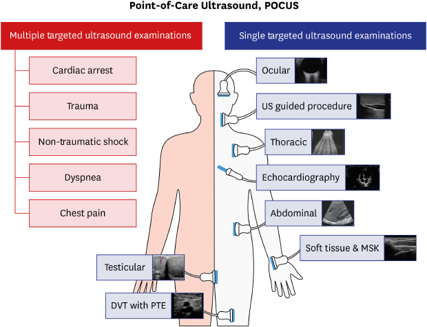
Emergency and intensive care physicians will perform basic ultrasonography using POCUS as designated by health insurance. Physicians also need to maintain a level of skills and attain the same scan proficiency.5 POCUS can be divided into single-targeted ultrasound examinations (STU, one or two scanning sites) and multiple-targeted ultrasound examinations (MTU) depending on the purpose. The key components of POCUS are considered and are described below (Fig. 1).
Fig. 1

Scope of practice for POCUS.

POCUS = point-of-care ultrasound, DVT = deep vein thrombosis, PTE = pulmonary thromboembolism, MSK = musculoskeletal.
jkms-35-e54-g001

SINGLE-TARGETED ULTRASOUND EXAMINATIONS (STU)

Point-of-care cardiac ultrasound

The point-of-care echocardiography (POCE) uses a limited number of standard echocardiography views to rapidly assess cardiac anatomy and function in the critically ill patient.6 Generally, the five standard views include the parasternal long-axis, parasternal short-axis, apical four-chamber, subcostal and suprasternal notch views that are scanned.7 The examination can be performed in a few minutes, and it is generally combined with other aspects of POCUS, such as thoracic ultrasound, Focused Assessment with Sonography for Trauma (FAST) and evaluation for abdominal aortic aneurysm (AAA) and deep vein thrombosis (DVT).8910
The POCE can be used to assess for pericardial effusion and tamponade, cardiac anatomy, pathology of the aortic root or the descending aorta, a global assessment of contractility and the hemodynamics for preload, cardiac activity and afterload.1112

Thoracic ultrasound

Thoracic ultrasound is performed on patients with dyspnea and pleuritic chest pain. Bedside thoracic ultrasound should be considered to be an image modality that can be used initially for patients who are in respiratory failure in the emergency or intensive care unit (ICU) because it is fast, safe, and informative compared with anteroposterior chest X-ray, and it demonstrates a similar performance compared with computed tomography.1314 Since Lichtenstein announced the Bedside Lung Ultrasound in Emergency protocol in 2008, numerous papers have shown the usefulness of lung ultrasound in many clinical scenarios.15 Thus, lung ultrasound is strongly recommended for detecting pneumothorax, alveolar-interstitial syndrome, pulmonary consolidation and pleural effusion.1617

Evaluation for DVT with pulmonary embolism

To differentiate the cause of pulmonary embolism, evaluation for DVT is needed. Hypotensive patients with pulmonary embolism have typical ultrasound findings, such as enlarged right ventricle, D-shaped left ventricle, and the presence of DVT. The POCUS DVT examination can be performed rapidly using the two-dimensional compression method without a Doppler study.1819

Abdominal ultrasound

The main applications in abdominal ultrasound include bowel, trauma, biliary, urinary tract, and AAA.20 A patient who has right upper quadrant (RUQ) pain in an emergency should be evaluated with abdominal POCUS to detect if the pain is of biliary origin. POCUS for acute cholecystitis is as accurate as ultrasound that is performed by a radiologist and cholescintigraphy.21 Use of POCUS for RUQ pain allows emergency physicians to determine if the origin of the pain is biliary colic. Sonographic tenderness in the gallbladder (GB) area, size, and presence of gallstone, GB wall thickening and pericholecystic fluid collection can be evaluated using abdominal POCUS, including common bile duct pathology. Renal POCUS helps to manage acute kidney injury and nephrolithiasis. In obstructive uropathy patients, hydronephrosis can be diagnosed with ultrasound.22 Emergency physicians can determine rapid and efficient disposition of patients with renal colic without radiation exposure using renal POCUS. Additionally, POCUS of the aorta includes measuring the shape and diameter of the abdominal aorta as well as inspection for a dissection flap. Emergency physicians can identify a ruptured AAA with a high sensitivity and specificity.23 In the emergency department, the primary concern for the women who are of their reproductive age and who have pelvic pain is ovarian torsion or ovarian cyst rupture. POCUS becomes the primary modality for its evaluation because it is difficult to diagnose based on symptoms and physical examination.24

Ultrasound-guided procedure

It is already well-known that ultrasound-guided procedures are accurate and safe methods.2526 It is especially very useful for critically ill patients in the emergency department and the ICU because physicians have to perform various procedures at the bedside.272829 The ultrasound-guided procedure can be used in central and peripheral venous catheterization, arterial catheterization, thoracentesis, paracentesis, pericardiocentesis, arthrocentesis, nerve block, confirming of endotracheal intubation, site marking for surgical airway, abscess drainage and foreign body removal.30

Testicular ultrasound

Acute scrotal pain accounts for approximately 0.5% of all patient complaints in the emergency department.31 In the emergency department, POCUS is used to diagnose testicular problems such as hydrocele, varicocele, testicular torsion, epididymitis, orchitis, and hernias, and also to evaluate scrotal trauma. Misdiagnosis of testicular torsion can lead to organ damage and compromise fertility, and testicular ultrasound is a useful rapid diagnostic tool.32

Soft tissue and musculoskeletal ultrasound

Soft tissue and musculoskeletal ultrasound is used to evaluate soft tissue infection, fracture, dislocation, muscle injury, foreign body, tendon injury, and joint effusions.3334353637 Ultrasound could diagnose some fractures including injuries that are difficult to diagnose using conventional radiography, such as scaphoid, sternum, occult rib, and some avulsion fractures. Additionally, ultrasound could identify radiolucent foreign materials such as glass or wood.33 Screening with ultrasound by an emergency physician could reduce costs, time requirements, and radiation exposure.

Ocular point-care-of ultrasound

Optic nerve sheath diameter is a well-known parameter of increased intracranial pressure.3839 Additionally, ocular POCUS would be useful for evaluation for traumatic eye injuries and visual disturbances. Emergency physicians can evaluate the anatomy of the eye via a dynamic POCUS study of the eyes, such as location of the lens, presence of a foreign body, and presence of vitreous detachment.4041

MULTIPLE TARGETED ULTRASOUND EXAMINATIONS (MTU)

Ultrasound in cardiac arrest

The transthoracic cardiac ultrasound in cardiac arrest patients can be performed within 10 seconds of stopping chest compressions for a pulse check, and this should not be associated with delays in chest compressions.42 If the heart cannot be visualized within seconds after the chest compressions are stopped, POCUS should be aborted. POCUS can be subsequently attempted using a different approach after five cycles of chest compressions. The subcostal approach is recommended first, and if the heart is not observed, the parasternal and apical approach can be the next option. 43 POCUS examinations of the thorax, abdomen, and inferior vena cava can be performed during chest compressions. In cardiac arrest patients, identifying the potential to reverse this condition cause might improve resuscitation outcomes. POCUS can provide guidance for better resuscitation using the information described below.44 We can differentiate true cardiac arrest rhythm45 such as pulseless electrical activity and false cardiac arrest rhythm using the sonographic cardiac activity in patients with no pulse. The prognosis for return of spontaneous circulation is improved when there is organized sonographic cardiac activity.46 If cardiac contractility is absent following a reasonable period of cardiopulmonary resuscitation, this indicates a limited probability for return of spontaneous circulation.45474849

POCUS for the evaluation of non-traumatic shock

POCUS provides real-time physiologic information in a patient with shock. Emergency and critical care physicians obtain guidance to differentiate between the type of shock, impact of management, and non-invasive monitoring by interpretation through POCUS. Shock has an estimated mortality rate of 36%–60%, depending on the type of shock.50 Rapid evaluations for etiology, monitoring, and rapid correction are important to decrease the mortality rate and improve patient safety. The categories of shock are quantitative shock (decreased flow: hypovolemic, cardiogenic, obstructive; decreased oxygen carrying capacity: anemia, hypoxemia) and distributive shock (sepsis, anaphylaxis). POCUS allows for rapid identification of the type of shock and it has been proposed as a first-line evaluation modality.51 In the assessment of patients with undifferentiated hypotension, hemodynamic assessment with POCUS for preload, cardiac function, and afterload has become an accepted diagnostic and monitoring tool.46525354 POCUS can be a risk reduction tool through its emergent diagnostic imaging capacity, shortening time to definitive therapy, and its use in therapeutics or in physiological monitoring.30

Ultrasound in chest and abdominal trauma

POCUS is useful as an initial diagnostic tool to identify free fluids in dependent locations within the peritoneal cavity in abdominal trauma patients.55 Immediate laparotomy is usually performed in unstable patients with intraperitoneal hemorrhage that is quickly identified by POCUS without CT scan.56 However, POCUS should not be used to replace more sensitive tests in patients with impaired abdominal or chest trauma symptoms. Multidetector computed tomography (MDCT) is highly sensitive for solid organ injury and intraperitoneal or intrathoracic bleeding and is the study of choice if the patients are hemodynamically stable. POCUS at the initial examination and resuscitation of trauma patients is known as FAST, which is a standard screening test that is performed on trauma patients.57 Evaluation of pericardial effusion, an intraperitoneal test, right and left flank and pelvic examinations are performed to locate free fluid in the abdominal cavity. In many cases, extended FAST (E-FAST) is performed to find a pneumothorax. FAST is most helpful when the results are positive, and when the time to definite treatment is decreased. FAST is generally performed during the “C” portion, which evaluates circulation and hemorrhage in a primary survey of Advanced Trauma Life Support. A low-frequency (2.5 to 5 MHz) curvilinear or phased array probe is appropriate. The standard sequence of FAST is pericardial, right flank (hepatorenal view or Morison's pouch), left flank (perisplenic view), pelvic (retrovesical views), and thoracic (pneumothorax and hemothorax evaluations).58 The use of MDCT in hemodynamically unstable patients should be determined by assessing the patient's degree of instability and the distance to the MDCT scanner.59 FAST is sensitive and specific in the determination of both traumatic pericardial effusion and intraperitoneal fluid that is indicative of injury, and thus, it effectively guides emergent surgical decision-making.60 However, limited sensitivity precludes the use of POCUS as a definitive test to rule out intraabdominal injury.61 Each FAST examination reflects the clinical situation at a single moment. The overall sensitivity of FAST for abdominal injury can be improved using serial ultrasound examinations. A second ultrasound examination improves sensitivity, negative predictive value, and accuracy for intra-abdominal injury.62 The pelvic region is the most dependent space in the supine patient, and therefore, it is an important site to look for free fluid. A determination of pneumothorax focuses on visualization of pleural sliding without relying on detecting free fluids.

Ultrasound in pediatrics

Pediatric patients are vulnerable to ionizing radiation, and therefore, ultrasound is the main diagnostic modality rather than computed tomography or radiography.6364 Ultrasonographic findings of viral pneumonia or bronchiolitis are B-line and/or subpleural consolidation, and those of bacterial pneumonia include B-lines and/or lung consolidation and sonographic air bronchogram.6566 In the emergency department, POCUS is used on pediatric patients with traumatic symptoms such as fractures, foreign bodies, or hip pain. The cause of hip effusion varies, but transient synovitis and septic arthritis are common in pediatric patients in the emergency department. Although ultrasound is not a confirmative diagnostic tool, it is important to confirm the presence of hip effusion with ultrasound. POCUS is also useful and valuable diagnostic imaging tool in children with nonspecific manifestations of intussusception.67

Ultrasound in the emergency department

Ultrasound in the emergency department has been used as an integral part of critical care and resuscitation in recent years, and the range of ultrasound use in the emergency department has greatly expanded. Emergency medicine (EM) physicians can use POCUS for critical care in the emergency department as follows: differentiating shock states; finding causes of respiratory failure; guiding fluid resuscitation; and accessing neurologic dysfunction.68 There are a variety of shock protocols using ultrasound but the use of the Rush protocol69 or the echo-guided life support protocol70 may be more efficient in our emergency department because it is feasible to evaluate the heart, the lung, and vessels in the abdomen together as a multi-directional assessment.6970
However, in addition to using ultrasound for critical care, ultrasound has a high use rate for diagnosis and procedures in various situations in which emergency department patients find themselves. EM physicians can use an extensive POCUS to examine abdominal, obstetric, testicular, musculoskeletal, and ocular lesions and to allow procedural guidance.
EM physicians should perform an exact evaluation using ultrasound, which can be used to make a definitive decision on the final disposition or discharge with a diagnosis that is determined by ultrasound. Therefore, assistance and advice should be sought from more experienced specialists if there are uncertain findings.3

Ultrasound in ICU

Ultrasound in the ICU is performed in an extensive field of diagnosis and management of critically ill patients who have a variety of disease features. As an extension of ultrasound in the emergency department, ultrasound in the ICU should focus on being comprehensive and complete rather than being rapid and essential.71 Comprehensive ultrasound studies to assess the full functional and structural status often revise the treatment strategy, and help to understand the disease status, and guide treatment. For example, routine comprehensive echocardiography in the general ICU revealed a clinically significant cardiac problem in one-third of patients with severe and critical cardiac disease that was previously unknown in 7.5% of patients.72 Because it is impossible for ICU physicians to perform comprehensive and professional ultrasound in diverse fields such as abdomen, thorax, and heart, ultrasound in the ICU has to be multidisciplinary and cooperative between clinical departments. ICU intensivists play a role in communication between the departments.
However, patients in the ICU are at high risk to experience disease exacerbation and develop complications. Therefore, clinicians in the ICU should be ready to perform POCUS to identify shock and assess fluid responsiveness.73 Additionally, procedures performed in the ICU such as insertion of an intravascular catheter, chest tube, effusion drainage from the thorax or abdomen, and tracheostomy can be performed under ultrasound guidance. Ultrasound guided procedures should always be considered first where available. ICU patients often experience clinical deterioration repeatedly from diverse causes, which requires comparison between current and previous ultrasound findings.

INSURANCE ISSUE

In Korea, the insurance coverage for ultrasound examination is gradually expanding in accordance with measures to enhance the national health insurance coverage, which began in 2017 and will continue until 2021, because the importance of ultrasound for on-site diagnosis has been emphasized. Full coverage of the insurance for POCUS in the emergency and critical care areas was implemented in July 2019.1 The National Health Insurance Act of Korea defined POCUS as a basic sonography tool that is required for differential diagnosis and treatment decisions in the emergency and critical care units. It is classified as STU (one or two scanning sites) and MTU (Table 1). The cost of POCUS can be covered by health insurance based on the conditions described below. First, it should be performed in the emergency department or ICU at the hospital. However, if the examination is performed in an emergency situation, the place where the examination can be conducted is not limited. Second, the physician should perform the ultrasound directly and record the findings (Table 2) in the patient's medical record (Tables 3 and 4). MTU examinations can be covered by insurance if the purpose (indication), personnel, and range of examination (Table 1) are all met. If these conditions are not met, it is regarded as a STU examination. The cost for payment of an ultrasound examination is different based on the Korean Triage and Acuity Scale in the emergency department. If the Korean Triage and Acuity Scale of the patients ranges from 1 to 3, 20% of the total cost will be charged to the patient. Additionally, 30% to 60% of the total cost will be charged to the patient depending on the types of medical institution (e.g., tertiary hospital, general hospital, or primary clinic).
Table 1

Classification of basic ultrasound examination: single targeted versus multiple targeted

jkms-35-e54-i001
Classification Single targeted ultrasound examinationa Multiple targeted ultrasound examination
Indication 1. Differential diagnosis of emergency situation, evaluation of acute lesion, decision of treatment plan, assistant for procedure (puncture and catheter insertion), etc. Trauma in chest, abdomen, and pelvis, cardiac arrest, shock, dyspnea, and chest pain
2. Ultrasound examination for fast decision making and accurate and safe treatment
Place Emergency department, intensive care unit Emergency department, intensive care, and other placesb
Equipment Ultrasound device should be installed in the emergency department or intensive care unit Ultrasound device should be installed in the emergency department or intensive care unit
Medical personnel Physician Resident physician or specialist in emergency medicine, specialist in critical care medicine, trauma surgery
Recording Medical record Medical record
Image storage No obligation No obligation
Scanning site Head and neck, chest, heart, abdomen (including urinary tract), genitalia, and extremities Chest, heart, and abdomen (including the urinary tract) should be examined. If necessary, the head and neck can be added
aExamination of one part or two or more parts among head and neck, chest, heart, abdomen (including urinary system), genitalia and extremities; bIf the examination is performed in the emergency situation, the place of examination is not limited.
Table 2

Essential component for medical records in single and multiple targeted ultrasound examination according to the scanning area

jkms-35-e54-i002
Site Findings
Chest Lung sliding
A-line
B-line
Presence of fluid in thoracic cavity
Heart Pericardial effusion
Left ventricular contractility (hypokinesia, normal, and hyperkinesia, etc.)
Right ventricle enlargement
Regional wall motion abnormality
Valve function
Aortic aneurysm rupture or aortic dissection
Intracardiac thrombosis
Abdomen Fluid collection in abdominal or pelvic cavity
Aortic aneurysm rupture or aortic dissection
Inferior vena cava dilatation or collapse
Gallbladder, bile duct
Head and neck Endotracheal intubation confirmation
Airway (trachea) confirmation
Internal jugular vein or subclavian vein confirmation
Extremity Deep vein thrombosis
Table 3

Examples of medical records for the single targeted ultrasound examination (point-of-care echocardiography)

jkms-35-e54-i003
Parameters Measurements
□ Left ventricle function □ Hyperdynamic □ Normal
□ Decreased □ Gross EF: ______ %
□ Left ventricle size □ Enlarged □ Normal
□ Right ventricle function □ Hyperdynamic □ Normal
□ Decreased
□ Right ventricle size □ Enlarged □ Normal
□ TR Max: ______ m/s
□ Pericardial effusion □ None/minimal □ Small ______ cm
□ Large without tamponade significance
□ Tamponade or impending tamponade state
□ IVC diameter □ Max. diameter ______ cm □ Variability: ______ %
□ Min. diameter ______ cm
□ Other diagnostic findings
EF = ejection fraction, TR = tricuspid regurgitation, IVC = inferior vena cava, Max. = maximum, Min. = minimum.
Table 4

Examples of medical records for the multiple targeted ultrasound examination (differential diagnosis for shock)

jkms-35-e54-i004
Organ Site Findings
Heart Pericardial effusion ○ Yes (Tamponade □ Yes □ No) ○ No
RV dilatation ○ Yes ○ No
Contractility ○ Hyperdynamic ○ Normal ○ Mild dysfunction
○ Moderate dysfunction ○ Severe dysfunction
LV kissing sign ○ Yes ○ No
Lung Loss of lung sliding ○ Yes (□ Rt. □ Lt. □ Lung point) ○ No
Pleural effusion ○ Yes (□ Rt. □ Lt.) ○ No
A-line ○ Yes (□ Rt. □ Lt.) ○ No
B-line ○ Yes (□ Rt. □ Lt.) ○ No
Abdomen Intraperitoneal fluid ○ Yes (□ Morrison's pouch □ Perisplenic □ Pelvic cavity) ○ No
Aorta Aneurysm ○ Yes (□ Thorax □ Abdomen) ○ No
Dissection ○ Yes (□ Thorax □ Abdomen) ○ No
IVC Positive pressure ventilation ○ Yes ○ No
Respiration response ○ > 50% collapse ○ < 50% collapse ○ Plethora
Thorax Compressibility
Rt. femoral vein ○ Yes ○ No
Lt. femoral vein ○ Yes ○ No
Rt. popliteal vein ○ Yes ○ No
Lt. popliteal vein ○ Yes ○ No
Impression
RV = right ventricle, LV = left ventricle, Rt. = right, Lt. = left, IVC = inferior vena cava.
There are several points to be noted regarding the type of medical institution. First, the insurance code system for diagnostic, guided, and special ultrasound examination has not been changed. Second, basic ultrasound examination classified as a STU or MTU examination can be performed whether or not the ultrasound is for diagnostic purpose based on the physician's decision. However, the Health Insurance Review and Assessment Service plans to monitor the prescription status on whether a basic or a diagnostic ultrasound examination is prescribed simultaneously and if it is repeated too many times. Third, the MTU examination is allowed only in cases of trauma, cardiac arrest, shock, chest pain, and dyspnea. In other cases, a STU examination over two or more sites should be prescribed, even though multiple sites will be scanned. Fourth, physicians should scan all parts of the chest, heart, abdomen, and pelvis when they prescribe an MTU examination. However, there are no obligations for recording all findings in the medical record. Recording one or more findings on each organ will be enough. Fifth, the cost of MTU can be covered on national health insurance when performed only by an authorized physician, such as: resident physician or specialist in EM, specialist in critical care medicine, or specialist in trauma surgery.

CONCLUSION

POCUS has become an important modality in the emergency and critical care setting. It will become an essential diagnostic component to treat patients appropriately and promptly or to make a differential diagnosis. More studies are needed to support various indications and critical situations in other fields.

Notes

Funding The Society of Emergency and Critical Care Imaging (SECCI) provided the funding needed to prepare and publish this manuscript in 2019.

Disclosure Prof. Bossng Kang is the chairman of the Society of Emergency and Critical Care Imaging (SECCI). The task force team (TFT) is organized in the SECCI to prepare full coverage of insurance for point-of-care ultrasound examinations in the emergency and critical care areas. All authors are the members of the TFT in the SECCI. The SECCI provided the fund needed to prepare and publish this manuscript. All authors declare that they have no competing interests.

Author Contributions

  • Conceptualization: Choi WJ, Ha YR, Oh JH.

  • Data curation: Choi WJ.

  • Funding acquisition: Choi WJ, Ha YR, Kang BS.

  • Resources: Choi WJ, Oh JH, Cho YS, Lee WW.

  • Software: Choi WJ, Lee WW.

  • Supervision: Ha YR, Oh JH, Kang BS.

  • Validation: Choi WJ, Ha YR, Cho YS, Kang BS.

  • Visualization: Choi WJ, Lee WW, Kang BS.

  • Writing - original draft: Choi WJ, Oh JH, Sohn YD, Cho GC, Koh CY, Do HH, Jeong WJ, Ryoo SM, Kwon JH, Kim HM, Kim SJ, Park CY, Lee JH,1 Lee JH,2 Lee DH, Park SY.

  • Writing - review & editing: Choi WJ, Ha YR, Oh JH.

Lee JH,1 Jin Hee Lee; Lee JH,2 Jae Hoon Lee.

References

1. Ministry of Health and Welfare (KR). Follow-up Measures to Strengthen Health Insurance Coverage. Sejong: Ministry of Health and Welfare;2019.
2. Dietrich CF, Goudie A, Chiorean L, Cui XW, Gilja OH, Dong Y, et al. Point of care ultrasound: a WFUMB position paper. Ultrasound Med Biol. 2017; 43(1):49–58.
crossref pmid
3. Whitson MR, Mayo PH. Ultrasonography in the emergency department. Crit Care. 2016; 20(1):227.
crossref pmid pmc
4. Ha YR, Toh HC. Clinically integrated multi-organ point-of-care ultrasound for undifferentiated respiratory difficulty, chest pain, or shock: a critical analytic review. J Intensive Care. 2016; 4:54.
crossref pmid pmc
5. Mayo PH, Beaulieu Y, Doelken P, Feller-Kopman D, Harrod C, Kaplan A, et al. American College of Chest Physicians/La Société de Réanimation de Langue Française statement on competence in critical care ultrasonography. Chest. 2009; 135(4):1050–1060.
crossref pmid
6. Walley PE, Walley KR, Goodgame B, Punjabi V, Sirounis D. A practical approach to goal-directed echocardiography in the critical care setting. Crit Care. 2014; 18(6):681.
crossref pmid pmc
7. Labovitz AJ, Noble VE, Bierig M, Goldstein SA, Jones R, Kort S, et al. Focused cardiac ultrasound in the emergent setting: a consensus statement of the American Society of Echocardiography and American College of Emergency Physicians. J Am Soc Echocardiogr. 2010; 23(12):1225–1230.
crossref pmid
8. Laursen CB, Sloth E, Lambrechtsen J, Lassen AT, Madsen PH, Henriksen DP, et al. Focused sonography of the heart, lungs, and deep veins identifies missed life-threatening conditions in admitted patients with acute respiratory symptoms. Chest. 2013; 144(6):1868–1875.
crossref pmid
9. Pivetta E, Goffi A, Lupia E, Tizzani M, Porrino G, Ferreri E, et al. Lung ultrasound-implemented diagnosis of acute decompensated heart failure in the ED: a SIMEU multicenter study. Chest. 2015; 148(1):202–210.
pmid
10. Shokoohi H, Boniface KS, Pourmand A, Liu YT, Davison DL, Hawkins KD, et al. Bedside ultrasound reduces diagnostic uncertainty and guides resuscitation in patients with undifferentiated hypotension. Crit Care Med. 2015; 43(12):2562–2569.
crossref pmid
11. Kovell LC, Ali MT, Hays AG, Metkus TS, Madrazo JA, Corretti MC, et al. Defining the role of point-of-care ultrasound in cardiovascular disease. Am J Cardiol. 2018; 122(8):1443–1450.
crossref pmid
12. Luong CL, Ong K, Kaila K, Pellikka PA, Gin K, Tsang TS. Focused cardiac ultrasonography: current applications and future directions. J Ultrasound Med. 2019; 38(4):865–876.
crossref pmid
13. Laursen CB, Sloth E, Lassen AT, Christensen R, Lambrechtsen J, Madsen PH, et al. Point-of-care ultrasonography in patients admitted with respiratory symptoms: a single-blind, randomised controlled trial. Lancet Respir Med. 2014; 2(8):638–646.
crossref pmid
14. Zanobetti M, Poggioni C, Pini R. Can chest ultrasonography replace standard chest radiography for evaluation of acute dyspnea in the ED? Chest. 2011; 139(5):1140–1147.
crossref pmid
15. Lichtenstein DA, Mezière GA. Relevance of lung ultrasound in the diagnosis of acute respiratory failure: the BLUE protocol. Chest. 2008; 134(1):117–125.
crossref
16. Volpicelli G, Elbarbary M, Blaivas M, Lichtenstein DA, Mathis G, Kirkpatrick AW, et al. International evidence-based recommendations for point-of-care lung ultrasound. Intensive Care Med. 2012; 38(4):577–591.
crossref pmid
17. Bouhemad B, Zhang M, Lu Q, Rouby JJ. Clinical review: bedside lung ultrasound in critical care practice. Crit Care. 2007; 11(1):205.
pmid pmc
18. Blaivas M, Lambert MJ, Harwood RA, Wood JP, Konicki J. Lower-extremity Doppler for deep venous thrombosis--can emergency physicians be accurate and fast? Acad Emerg Med. 2000; 7(2):120–126.
crossref pmid
19. Lensing AW, Doris CI, McGrath FP, Cogo A, Sabine MJ, Ginsberg J, et al. A comparison of compression ultrasound with color Doppler ultrasound for the diagnosis of symptomless postoperative deep vein thrombosis. Arch Intern Med. 1997; 157(7):765–768.
crossref pmid
20. Boniface KS, Calabrese KY. Intensive care ultrasound: IV. Abdominal ultrasound in critical care. Ann Am Thorac Soc. 2013; 10(6):713–724.
crossref pmid
21. Kendall JL, Shimp RJ. Performance and interpretation of focused right upper quadrant ultrasound by emergency physicians. J Emerg Med. 2001; 21(1):7–13.
crossref pmid
22. Noble VE, Brown DF. Renal ultrasound. Emerg Med Clin North Am. 2004; 22(3):641–659.
crossref pmid
23. Rubano E, Mehta N, Caputo W, Paladino L, Sinert R. Systematic review: emergency department bedside ultrasonography for diagnosing suspected abdominal aortic aneurysm. Acad Emerg Med. 2013; 20(2):128–138.
crossref pmid
24. Nicola R, Dogra V. Ultrasound: the triage tool in the emergency department: using ultrasound first. Br J Radiol. 2016; 89(1061):20150790.
crossref
25. Barr L, Hatch N, Roque PJ, Wu TS. Basic ultrasound-guided procedures. Crit Care Clin. 2014; 30(2):275–304. vi
crossref pmid
26. Bauman M, Braude D, Crandall C. Ultrasound-guidance vs. standard technique in difficult vascular access patients by ED technicians. Am J Emerg Med. 2009; 27(2):135–140.
crossref pmid
27. Karakitsos D, Labropoulos N, De Groot E, Patrianakos AP, Kouraklis G, Poularas J, et al. Real-time ultrasound-guided catheterisation of the internal jugular vein: a prospective comparison with the landmark technique in critical care patients. Crit Care. 2006; 10(6):R162.
28. Riley DC, Garcia S. Emergency department ultrasonography guided long-axis antecubital intravenous cannulation: How to do it. Crit Ultrasound J. 2012; 4(1):3.
crossref pmid pmc
29. Jo MS, Doh HH, Seo JS, Lee SH, Kim HY, Ha YR. A point of care ultrasound during catheterization of internal jugular vein; sonographic assessment of the venous excavation (SAVE) protocol. J Korean Soc Emerg Med. 2014; 25(3):291–298.
30. Ultrasound guidelines: emergency, point-of-care and clinical ultrasound guidelines in medicine. Ann Emerg Med. 2017; 69(5):e27–54.
31. Lewis AG, Bukowski TP, Jarvis PD, Wacksman J, Sheldon CA. Evaluation of acute scrotum in the emergency department. J Pediatr Surg. 1995; 30(2):277–281.
crossref pmid
32. Dunne PJ, O'Loughlin BS. Testicular torsion: time is the enemy. Aust N Z J Surg. 2000; 70(6):441–442.
crossref pmid
33. Situ-LaCasse E, Grieger RW, Crabbe S, Waterbrook AL, Friedman L, Adhikari S. Utility of point-of-care musculoskeletal ultrasound in the evaluation of emergency department musculoskeletal pathology. World J Emerg Med. 2018; 9(4):262–266.
crossref pmid pmc
34. Gottlieb M, Holladay D, Peksa GD. Point-of-care ultrasound for the diagnosis of shoulder dislocation: a systematic review and meta-analysis. Am J Emerg Med. 2019; 37(4):757–761.
crossref pmid
35. Lee SH, Yun SJ. Diagnostic performance of ultrasonography for detection of pediatric elbow fracture: a meta-analysis. Ann Emerg Med. 2019; 74(4):493–502.
crossref pmid
36. Nesselroade RD, Nickels LC. Ultrasound diagnosis of bilateral quadriceps tendon rupture after statin use. West J Emerg Med. 2010; 11(4):306–309.
pmid pmc
37. Freeman K, Dewitz A, Baker WE. Ultrasound-guided hip arthrocentesis in the ED. Am J Emerg Med. 2007; 25(1):80–86.
crossref pmid
38. Soldatos T, Karakitsos D, Chatzimichail K, Papathanasiou M, Gouliamos A, Karabinis A. Optic nerve sonography in the diagnostic evaluation of adult brain injury. Crit Care. 2008; 12(3):R67.
crossref
39. Kimberly HH, Shah S, Marill K, Noble V. Correlation of optic nerve sheath diameter with direct measurement of intracranial pressure. Acad Emerg Med. 2008; 15(2):201–204.
crossref pmid
40. Lahham S, Shniter I, Thompson M, Le D, Chadha T, Mailhot T, et al. Point-of-care ultrasonography in the diagnosis of retinal detachment, vitreous hemorrhage, and vitreous detachment in the emergency department. JAMA Netw Open. 2019; 2(4):e192162.
crossref
41. Gandhi K, Shyy W, Knight S, Teismann N. Point-of-care ultrasound for the evaluation of non-traumatic visual disturbances in the emergency department: the VIGMO protocol. Am J Emerg Med. 2019; 37(8):1547–1553.
crossref pmid
42. Huis In 't Veld MA, Allison MG, Bostick DS, Fisher KR, Goloubeva OG, Witting MD, et al. Ultrasound use during cardiopulmonary resuscitation is associated with delays in chest compressions. Resuscitation. 2017; 119:95–98.
pmid
43. Breitkreutz R, Walcher F, Seeger FH. Focused echocardiographic evaluation in resuscitation management: concept of an advanced life support-conformed algorithm. Crit Care Med. 2007; 35(5):Suppl. S150–S161.
crossref
44. Hernandez C, Shuler K, Hannan H, Sonyika C, Likourezos A, Marshall J. C.A.U.S.E.: cardiac arrest ultra-sound exam--a better approach to managing patients in primary non-arrhythmogenic cardiac arrest. Resuscitation. 2008; 76(2):198–206.
crossref pmid
45. Flato UA, Paiva EF, Carballo MT, Buehler AM, Marco R, Timerman A. Echocardiography for prognostication during the resuscitation of intensive care unit patients with non-shockable rhythm cardiac arrest. Resuscitation. 2015; 92:1–6.
crossref pmid
46. Breitkreutz R, Price S, Steiger HV, Seeger FH, Ilper H, Ackermann H, et al. Focused echocardiographic evaluation in life support and peri-resuscitation of emergency patients: a prospective trial. Resuscitation. 2010; 81(11):1527–1533.
crossref pmid
47. Blaivas M, Fox JC. Outcome in cardiac arrest patients found to have cardiac standstill on the bedside emergency department echocardiogram. Acad Emerg Med. 2001; 8(6):616–621.
crossref pmid
48. Cureton EL, Yeung LY, Kwan RO, Miraflor EJ, Sadjadi J, Price DD, et al. The heart of the matter: utility of ultrasound of cardiac activity during traumatic arrest. J Trauma Acute Care Surg. 2012; 73(1):102–110.
49. Gaspari R, Weekes A, Adhikari S, Noble VE, Nomura JT, Theodoro D, et al. Emergency department point-of-care ultrasound in out-of-hospital and in-ED cardiac arrest. Resuscitation. 2016; 109:33–39.
crossref pmid
50. Tintinalli JE, Stapczynski JS, Ma OJ, Cline DM, Meckler GD. Tintinalli's Emergency Medicine: a Comprehensive Study Guide. 8th ed. New York, NY: McGraw-Hill Education;2016.
51. Vincent JL, Fink MP, Abraham E, Kochanek P, Moore FA. Textbook of Critical Care. 7th ed. Philadelphia, PA: Elsevier;2017.
52. Kanji HD, McCallum J, Sirounis D, MacRedmond R, Moss R, Boyd JH. Limited echocardiography-guided therapy in subacute shock is associated with change in management and improved outcomes. J Crit Care. 2014; 29(5):700–705.
crossref pmid
53. Barbier C, Loubières Y, Schmit C, Hayon J, Ricôme JL, Jardin F, et al. Respiratory changes in inferior vena cava diameter are helpful in predicting fluid responsiveness in ventilated septic patients. Intensive Care Med. 2004; 30(9):1740–1746.
crossref pmid
54. Weekes AJ, Thacker G, Troha D, Johnson AK, Chanler-Berat J, Norton HJ, et al. Diagnostic accuracy of right ventricular dysfunction markers in normotensive emergency department patients with acute pulmonary embolism. Ann Emerg Med. 2016; 68(3):277–291.
pmid
55. Radwan MM, Abu-Zidan FM. Focussed assessment sonograph trauma (FAST) and CT scan in blunt abdominal trauma: surgeon's perspective. Afr Health Sci. 2006; 6(3):187–190.
pmid pmc
56. Henry S. Advanced Trauma Life Support. 10th ed. Chicago, IL: American College of Surgeons;2019.
57. Bahner D, Blaivas M, Cohen HL, Fox JC, Hoffenberg S, Kendall J, et al. AIUM practice guideline for the performance of the focused assessment with sonography for trauma (FAST) examination. J Ultrasound Med. 2008; 27(2):313–318.
pmid
58. Ha YR. Initial evaluation of a trauma patient using an ultrasound. J Korean Med Assoc. 2012; 55(11):1097–1112.
crossref
59. Fornell Pérez R. Focused assessment with sonography for trauma (FAST) versus multidetector computed tomography in hemodynamically unstable emergency patients. Radiologia. 2017; 59(6):531–534.
pmid
60. Tayal VS, Beatty MA, Marx JA, Tomaszewski CA, Thomason MH. FAST (focused assessment with sonography in trauma) accurate for cardiac and intraperitoneal injury in penetrating anterior chest trauma. J Ultrasound Med. 2004; 23(4):467–472.
crossref pmid
61. Shanmuganathan K, Mirvis SE, Sherbourne CD, Chiu WC, Rodriguez A. Hemoperitoneum as the sole indicator of abdominal visceral injuries: a potential limitation of screening abdominal US for trauma. Radiology. 1999; 212(2):423–430.
crossref pmid
62. Blackbourne LH, Soffer D, McKenney M, Amortegui J, Schulman CI, Crookes B, et al. Secondary ultrasound examination increases the sensitivity of the FAST exam in blunt trauma. J Trauma. 2004; 57(5):934–938.
crossref
63. Pearce MS, Salotti JA, Little MP, McHugh K, Lee C, Kim KP, et al. Radiation exposure from CT scans in childhood and subsequent risk of leukaemia and brain tumours: a retrospective cohort study. Lancet. 2012; 380(9840):499–505.
crossref pmid pmc
64. Brenner DJ, Hall EJ. Computed tomography--an increasing source of radiation exposure. N Engl J Med. 2007; 357(22):2277–2284.
pmid
65. Tsung JW, Kessler DO, Shah VP. Prospective application of clinician-performed lung ultrasonography during the 2009 H1N1 influenza A pandemic: distinguishing viral from bacterial pneumonia. Crit Ultrasound J. 2012; 4(1):16.
crossref pmid pmc
66. Vieira RL, Hsu D, Nagler J, Chen L, Gallagher R, Levy JA, et al. Pediatric emergency medicine fellow training in ultrasound: consensus educational guidelines. Acad Emerg Med. 2013; 20(3):300–306.
crossref pmid
67. Han SM, Kim JH, Lee JS. Point-of-care ultrasound may reduce emergency department length of stay in children with nonspecific manifestations of intussusception. Pediatr Emerg Med J. 2016; 3(1):15–23.
crossref
68. Peterson D, Arntfield RT. Critical care ultrasonography. Emerg Med Clin North Am. 2014; 32(4):907–926.
crossref pmid
69. Perera P, Mailhot T, Riley D, Mandavia D. The RUSH exam: Rapid Ultrasound in SHock in the evaluation of the critically lll. Emerg Med Clin North Am. 2010; 28(1):29–56.
crossref pmid
70. Lanctôt JF, Valois M, Beaulieu Y. EGLS: echo-guided life support. Crit Ultrasound J. 2011; 3(3):123–129.
crossref
71. Frankel HL, Kirkpatrick AW, Elbarbary M, Blaivas M, Desai H, Evans D, et al. Guidelines for the appropriate use of bedside general and cardiac ultrasonography in the evaluation of critically ill patients—part I: general ultrasonography. Crit Care Med. 2015; 43(11):2479–2502.
pmid
72. Marcelino PA, Marum SM, Fernandes AP, Germano N, Lopes MG. Routine transthoracic echocardiography in a general intensive care unit: an 18 month survey in 704 patients. Eur J Intern Med. 2009; 20(3):e37–42.
73. Levitov A, Frankel HL, Blaivas M, Kirkpatrick AW, Su E, Evans D, et al. Guidelines for the appropriate use of bedside general and cardiac ultrasonography in the evaluation of critically ill patients—part II: cardiac ultrasonography. Crit Care Med. 2016; 44(6):1206–1227.
pmid
TOOLS
ORCID iDs

Wook Jin Choi
https://orcid.org/0000-0001-8779-0081

Young-Rock Ha
https://orcid.org/0000-0002-4889-6550

Je Hyeok Oh
https://orcid.org/0000-0002-5211-3838

Young Soon Cho
https://orcid.org/0000-0002-6843-9517

Won Woong Lee
https://orcid.org/0000-0003-0248-9172

You Dong Sohn
https://orcid.org/0000-0001-8789-0090

Gyu Chong Cho
https://orcid.org/0000-0001-9228-3674

Chan-Young Koh
https://orcid.org/0000-0003-0967-8208

Han Ho Do
https://orcid.org/0000-0001-6950-9137

Won Joon Jeong
https://orcid.org/0000-0002-6320-230X

Seung Mok Ryoo
https://orcid.org/0000-0002-2436-3311

Jae Hyun Kwon
https://orcid.org/0000-0003-3927-380X

Hyung Min Kim
https://orcid.org/0000-0002-1738-0922

Su Jin Kim
https://orcid.org/0000-0003-3769-9647

Chan Yong Park
https://orcid.org/0000-0002-5111-3270

Jin Hee Lee
https://orcid.org/0000-0002-2385-2834

Jae Hoon Lee
https://orcid.org/0000-0002-5815-6994

Dong Hyun Lee
https://orcid.org/0000-0001-6253-3396

Sin-Youl Park
https://orcid.org/0000-0003-4005-1956

Bo Seung Kang
https://orcid.org/0000-0002-0792-0198

Similar articles